Original Research Article
Multidrug resistant, extensively drug resistant and pan drug resistant
gram negative bacteria at a tertiary care centre in Bhubaneswar
Dipti Pattnaik
1, Subhra Snigdha Panda
1*, Nipa Singh
1, Smrutilata Sahoo
1,
Ipsa Mohapatra
2, Jagadananda Jena
1INTRODUCTION
Discovery of antimicrobials is one of the most important milestones achieved in the history of medical science. They behaved as wonder drugs in the treatment of infectious diseases till the advent of antimicrobial resistance. The burden of drug resistance has increased so
much that it has emerged as a major challenge of 21st century in health care system.1 In developing countries like India the problem of antibiotic resistance is quite higher compared to developed countries.2 Recently, the emergence of multi drug resistant strains of bacteria has been considered as a major public health problem by World Health Organisation (WHO).3 Again the
ABSTRACT
Background: Multidrug resistance has emerged as a challenge in health care settings. Again increasing prevalence of
multidrug resistant (MDR), extensively drug resistant (XDR) and pan drug resistant (PDR) gram negative bacteria is making the condition more critical because of limited options of antibiotics, increasing morbidity, mortality and hospital stay of the patients. The present study is carried out with an aim to estimate the prevalence of MDR, XDR, PDR gram negative bacteria in a tertiary care hospital.
Methods: Total of 912 gram negative bacterial isolates obtained from various samples of indoor patients in a tertiary
care hospital, were studied over a period of six months. The bacteria were identified by conventional methods. Antibiotic sensitivity testing was done by Kirby Bauer disc diffusion method. Minimum inhibitory concentration (MIC) of antibiotics for the resistant isolates were detected by Vitek-2 automated method. MDR, XDR and PDR were determined according to the definitions suggested by European Centre for Disease Prevention and Control (ECDC), and Centers for Disease Control and Prevention (CDC). Prevalence of extended spectrum beta lactamase (ESBL) producers was estimated.
Results: Out of 912 isolates, prevalence of MDR, XDR and PDR were 66.12%, 34.32% and 0.98% respectively.
Prevalence of MDR and XDR were higher in ICUs than clinical wards (p<0.0001). Prevalence of ESBL producers was 48.4%.
Conclusions: The study highlights increased prevalence of multidrug resistant and extensively drug resistant strains
in our hospital. Stringent surveillance, proper implementation of hospital infection control practices and antimicrobial stewardship will help in limiting the emergence and spread of drug resistant strains.
Keywords: Gram negative bacilli, Multi drug resistant, Extensively drug resistant, Pan drug resistant
1
Department of Microbiology, 2Department of Community Medicine, Kalinga Institute of Medical Sciences, Bhubaneswar, Odisha, India
Received: 05 January 2019
Revised: 19 January 2019
Accepted: 21 January 2019
*Correspondence:
Dr. Subhra Snigdha Panda, E-mail: [email protected]
Copyright: © the author(s), publisher and licensee Medip Academy. This is an open-access article distributed under
the terms of the Creative Commons Attribution Non-Commercial License, which permits unrestricted non-commercial use, distribution, and reproduction in any medium, provided the original work is properly cited.
prevalence of multidrug resistant gram negative bacilli is increasing significantly.4 Over last two decades the infections caused by these multidrug resistant gram negative strains have increased morbidity, mortality and the duration of hospital stay, particularly in developing countries.5 Among gram negative bacteria, members of
Enterobacteriaceae such as Escherichia coli, Klebsiella spp., Enterobacter spp., Proteus spp. and among non lactose fermenters, Pseudomonas spp., Acinetobacter
spp., have been identified as predominant cause of multidrug resistant bacterial infections.6-8 European Centre for Disease Control (ECDC) and Centre for Disease Control and Prevention (CDC), Atlanta have proposed standardised definitions for the multidrug-resistant (MDR), extensively drug multidrug-resistant (XDR), and pan drug resistant (PDR) bacteria.9 Multidrug resistant (MDR) was defined as acquired non susceptibility to at least one agent in three or more antimicrobial categories as per guidelines. Extensively drug resistant (XDR) was defined as non susceptibility to at least one agent in all but two or fewer antimicrobial categories listed and pandrug resistant (PDR) was defined as non susceptibility to all agents in all antimicrobial categories.9
Various studies have found out that there are differences in resistance pattern of the bacteria depending on different regions even different institutions.10,11 Hence this study was carried out with an aim to know the prevalence of MDR, XDR and PDR gram negative bacteria at a tertiary care hospital in Odisha and to help implementing an effective antibiotic policy.
METHODS
This was a cross sectional study carried out between 1st of January to 30th of June 2017 at Kalinga Institute of Medical Sciences (KIMS), a tertiary care teaching hospital in Bhubaneswar, Odisha.
Inclusion and exclusion criteria
A total of 912 non repetitive and consecutive bacterial strains isolated from various samples (respiratory samples, urine, blood, body fluids, wound swab, pus, tissue samples etc.) of indoor patient of wards and different ICUs were included in the study. Clinical samples from outdoor patients were not included.
The bacterial strains were identified by conventional phenotypic methods, using gram’s staining and different biochemical reactions.12 Antibiotic susceptibility testing was done by Kirby Bauer disc diffusion method on Mueller-Hinton agar and results were interpreted as per CLSI guidelines.13,14 Dehydrated media and antibiotic discs procured from Hi‑Media (Mumbai) were used. In case of bacterial isolates those showed resistance to all antibiotics by disc diffusion method, minimum inhibitory concentration (MIC) value was determined for antibiotics like colistin, polymixin B, tigecycline by automated method (Vitek2 compact; bioMerieux, France). Intrinsic
resistance of bacteria for concerned antibiotics was considered while detecting resistant strains. MDR, XDR and PDR bacterial strains were detected according to guidelines proposed by ECDC and CDC.9 Extended spectrum beta lactamase (ESBL) producing strains were detected amongst members of Enterobacteriaceae by disc diffusion method using ceftazidime (30µg), cefotaxime (30µg), ceftazidime plus clavulanic acid ( 30/10µg) and cefotaxime plus clavulanic acid (30/10µg) combination. For a bacterial isolate, an increase in zone diameter by ≥5 mm with ceftazidime and clavulanic acid combination disc in comparison to ceftazidime disc alone and or ≥5 mm increase in the zone diameter of cefotaxime/ clavulanic acid disc and that of cefotaxime disc alone was considered to be phenotypic confirmatory test for that strain.14 For quality control purpose Escherichia coli
ATCC 25922, Klebsiella pneumonae ATCC 700603,
Pseudomonas aeruginosa ATCC 27853 were taken as
control strains.14
The data was entered in Microsoft excel 2010 version and compared. Statistical significance was calculated using chi-square test. P<0.05 was considered to be statistically significant.
RESULTS
A total number of 912 gram negative bacterial isolates isolated from 784 various samples were studied. Out of these, 375 isolates were obtained from samples of intensive care units and 537 isolated were from samples of other clinical wards. Maximum number of isolates were obtained from urine samples (387/42.43%), followed by sputum (135/14.8%) and tracheal secretions (99/10.85%) (Table 1).
Table 1: Number of gram negative bacteria isolated from various clinical samples.
Clinical samples (number) No. of GNB isolated N (%)
Urine (362) 387 (42.43)
Sputum (112) 135 (14.8)
Tracheal secretion (78) 99 (10.85)
Pus (74) 96 (10.53)
Blood (76) 96 (10.53)
Body fluid (6) 9 (0.99)
Stool (24) 24 (2.63)
Others (52) 66 (7.24)
Total sample (784) n=912
Amongst 912 gram negative bacterial isolates most common was Escherichiacoli (267/29.3%), followed by
Table 2: Distribution of bacterial isolates in clinical samples (total no. of isolates n=912).
Sample E. coli Klebsiella sp.
Enterobacter sp.
Citrobacter sp.
Proteus sp.
Pseudomonas sp.
Acinetobacter
sp. Others
Urine 201 81 9 9 24 24 21 18
Pus 9 33 6 3 --- 15 21 9
Blood 9 30 --- --- ---- 9 18 30
Tracheal
secretion 3 27 --- --- 3 21 39 6
Body fluid 3 6 --- --- --- --- ---- ---
Stool 24 --- --- --- --- --- --- ---
Sputum 3 75 3 --- --- 27 27 ---
Others 15 3 6 --- --- 21 15 6
Total no. (%)
267 (29.3)
255 (27.9)
24 (2.63)
12 (1.31)
27 (2.96)
117 (12.82)
141 (15.5)
69 (7.6)
ESBL producers (%)
154 (57.6) 111 (43.5) 9 (37.5) 3 (25) 6 (22.2) --- --- 11 (16)
Table 3: Distribution of MDR, XDR and PDR isolates in ICUs and clinical wards.
Types of isolates
ICUs (total no. of isolates = 375) N (%)
Clinical wards (total no. of isolates = 537) N (%)
Total P value
MDR 281 (75 ) 322 (60 ) 603 <0.0001
XDR 171 (45.6 ) 142 (26.4 ) 313 <0.0001
PDR 6 (1.6 ) 3 (0.56 ) 9 0.22
Table 4: Prevalence of MDR, XDR& PDR strains among gram negative bacterial isolates.
Bacteria Total numbers MDR No.
XDR No.
PDR No.
N (%) N (%) N (%)
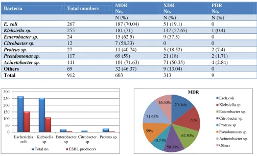
E. coli 267 187 (70.04) 51 (19.1) 0
Klebsiella sp. 255 181 (71) 147 (57.65) 1 (0.4)
Enterobacter sp. 24 15 (62.5) 9 (37.5) 0
Citrobacter sp. 12 7 (58.33) 0 0
Proteus sp. 27 11 (40.74) 5 (18.52) 2 (7.4)
Pseudomonas sp. 117 69 (59) 21 (18) 2 (1.71)
Acinetobacter sp. 141 101 (71.63) 71 (50.35) 4 (2.84)
Others 69 32 (46.37) 9 (13.04) 0
Total 912 603 313 9
Figure 1: Distribution of ESBL producers among bacterial isolates.
Figure 2: Prevalence of MDR strains among bacterial isolates.
0 50 100 150 200 250 300
Escherichia coli
Klebsiella sp.
Enterobacter sp
Citrobacter sp
Proteus sp.
Total no. ESBL producers
70.04%
71%
62.50% 58.33% 40.74% 59% 71.63%
46.40%
MDR
Out of 912 bacterial isolates, 603(66.12%) were MDR strains, 313 (34.32%) were XDR and 9 (0.98%) were PDR strains. Number of MDR, XDR and PDR strains in intensive care units (ICUs) were 281 (75%), 171 (45.6%) and 6 (1.6%) respectively whereas the same in other clinical wards were 322 (60%), 142 (26.4%) and 3 (0.56%) respectively (Table 3).
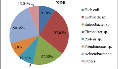
Figure 3: Prevalence of XDR strains among bacterial isolates.
In the present study most frequent MDR bacteria was
Acinetobacter spp. 71.63% (101/141) followed by
Klebsiella spp. 71% (181/255) and E. coli 70.04% (187/267) (Figure 2). Among XDR strains most common
was Klebsiella spp. 57.65% (147/255) followed by
Acinetobacter spp. 50.35% (71/141) Figure 3. Total of 9
PDR gram negative bacteria were observed in our study which consists of 4 (2.84%) Acinetobacter spp. strains, 2 (1.71%) Pseudomonas spp. strains (1.71%), 2 (7.4%)
Proteus spp. strains, and 1 (0.4%) Klebsiella spp. strains (Table 4).
DISCUSSION
Increasing incidence of infections by MDR bacteria or superbugs has become a matter of grave concern in health care sectors as it accelerates both morbidity and mortality of the patients. Inappropriate use of antibiotics, rising number of immunocompromised patients and poor infection control practices contribute to emergence and further spread of MDR strains.15 According to a WHO survey, expanding global trade and tourism also help in spreading of drug resistant bacteria between continents. In addition lower pace of development of newer antibiotics in comparison to rapidly emerging MDR strains is making the condition more critical.16,17 Several studies have indicated increased incidence of multidrug resistance gram negative bacterial infections in hospitalised patients for last few years.18,19 For last few decades more emphasis is given on MDR gram positive bacteria compared to gram negative bacteria to combat the problem of drug resistance.17 This study was carried out with an aim to quantify the burden of MDR, XDR and PDR gram negative bacteria in our hospital.
In the present study urine was the predominant sample from which maximum number of gram negative bacteria (387/42.43%) were isolated (Table 1). Similar findings was seen in the study by Agyepong et al where maximum number of isolates (94/47%) were from urine sample.20 It could be due to the fact that urine was the major sample in our study.
According to our study, most frequent gram negative bacteria was E. coli (267/29.3%) followed by Klebsiella
spp. (255/27.9%) and Acinetobacter spp. (141/15.5%) (Table 2). Our finding correlates with some other studies like Agyepong et al (24.5%), Basak et al (35%), Folgori et al (67.6%), where most common gram negative bacteria isolated was E.coli.20-22
ESBL producing bacteria often contain MDR strains by exhibiting resistance to other classes of antibiotics in addition to penicillins, first, second and third generation cephalosporins and aztreonam.7,23 Prevalence of ESBL producers in our study was 48.4% (283/585). Different authors throughout India showed varying prevalence of ESBL producers like Basak et al (18.4%), Rodrigues et al (53%), Singhal et al (64%), Mathur et al (68%) etc.21,24-26
In this study most common ESBL producer was E. coli
(154/57.6%) followed by Klebsiella spp. (111/43.5%) (Table 2/Figure 1). Our finding correlated well with other studies like Rodrigues (65.8%), Dalela (73.5%) where
Escherichia coli was the most common ESBL
producer.24,27
The prevalence of MDR, XDR and PDR strains in our study are 66.12% (603/912), 34.32% (313/912) and 0.98% (9/912) respectively. Study by Basak et al showed 33.5% MDR strains, 12.1% XDR and no PDR strains which is lower than our findings.21 Oliveria et al observed MDR prevalence to be 36%.10 Bhatt et al in their study found out prevalence of XDR and PDR strains as 8.1% (101/1240) and 0.9%(11/1240) respectively.28 Adrizain et al showed MDR and XDR prevalence as 28.7% (86/299) and 4.7% (14/299) among paediatric patients from blood culture pathogens.29
In the study done by Agyepong et al in Ghana, prevalence of MDR strains was 89.5% which is higher than prevalence of our study.20 In the study by Bajpai et al percentage of MDR strains (75.8%) and PDR strains (2.1%) among uropathogens are higher than our observations for MDR and PDR strains whereas percentage of XDR strains (12%) is lower than our finding.30
The higher prevalence in our study could be due to the fact that our institute is a tertiary care hospital where most of the patients are referred ones having prior exposure of antibiotics which could be a predictor factor for development of multi drug resistance.31
19.10%
57.65%
37.50% 0
18.52% 18% 50.35%
13.04% XDR
Percentage of isolation of MDR and XDR strains from ICU in our study were 75% (281/375) and 45.6% (171/375) which are higher than prevalence of MDR strains (60%, 322/537) and XDR strains (26.4%, 142/537) isolated from different clinical wards. This differences were also found to be statistically significant with p<0.0001. Similar finding observed by Basak et al where percentage of MDR (52.2%) and XDR isolates (18.8%) from ICUs were more than MDR isolates (43.3%) and XDR isolates (16.2%) obtained from clinical wards.21 Oliveria et al also observed higher MDR isolation rate (67%) from ICUs than wards (33%).10 Higher prevalence of drug resistant strains in ICUs may be due to frequent exposure of critically ill patients to antibiotics, longer stay and interventional instrumentations such as use of ventilators, central lines, urinary catheters.31,32
Prevalence of PDR in ICU (1.6%, 6/37) is more than that of clinical wards (0.56%, 3/537). This difference is not found to be statistically significant which could not be evaluated due to less number of PDR isolates (9) in our study.
Amongst all the MDR strains most common was
Acinetobacter spp. 71.63% (101/141) followed by
Klebsiella spp. 71% (181/255) and E. coli 70.04% (187/267). (Table 4, Figure 2). In the study by Agyepong et al most common MDR bacteria were Acinetobacter
spp. (100%) and Pseudomonas sp. (100%) whereas Basak et al (31.6%) and Tohamy et al (38.6%) in their studies showed E. coli as the most common MDR strain.20,21,33
Most common XDR strains were Klebsiella spp 57.65% (147/255) followed by Acinetobacter spp. 50.35% (71/141) (Table 4/Figure 3). In the study of Basak et al
Pseudomonas spp. (32.2%) were the most common XDR
strains.21
Out of total 9 PDR (0.98%) strains, four were
Acinetobacter spp., two strains of Pseudomonas spp., two strains of Proteus spp. and one strain of Klebsiella spp. Our finding is similar to that of Bhatt et al where the prevalence of PDR was 0.9%.28 In the study of Bajpai et al the prevalence of PDR (2.1%) is higher than our observation.30 All these isolates were resistant to all antibiotics used as per CDC and ECDC recommendations including colistin, polymixin B and tigecycline. Proteus
spp. strains were not tested against colistin and tigecycline as they are intrinsically resistant to these antibiotics. Resistant to colistin is the matter of grave concern as it is used as last resort of treatment in gram negative bacterial infections.28 However prevalence of PDR bacteria was still low, giving a hope for adopting appropriate measures to check the emergence and spread of drug resistant strains. We have not tested the isolates for fosfomycin which could be a limitation in our study.
CONCLUSION
This study highlights higher prevalence of MDR (66.12%) with a considerable number of XDR bacteria (34.32%) in our set up which is alarming. It is recommended to have knowledge of own epidemiology of drug resistant bacteria in each hospital settings by continuous surveillance which will help in implementing proper hospital infection control measures and prescribing proper antibiotics. Overuse or misuse of antibiotics must be checked through antimicrobial stewardship which is the need of hour.
ACKNOWLEDGEMENTS
We are thankful to the Principal, the head of department, all the faculty members and the laboratory staff of Microbiology department who have helped us in completing this research work.
Funding: No funding sources Conflict of interest: None declared Ethical approval: Not required
REFERENCES
1. Odonkor ST, Addo KK. Bacteria resistance to antibiotics: recent trends and challenges. Int J Biol Med Res. 2011;2(4):1204-10.
2. Kumar S, Adithan C, Harish B, Roy G, Malini A, Sujatha S. Antimicrobial resistance in India: A review. J Natural Sci Biol Med. 2013;4(2):286. 3. The Lancet. Urgently needed: new antibiotics.
Lancet. 2009;374(9705):1868.
4. D’Agata EMC. Rapidly rising prevalence of nosocomial multidrug resistant, gram-negative bacilli: a 9-year surveillance study. Infect Control Hosp Epidemiol. 2004;25:842–6.
5. Singh N, Manchanda V. Control of multidrug-resistant gram-negative bacteria in low-and middle-income countries—high impact interventions without much resources. Clin Microbiol Infect. 2017;23:216–8.
6. De Angelis G, D’Inzeo T, Fiori B, Spanu T, Sganga G. Burden of antibiotic resistant gram negative bacterial infections: evidence and limits. J Med Microbiol Diagn. 2014;3:132–8.
7. Rossolini GM, Mantengoli E, Docquier J, Musmanno RA, Coratza G. Epidemiology of infections caused by multiresistant gram-negatives: ESBLs, MBLs, panresistant strains. New Microbiol. 2007;30:332.
8. Oduro-Mensah D, Obeng-Nkrumah N, Bonney EY, Oduro-Mensah E, TwumDanso K, Osei YD, et al. Genetic characterization of TEM-type ESBL associated antibacterial resistance in Enterobacteriaceae in a tertiary hospital in Ghana. Ann Clin Microbiol Antimicrob. 2016;15:29–38. 9. Magiorakos AP, Srinivasan A, Careyetal RB.
pandrug- resistant bacteria: an international expert proposal for interim standard definitions for acquired resistance,” Clinical Microbiology and Infection. 2012;18(3):268–81.
10. Oliveira VDC, Rubio FG, Almeida MTG, Nogueira, MCL, Pignatari ACC. Trends of 9,416 multidrug-resistant Gram-negative bacteria. Revista da Associação Médica Brasileira, 2015;61(3):244–9. 11. Raghunath D. Emerging antibiotic resistance in
bacteria with special reference to India. J Biosci. 2008;33:593-603.
12. Washington Jr CW, Stephen DA, William MJ. Koneman’s Color Atlas and Textbook of Diagnostic Microbiology. 7th edition. USA, Philadelphia, Pa: Lippincott Williams & Wilkins; 2016.
13. Bauer AW, Kirby QMM, Sherns JC, Turik M. Antibiotic susceptibility testing by standardized single disk method. Am J Clin Path. 1966;45:493-6. 14. Clinical Laboratory Standards Institute.
Performance standard for antimicrobial susceptibility testing. 23rd Information supplement. NCCLS Document M100‑S23; 2013.
15. Tanwar J, Das S, Fatima Z, Hameed S. Multidrug Resistance: An Emerging Crisis. Interdisciplinary Perspectives on Infectious Diseases. 2014;2014:1–7. 16. Souli M, Galani I, Giamarellou H. Emergence of extensively drug‑resistant and pan drug‑resistant gram‑negative bacilli in Europe. Euro Surveill. 2008;13.
17. Sharma J, Gulati N, Chander J. Drug resistant urinary isolates of Pseudomonas aeruginosa and Acinetobacter species. J Glob Infect Dis. 2010;2:315‑7.
18. Gaynes R, Edwards JR. National Nosocomial Infections Surveillance System. Overview of nosocomial infections caused by gram-nagative bacilli. Clin Infect Dis. 2005;41(6):848-54.
19. National Nosocomial Infections Surveillance (NNIS) System Report, data summary from January 1992 through June 2004, issued October 2004. Am J Infect Control. 2004;32(8):470-85.
20. Agyepong N, Govinden U, Owusu-Ofori A, Essack SY. Multidrug-resistant gram-negative bacterial infections in a teaching hospital in Ghana. Antimicrobial Resistance Infection Control. 2018;7(1).
21. Basak S, Singh P, Rajurkar M. Multidrug Resistant and Extensively Drug Resistant Bacteria: A Study. J Pathogens. 2016;2016:1–5.
22. Folgori L, Livadiotti S, Carletti M, Bielicki J, Pontrelli G. Epidemiology and clinical outcomes of multidrug-resistant, gram-negative bloodstream infections in a european tertiary pediatric hospital during a 12-month period. Pediatr Infect Dis J. 2014;33(9):929-32.
23. Singh N, Pattnaik D, Neogi DK, Jena J, Mallick B. Prevalence of ESBL in Escherichia coli Isolates
Among ICU Patients in a Tertiary Care Hospital. J Clin Diagnos Res. 2016;10(9):19-22.
24. Rodrigues C, Joshi P, Jani SH, Alphonse M, Radhakrishnan R, Mehta A. Detection of β-lactamases in nosocomial gram negative clinical isolates. Indian J Med Microbiol. 2004;22(4):247-50.
25. Singhal S, Mathur T, Khan S, Upadhyay DJ, Chugh S, Gaind R, et al. Evaluation of the methods for AmpC β-lactamase in gram negative clinical isolates from tertiary care hospitals. Indian J Med Microbiol. 2005;23(2):120-4.
26. Mathur P, Kapil A, Das B, Dhawan B. Prevalence of extended spectrum β-lactamase producing gram negative bacteria in a tertiary care hospital. Indian J Med Res. 2002;115:153-7.
27. Dalela G. Prevalence of Extended Spectrum Beta Lactamase (ESBL) Producers among Gram Negative Bacilli from Various Clinical Isolates in a Tertiary Care Hospital at Jhalawar, Rajasthan, India. J Clin Diagnos Res. 2012;6(2):182-7.
28. Bhatt P, Tandel K, Shete V, Rathi K. Burden of extensively drug-resistant and pandrug-resistant Gram-negative bacteria at a tertiary-care centre. New Microbes and New Infections. 2015;8:166–70. 29. Adrizain R, Suryaningrat F, Alam A, Setiabudi D.
Incidence of multiresistant, extensively drug-resistant and pan-drug-drug-resistant bacteria in children hospitalized at Dr. Hasan Sadikin general hospital Bandung Indonesia. IOP Conference Series: Earth Environ Sci. 2018;125:012077.
30. Bajpai T, Bhatambare GS, Pandey M, Varma M. Prevalence of multi, extensively and pan drug resistant uropathogens among the women patients visiting a tertiary care hospital in central India. Int J Health Syst Disaster Manage. 2014;2:38-43. 31. Patolia S, Abate G, Patel N, Patolia S, Frey S. Risk
factors and outcomes for multidrug-resistant Gram-negative bacilli bacteremia. Therap Adv Infect Dis. 2017;5(1):11–8.
32. Ulu-Kilic A, Ahmed S, Alp E, Doğanay M. Challenge of intensive care unit-acquired infections and Acinetobacter baumannii in developing countries. OA Critical Care. 2013;1(1).
33. Tohamy S, Aboshanab K, Mahallawy H, El-Ansary MR, Afifi S. Prevalence of multidrug-resistant Gram-negative pathogens isolated from febrile neutropenic cancer patients with bloodstream infections in Egypt and new synergistic antibiotic combinations. Infection Drug Resist. 2018;11:791– 803.
Cite this article as: Pattnaik D, Panda SS, Singh N,
Sahoo S, Mohapatra I, Jena J.Multidrug resistant, extensively drug resistant and pan drug resistant gram negative bacteria at a tertiary care centre in