Asian Journal of Pharmaceutical Research and Development
(An International Peer-Reviewed Journal of Pharmaceutical Research and Development)www.ajprd.com
ISSN 2320-4850
Research Article
IMPACT OF ANTIDEPRESSANT TREATMENT ON STRESS
RESPONSIVITY AND BEHAVIOUR IN RATS PREVIOUSLY
SUBJECTED TO A PHYSIOLOGICAL ADVERSITY
Sahil Abbas*, Ambrin Fatima
Department of Pharmacology, Faculty of Pharmacy,Jamia Hamdard (Hamdard University), New Delhi 110062, India
Received: April 2015 Revised and Accepted: April 2015
ABSTRACT
Most of today’s medications are based on the tricyclic antidepressants, which are believed to act by inhibiting the plasma membrane transporters for serotonin and/or noradrenalin. In our study we selected Paroxetine and Astressin, a novel corticotropin releasing factor antagonist, has been found to be particularly potent at inhibiting the hypothalamo-pituitary-adrenal axis. The obsevation of the study was based on impact of antidepressant on the behavioral frame observed in rats. We have chosen hypernatremia for physiological adversity and EPM for psychological stress. We have determined plasma glucose and epinephrine, and brain norepinephrine in our study. Glucose represents a good marker of homeostasis whereas epinephrine and norepinephrine are mediators of allostasis. Both astressin and paroxetine perse showed similar effects on behaviour and stress mediator in our experiments. This indirectly suggests that at least in parts they share common physiology in stress integration. Also, the combination of paroxetine and astressin showed a dichotomy in terms that it has improved the behaviour on EPM
Keywords: Astressin, elevated plus maze, Epinephrine, Hypernatremia, hypothalamo-pituitary-adrenal axis, Norepinephrine, Paroxetine.
INTRODUCTION
orld Health Organization predicts that depression, although a neuropsychiatric illness will be the second leading cause of death by the year 2020 due to complications arising from stress and the cardiovascular system [1]. Newer classes of antidepressants, include SSRIs (selective serotonin reuptake inhibitors), NRIs (noradrenaline reuptake inhibitors) and SNRIs (serotonin and noradrenalin reuptake inhibitors).
*For Correspondence Sahil Abbas
College of Pharmacy-Agra
Gwalior Road, Rohta ki Neher, Jakhoda, Agra (U.P.) Email: [email protected]
Mobile: 09997752771
Despite their relatively effective use in depression for several decades, there is a need for research in the field of neurobiology of depression because of many issues associated with these treatments. The main issues are: need to administer the drugs remission with optimized treatment, including trials on numerous medications with and without for weeks or months to see clinical benefit, side effects are still a serious problem even with the newer medications. And, most importantly, fewer than 50% of all patients with depression show full concurrent psychotherapy [1]. As a result, a host of fundamentally new targets has been identified in neurobiology of depression. Among several, the most promising targets are the neuropeptides involved in modulation of hypothalamic pituitary-adrenal axis (HPA) axis.
Abnormalities of the HPA axis due to persistent enhancement of stress reactivity in depressed patients are well-described [2]. In fact, preclinical and clinical data with corticotrophin releasing factor (CRF) receptor antagonist has been found to be promising in reducing anxiety and depression scores [3]. Stress is defined as a threat, real or implied, to homeostasis often in the form of “distress” [4]. It is often used to mean the event (stressor) or, sometimes, the response (stress response). In contrast, perceived threats or psychogenic stressors create signals that arise within the brain itself as it interprets stimuli in the external environment as potential insults [5].
There has been demonstrated a causal interaction between a specific polymorphic variant of the serotonin transporter (5HT-T), early and late adversity, and the occurrence of depression [6]. There are few studies done in the past which have tested the neural mechanisms underlying stress integration within the context of homeostatic adversity by evaluating the impact of a pronounced physiological (hypernatremia) challenge to an acute psychogenic stress. It is found that two different stressors can interact with each other and can influence the perception of each other by shifting the Increased plasma tonicity (hypernatremia) is a physical stressor that stimulates osmoreceptors that communicate hydromineral imbalance and ultimately activate magnocellular neurons in the hypothalamic paraventricular nucleus (PVN), which coordinate autonomic and endocrine responses that work to restore body fluid homeostasis [7,8].
Osmotic dehydration
activates AVP-containing neurons but
inhibits activation of parvocellular
CRH-containing neurons in the hypothalamic
paraventricular nucleus (PVN). It has been
reported that chronic paroxetine treatment,
a selective serotonin reuptake inhibitor,
has
been
reported
to
reduce
vasopressinergic hyperdrive in PVN
[9].Moreover, several studies have confirmed that SSRI treatment can alter activity of the HPA axis [10-13]. Given the wide use of SSRI‟s in depression, a better understanding of HPA responsiveness and function following
exposure to SSRI‟s, may provide an improved knowledge of how SSRI‟s may contribute to clinical improvement in various patients, issues associated with their treatment and development of new targets for drug discovery.
Paroxetine is one of the most potent inhibitor of the reuptake of serotonin (5-HT) of all the currently available antidepressants including the class of SSRIs. It is a very weak inhibitor of norepinephrine (NE) uptake but it is still more potent at this site than the other SSRIs. The selectivity of paroxetine, i.e., the ratio of inhibition of uptake of norepinephrine to serotonin (NE/5-HT) is amongst the highest of the SSRIs. Paroxetine has little affinity for catecholaminergic, dopaminergic or histaminergic systems and by comparison with tricyclic antidepressants (TCAs) has, therefore, a reduced propensity to cause central and autonomic side effects.
Currently, the development of CRF receptor antagonists has become an important task in the field of CRF research, to investigate the potential involvement of CRF receptors in stress, anxiety and depressive disorders [14, 15]. The most commonly used peptidic CRF receptor antagonists are α-helical CRF9–41 (α-helCRF) [16] and the 30 residue peptide astressin.
Astressin, a novel CRF receptor antagonist, has been found to be particularly potent at inhibiting the HPA axis [17]. However, it also includes a bridge between the 30 and 33 amino acid within the sequence, which may account for the higher affinity for CRF receptors [18].
reflected in a decrease in exploratory behavior of the open arms of the elevated plusmaze.
MATERIAL AND METHODS Animals and Drugs
The study was carried out under controlled condition on Wistar albino rats 250-300g procured from the Central Animal House Facility of Jamia Hamdard, New Delhi. All the experiments were carried out in strict accordance with CPCSEA guidelines. They were allowed one week period of acclimation and handled 3 times a week before starting of experiment. Paroxetine was obtained from Ranbaxy Research Labs, Gurgaon, Haryana. (India).whereas Astressin was generously gifted by Dr. W. Vale and Dr J. Rivier Salk Institute, California; USA both the drugs were freshly dissolved in 0.9% saline and injected intraperitoneally (i.p.)
Methodology
Treatment schedule
In group I and II, rats were subjected to psychogenic stress by placing them on elevated plus maze (EPM) for five minutes. Rats of group III, IV, V, VI and VII were first subjected to a physiological adversity by injecting 1 ml of 2.0 molar saline s.c. and 1 hour after that they were placed on EPM for 5 minutes. Five min post-EPM rats were killed and blood was withdrawn to estimate plasma epinephrine, glucose and sodium. Brains were also removed and norepinephrine levels were determined. High dose and low doses of paroxetine were administered daily for 15 days (including the test day). Astressin was administered on the test day only. On the test day, drug treatment was done half an hour prior to 2.0 molar NaCl injection (Table-1).
Table-1: Treatment schedule
Group (n=7) Drug Treatment Dosage(mg/kg), Route of Administration
I Control (EPM+NS) NS
II AST+EPM AST 10µg/kg,i.p
III HTc+ EPM Htc 2 molar NaCl, s.c
IV AST +HTc +EPM AST 10 ug/kg, i.p. + 2 molar NaCl, s.c
V LdPXT + HTc + EPM PXT 3 mg/kg , i.p + 2 molar NaCl, s.c
VI HdPXT +HTc + EPM PXT 5 mg/kg , i.p. + 2 molar NaCl, s.c
VII PXT+AST+HTc+EPM PXT+AST 3 mg/kg , i.p. + 10ug/kg, i.p. + 2 molar NaCl,
s.c
NS=Normal saline, EPM= Elevated plus maze,PXT= Paroxetine, AST= Astressin, LdPXT= Low dose of PXT,
HdPXT= High dose of PXT, HTc =Hypertonicity ,
Animal Models:
In Rodent model for physiological stress:
Rats were be renderd hypernatraemic by acute administration of 2 molar sodium chloride [5]
In Behavioural model for psychogenic stress:
EPM test is based on creating a conflict between the rat‟s exploratory drive and its
apparatus (facing an enclosed arm) and allowing it to freely explore. The number of entries into the open/closed arms and the time spent in these arms were scored for a 5-min test period. An entry was defined as placing all four paws within the boundaries of the arm. Total number of arm entries, open arm entries, closed arm entries, time spent in open and closed arm were observed.
Biochemical parameters
Estimation of Blood glucose
Glucose-Oxidase acts on glucose in the presence of oxygen to give gluconic acid and hydrogen peroxide. Peroxidase breaks hydrogen peroxide into water and reactive oxygen. The reactive oxygen liberated is accepted by the chromogen system that gives a quinine-imine red coloured compound. The red colour so developed is proportional to the glucose concentration and is measured photometrically at 520 nm (500-540 nm) or with a green filter [21].
Esttimation of sodium:
Systronics flame photometer “Mediflame” 127 is a dual channel instrument, capable of quick and simultaneous estimation of sodium (Na) and potassium (K). The respective channels digitally display-estimated results. Once the burner was ignited and set mains supply to the unit was switched on. The „Set F. S. Coarse‟ and „Fine Controls‟ were turned in the maximum clockwise position. Appropriate filter was selected with the help of the filter selector wheel. Then Distilled water was fed to the atomizer for at least 30 seconds. The „Set ref. coarse‟ and „fine controls‟ were set to display readout of zero for K only. 1mEq per liter of Na solution (or the standard 1.0/0.01 mEq per liter of Na/K solution) was aspired. After 30 seconds of aspiration, the „SET REF COARSE‟ and „FINE CONTROLS‟ were adjusted to give out a reading of 100 on the Na display. 1.7/0.08 mEq per litre of Na/K solution was aspired. After 30 seconds of aspiration , the „Set F. S. control‟ of Na-side was adjusted to give the readout of 170 0. The unit was calibrated in this manner. Then the unknown sample was then fed to the atomizer
for at least 30 seconds to give out a display of the concentration of Na.
Determination of epinephrine concentration
Blood was withdrawn from tail vein of rat in EDTA tube under light anesthesia and subjected to estimation of plasma epinephrine level. To 100 µl of plasma, 1M glycine buffer (pH 2.8) was added and volume was made up to 5.4 ml with distilled water and vortexed. After this, 1.2 ml of sample was withdrawn into another tube and 0.1 ml of HCl (0.01N) was added. In this tube 0.1ml of cuperic acetate (0.01 M) and 0.1 ml of potassium ferric cyanide (0.25%) were added and vortexed. After 5 minutes 0.3 ml of 10 N sodium hydroxide-dithioerythritol solution was added and vortexed. After another 5 min 0.3 ml 10 N of acetic acid was added and vortexed. In the blank tube, reagents were added in reverse order. After about 10 min the samples were read for the fluorescence at 415 (excitation)/500 (emission) nanometer. Standard curves were constructed by plotting fluorescence intensity of the standards against the spiked concentration of epinephrine. Epinephrine concentrations of the 10 ml plasma sample were read from the graph (Fig.1) (Table-2)
Estimation of norepinephrine in brain
Extraction of norepinephrine
of potassium perchlorate and aggregation of any unprecipitated proteinaceous matter. The sample will thaw, centrifuged at 4 º C for 7 min at 4,300 g, the supernatant removed. The supernatant were extracted with 2 ml of n butanol by vigorous shaking for 10 min and centrifuged for 10 min. The supernatants were removed and again the sediment was rextracted with 2 ml of n butanol. The pooled butanol extract was further extracted 3 times with 0.05N-HCI (2 ml. / extraction and centrifugation for 10 min)[22].
Fluorimetric estimation of norepinephrine
Titrate extracted brain homogenate with 1M MES buffer to a pH 6.0 and volume was made to 8 ml with distilled water and vortexed. Now, these sample will be divide by putting 1.2 ml into each of three tubes and labeled them a,b,c: tube of label a and b is replicate
and tube c will consider as blank. After this in tube a and b, 0.1 ml of potassium ferric cyanide (0.25%) was added and vortexed. After 5 minute, 0.3 ml of 10 N sodium hydroxide- dithioerythritol solution was added and vortexed. After another 5 minutes 0.2 ml 10 N of acetic acid was added and vortexed. In blank tube, reagents were added in reverse order. A Spin of low speed (~600 G x 5 10') was done to pellet any flocculent material. After 10 min the samples were read for the fluorescence at 395 (excitation)/475(emission) nanometres. No corrections were applied to the data for losses in the extraction procedures. Standard curves were constructed by plotting fluoresence of the standards against the spiked concentration of norepinephrine. Norepinephrine concentrations of the plasma sample were read from the graph (Fig.2), (Table-2).
Table-2: Results of Regression Analysis
Range(µg/ml) LRa Sx Sy R2
0.06-50* y=0.077x+0.05 0.01 0.23 0.95
0.06-100# y=0.012x+0.03 0.003 0.01 0.99
*epinephrine based on five calibration standards,# for norepinephrine based on six calibration standards, LR: Linear Regression, Sx: Standard deviation of intercept of regression line, Sy: Standard deviation of slope of regression line, R2: Coefficient of correlation
0 5 10 15 20 25 30 35 40 45 50 55 60 0
1 2 3 4 5
Conc (µg/ml)
F
lo
u
re
s
c
e
n
c
e
I
n
te
n
s
it
y
0 25 50 75 100 125
0.0 0.1 0.2 0.3 0.4 0.5 0.6 0.7 0.8 0.9 1.0 1.1 1.2 1.3 1.4
Conc (µg ml)
Fl
ou
re
sc
en
ce
In
te
ns
ity
Figure1: Standard curve of epinephrine Figure 2: Standard curve of norepinephrine
Statistical Analysis
Statistical analysis was performed using the Graphpad Prism (v. 4.0). All values are expressed as mean ± SEM. A one way
comparison. A p value less than 0.05 was considered significant.
RESULTS AND DISCUSSION
We have determined plasma glucose and epinephrine, and brain norepinephrine in our study. Glucose levels represent a good marker of homeostasis whereas epinephrine and norepinephrine are mediators of allostasis. Acute stressful stimuli are generally accepted to cause a sympathoadrenal hyperglycemic response via CNS. Adrenergic stimulation of pancreatic islet cells can lead to either facilitation or inhibition of insulin secretion. Beta adrenergic stimulation is facilitatory to insulin output while alpha-2 adrenergic stimulation is inhibitory. Beta adrenergic stimulation also stimulates glucagon release from the pancreatic alpha cells. Glucagon, in turn, stimulates glucose production in the liver as well as fat to free fatty acids in adipose tissue. Minute amounts of NE, when administered into the hypothalamus, have been reported to cause an increase in blood glucose.
The release of catecholamines from the adrenal medulla is among the most rapid responses to stress. The major source of peripheral epinephrine is the adrenal medulla
and specifically, medullary chromaffin cells with large, medium-density vesicles containing the releasable pool of epinephrine. Chromaffin cells expressing NE can readily be distinguished from the latter by the presence of the NE containing small, dense core vesicles.
We have used the spectrofluorimetric method of Weil-Malherbe (1971) for determination of catecholamines in biological fluids. Chemically, catecholamines are monoamines linked to a benzene ring with two vicinal hydroxyl groups (catechol). The ring structure not only makes the compounds naturally fluorescent but also sensitive to light and easily oxidized.
Behavioural parameters
Frequency to visit open arm
A one way ANOVA revealed that none of the treatment pair differ significantly from each other in terms of number of to visits open arm of EPM (F(6,38)= 1.078; p < 0.05; Fig.3).
Treatment with astressin non-significantly increased the number of visit to open arm in EPM alone or when coupled with prior hypertonic adversity.
0 1 2 3 4 5
Control Ast Htc Ast+Htc LdPxt HdPxt Ast+Pxt
EPM Htc
+ + + + + + +
+ + + + +
-
-N
u
m
b
er
o
f
vi
si
ts
to
o
p
en
a
rm
Figure 3: Number of visits to the open arms during a 5-min test on a plus-maze. Rats were naive to the
apparatus. Data are expressed as means + SEM (n = 6-7).
Time spent in open arm
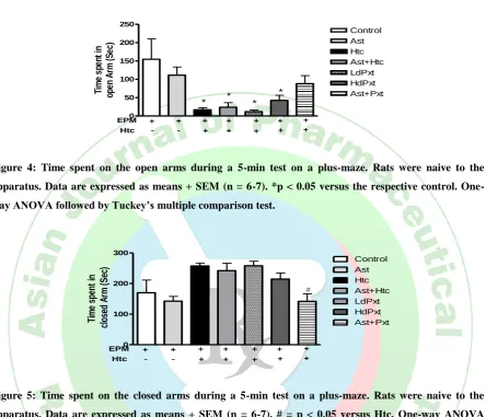
Fig.4. depicts that in comparison to control group, administration of astressin didn‟t significantly affect the time spent by the rats in open arm of EPM. Injection of 2.0 Molar NaCl prior to EPM significantly decreased the time spent in the open arm compared with controls
(p < 0.01). Treatment with astressin/low dose paroxetine/high dose paroxetine/astressin plus low dose paroxetine was not able to reverse this effect.
Time spent in closed arm
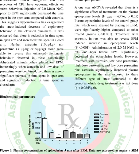
differ significantly (F(6,38)= 5.29; p < 0.05;
Fig.4,5). Posthoc analysis revealed that none of the treatment groups differ significantly from the control group. No statistical differences were found between LdPXT,
HdPXT, Ast + Htc and Htc group. There is a significant decrease in time spent in the closed arm in Ast+Pxt group compared to the group in which the rats were administered 2.0 M NaCl prior to EPM (p<0.05)
0 50 100 150 200 250
Control Ast Htc Ast+Htc LdPxt HdPxt Ast+Pxt
EPM Htc
+ + + + + + +
+ + + + +
-
-* * *
*
Ti
m
e
sp
en
t
in
op
en
A
rm
(S
ec)
Figure 4: Time spent on the open arms during a 5-min test on a plus-maze. Rats were naive to the
apparatus. Data are expressed as means + SEM (n = 6-7). *p < 0.05 versus the respective control.
One-way ANOVA followed by Tuckey’s multiple comparison test.
0 100 200 300
Control Ast Htc Ast+Htc LdPxt HdPxt Ast+Pxt
EPM Htc
+ + + + + + +
+ + + + +
-
-#
Ti
m
e
sp
en
t
in
c
lo
se
d
A
rm
(
S
ec
)
Figure 5: Time spent on the closed arms during a 5-min test on a plus-maze. Rats were naive to the
apparatus. Data are expressed as means + SEM (n = 6-7). # = p < 0.05 versus Htc. One-way ANOVA
followed by Tuckey’s multiple comparison test.
Influence of hypernatremia on elevated plus maze induced psychological stress
Effect on behaviour:
EPM has been frequently used as a tool to screen anxioselective effects of drugs. . In fact, the EPM test has been widely used in a post-hoc fashion. Hence based on the information available in literature, we have used it posthoc to induction of hypernatremia. It is an ethological model that involves spontaneous exploration by rodents of the environment, in the absence of explicit reward or consummatory behaviour. The unconditioned response characteristics seen in the EPM task are attributed to the spontaneous fear which the EPM elicits, given that during the regular 5
min session there is a clear preference to be in the enclosed arms rather than the open arms. There is also substantial evidence showing that drugs which increase open arms activity are anxiolytic compounds in other animal models while drugs which reduce open arms activity are anxiogenic.
nonselective CRFR antagonist and two receptors of CRF have opposing effects on stress behaviour. Injection of 2.0 Molar NaCl prior to EPM significantly decreased the time spent in the open arm compared with controls. This suggests hypernatremia has exaggerated the stress-induced decrease of exploratory behavior in the elevated plus-maze. It was observed that there is reduction in time spent in open arm and increased time spent in closed arm. Neither astressin (10µg/kg) nor paroxetine (3 µg/kg or 5µg/kg) alone were able to reverse this enhanced anxiogenic behaviour observed in these osmotically dehydrated animals when placed on EPM. Interestingly when astressin and low dose of paroxetine were combined, then there is a non- significant increase in time spent in open arm and significant reduction in time spent in closed arm.
Biochemical parameters
Blood epinephrine:
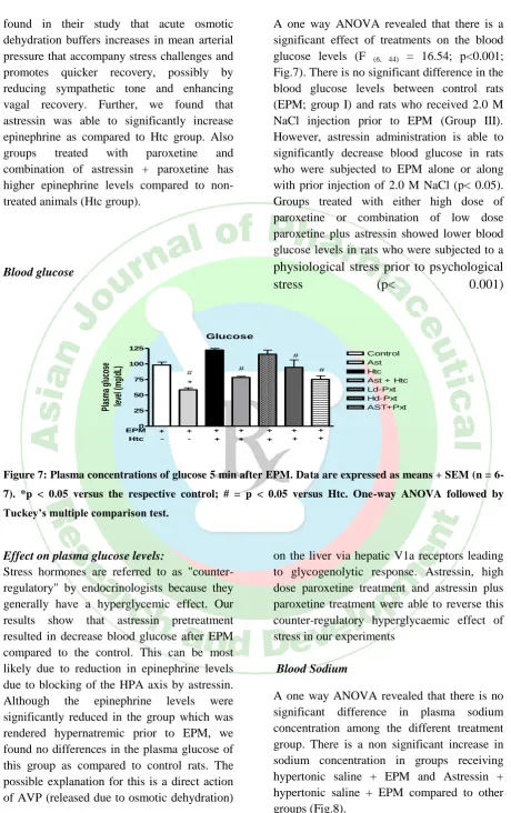
A one way ANOVA revealed that there is a significant effect of treatments on the plasma epinephrine levels (F (6,40) = 63.96; p<0.05)
Plasma epinephrine levels of the control group rats, which were stressed by placing on EPM, were significantly higher compared to other treated groups (P<0.001). Treatment with astressin, in rats was able to reverse EPM induced increase in epinephrine levels (P <0.001). Administration of 2.0 M NaCl to rats one hour before EPM, significantly decreased the epinephrine levels. However, treatment with astressin, low dose paroxetine, high dose paroxetine, and low dose paroxetine plus astressin significantly increased plasma epinephrine in the rats exposed to these different type of stress compared to the group in which drug treatment was not done (p < 0.05:Fig.6).
0.00 0.25 0.50 0.75 1.00 1.25
Control Ast Htc Ast + Htc Ld-Pxt Hd-Pxt AST+Pxt
EPM Htc
+ + + + + + +
+ + + + +
-
-*
* *
* * *
#
# # #
P
la
sm
a
ep
in
ep
h
ri
n
e
(µ
g
/m
l)
Figure 6: Plasma concentrations of epinephrine 5 min after EPM. Data are expressed as means + SEM
(n = 6-7). *p < 0.05 versus the respective control; # = p < 0.05 versus Htc. One-way ANOVA followed by
Tuckey’s multiple comparison test.
Effect on plasma epinephrine levels:
Epinephrine is a short-term neuromodulator of behavioral and physiological responses activated by stress to counter the stress and restore homeostasis. In our study we found that rats treated with peripheral CRFR antagonist had lower epinephrine levels after EPM test as compared to the control. A variety of stressors cause an increased activity of the sympathetic nervous system and adrenal medulla. This leads to increase epinephrine and norepinephrine levels into the blood stream and changes in the activity of enzymes that synthesize catecholamines and in the
found in their study that acute osmotic dehydration buffers increases in mean arterial pressure that accompany stress challenges and promotes quicker recovery, possibly by reducing sympathetic tone and enhancing vagal recovery. Further, we found that astressin was able to significantly increase epinephrine as compared to Htc group. Also groups treated with paroxetine and combination of astressin + paroxetine has higher epinephrine levels compared to non-treated animals (Htc group).
Blood glucose
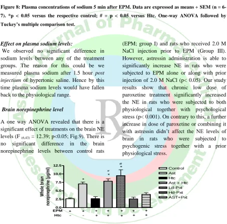
A one way ANOVA revealed that there is a significant effect of treatments on the blood glucose levels (F (6, 44) = 16.54; p<0.001;
Fig.7). There is no significant difference in the blood glucose levels between control rats (EPM; group I) and rats who received 2.0 M NaCl injection prior to EPM (Group III). However, astressin administration is able to significantly decrease blood glucose in rats who were subjected to EPM alone or along with prior injection of 2.0 M NaCl (p< 0.05). Groups treated with either high dose of paroxetine or combination of low dose paroxetine plus astressin showed lower blood glucose levels in rats who were subjected to a
physiological stress prior to psychological
stress
(p<
0.001)
Glucose
0 25 50 75 100 125
Control Ast Htc Ast + Htc Ld-Pxt Hd-Pxt AST+Pxt
Pl
as
m
a
gl
uc
os
e
le
ve
l (
m
g/
dL
)
EPM Htc
+ + + + + + +
+ + + + +
-
-*
# #
#
#
Figure 7: Plasma concentrations of glucose 5 min after EPM. Data are expressed as means + SEM (n =
6-7). *p < 0.05 versus the respective control; # = p < 0.05 versus Htc. One-way ANOVA followed by
Tuckey’s multiple comparison test.
Effect on plasma glucose levels:
Stress hormones are referred to as "counter-regulatory" by endocrinologists because they generally have a hyperglycemic effect. Our results show that astressin pretreatment resulted in decrease blood glucose after EPM compared to the control. This can be most likely due to reduction in epinephrine levels due to blocking of the HPA axis by astressin. Although the epinephrine levels were significantly reduced in the group which was rendered hypernatremic prior to EPM, we found no differences in the plasma glucose of this group as compared to control rats. The possible explanation for this is a direct action of AVP (released due to osmotic dehydration)
on the liver via hepatic V1a receptors leading to glycogenolytic response. Astressin, high dose paroxetine treatment and astressin plus paroxetine treatment were able to reverse this counter-regulatory hyperglycaemic effect of stress in our experiments
Blood Sodium
0 100 200
control Ast Htc Ast+htc Ldpxt Hdpxt Ast+Pxt+Htc
EPM Htc
+ + + + + + +
+ + + + +
-
-B
lo
o
d
s
o
d
iu
m
c
o
n
c
e
n
tr
a
ti
o
n
(m
e
q
L
-1 )
Figure 8: Plasma concentrations of sodium 5 min after EPM. Data are expressed as means + SEM (n =
6-7). *p < 0.05 versus the respective control; # = p < 0.05 versus Htc. One-way ANOVA followed by
Tuckey’s multiple comparison test.
Effect on plasma sodium levels:
We observed no significant difference in sodium levels between any of the treatment groups. The reason for this could be we measured plasma sodium after 1.5 hour post
injectium of hypertonic saline. Hence by this
time plasma sodium levels would have fallen back to the physiological range.
Brain norepinephrine level
A one way ANOVA revealed that there is a significant effect of treatments on the brain NE levels (F (6,43) = 12.39; p<0.05; Fig.9). There is
no significant difference in the brain norepinephrine levels between control rats
(EPM; group I) and rats who received 2.0 M NaCl injection prior to EPM (Group III). However, astressin administration is able to significantly increase NE in rats who were subjected to EPM alone or along with prior injection of 2.0 M NaCl (p< 0.05). Our study results show that chronic low dose of paroxetine treatment significantly increased the NE in rats who were subjected to both physiological together with psychological stress (p< 0.001). On contrary to this, a further increase in dose of paroxetine or combining it with astressin didn‟t affect the NE levels of brain in rats who were subjected to psychogenic stress together with a prior physiological stress.
0.0 2.5 5.0 7.5 10.0 12.5
Control
Htc Ast
Ast + Htc Ld-Pxt Hd-Pxt AST+Pxt
EPM Htc
+ + + + + + +
+ + + + +
-
-*
* *
#
#
W
ho
le
b
ra
in
no
re
pi
ne
ph
ri
ne
(µ
g/
m
l)
Figure 9: Concentrations of norepinephrine in whole brain homogenate 5 min after EPM. Data are
expressed as means + SEM (n = 6-7). *p < 0.05 versus the respective control; # = p < 0.05 versus Htc.
One-way ANOVA followed by Tuckey’s multiple comparison test.
Effect on brain norepinephrine levels:
NE has been implicated as a primary neuro-transmitter of central autonomic regulation. Initial literature on stress reported that rats subjected to a variety of stressors show considerably reduced concentration of brain
demonstrated that centrally administered CRF stimulates the region specific release of NE in the brain. According to our results CRFR antagonist pretreatment increased the brain epinephrine levels in the rats subjected to EPM. This is contrary to recent reports which say that administration of CRF antagonist decreased NE levels in hippocampus. It is reported that acute stress releases NA in the terminal fields of neurons localized in the LC, which leads to depletion of NE and an increase of its metabolites. Hence it is possible that pretreatment with astressin led to inhibition of CRF mediated increase in the metabolism of NE in the forebrain (increases in 3-methoxy, 4-hydroxyphenylethylene glycol MHPG). In our study, hypernatremia posed no influence on NE concentrations in rats who were subsequently subjected to EPM stress. This could be due to the noradrenergic responses to repeated stressful treatments exhibit desensitization and cross-desensitization has been demonstrated between CRF and stressors. Astressin and low dose of paroxetine were able to increase NE levels in rats that were osmotically dehydrated and subjected EPM test
CONCLUSION
Our findings suggest that hypernatremia has exaggerated the stress-induced behavior in the elevated plus-maze test. Neither astressin nor paroxetine alone were able to reverse this enhanced anxiogenic behaviour observed in these osmotically dehydrated animals when placed on EPM. Interestingly when astressin and low dose of paroxetine were combined, then there is a non- significant increase in time spent in open arm and significant reduction in time spent in closed arm. We observed epinephrine levels were significantly reduced in the group which was rendered hypernatremic prior to EPM. Treatment of astressin, paroxetine and combination of both caused a significant increase in epinephrine. High dose paroxetine treatment and astressin plus paroxetine treatment were able to reverse this counter-regulatory hyperglycaemic effect of stress in our experiments. Astressin and low dose of paroxetine increased NE levels in rats that were osmotically dehydrated and subjected to EPM test. Both astressin and
paroxetine perse showed similar effects on behaviour and stress mediator in our experiments. This indirectly suggests that at least in parts they share common physiology in stress integration. Also, the combination of paroxetine and astressin showed a dichotomy in terms that it has improved the behaviour on EPM but has no advantage over the perse
treatment in terms of stress mediators
ACKNOWLEDGEMENT:
The authors wish to thank Department of Pharmacology, Jamia Hamdard for providing facilities at Pharmacology Laboratory.
REFERENCES
1. Berton O., Nestler EJ. New approaches to antidepressant drug discovery: beyond monoamines, Nature Reviews Neuroscience, 2006; 7:2:137-51. 2. Shelton RC., The Molecular Neurobiology of
Depression, Psychiatric Clinics of North America, 2007; 30 1:1–11.
3. Madaan V and Wilson DR. Neuropeptides: relevance in treatment of depression and anxiety disorders., Drug New Perspect, 2009; 22 :6:319-24.
4. McEwen BS., Wingfield JC., The concept of allostasis osmoregulation in biology and biomedicine, Hormones and Behavior, 2003; 43:1:2-15.
5. Krause EG., Kloet AD., Flak JN., Smeltzer MD., Solomon MB., Evanson NK., Woods SC., Sakai RR., and Herman JP., Hydration State Controls Stress Responsiveness and Social Behaviour, The Journal of Neuroscience, 2011; 31:14:5470 –76.
6. Avashalom C., Karen S., Terrie E., Allan T., Taylor A. ,Ian WG., HonaLee H., Joseph MC., Jonathan M., Judy M., Antony B., Richie P., Influence of life stress on depression: Moderation by a polymorphism in the 5-HTT Gene, The Journal of Life Long Learning in Psychiatry(FOCUS), 2005; 3:1:156-60.
7. Bourque CW., Central mechanisms of osmosensation and systemic osmoregulation, Nature Review Neuroscience, 2008; 9:519 –31.
8. Toney GM., Stocker SD., Hyperosmotic activation of CNS sympathetic drive: implications for cardiovascular disease., Journal of Physiology, 2010; 588:3375–84.
9. Wigger A., Sánchez MM., Mathys KC., Ebner K., Frank E., Liu D., Kresse A., Neumann ID., Holsboer F., Plotsky PM., Landgraf R.., Alterations in central neuropeptide expression, release and receptor binding in rats bred for high anxiety: critical role of vasopressin., Neuropsychopharmacology, 2004; 29(1):1-14.
10.Dani KR., Vandekar LD., Selective serotonin reuptake inhibitors and neuroendocrine function., Life Sciences, 1999; 65:12:1217-35.
11.Jensen J., Mork A., Mikkelsen J., Chronic antidepressant treatments decrease pro-opiomelanocortin mRNA expression in the pituitary gland: Effects of acute stress and 5HT1A receptor activation, Journal of Neuroendocrinology, 2001; 131, 887–93.
inhibitor citalopram modulate the HPA axis activity at different levels in male rats, Journal of Neuroendocrinology, 1999; 11:6:465-71.
13.Wieczorek I., Schulz C., Jarry H., Lehnert H., The effects of the selective serotonin reuptake-inhibitor fluvoxamine on body weight in Zucker rats are mediated by corticotropin-releasing hormone, International Journal of Metabolic Obesity and Related Metabolic Disorders, 2001; 25:10:1566-69. 14.Arborelius L., Owens MJ., Plotsky PM., Nemeroff
CB., The role of corticotropin-releasing factor in depression and anxiety disorders, Journal of Endocrinology, 1999; 160:1:1-12.
15.Radulovic J., Ruhmann A., Liepold T., Spiess J., Modulation of Learning and Anxiety by Corticotropin-Releasing Factor (CRF) and Stress: Differential Roles of CRF Receptors 1 and 2. The Journal of Neuroscience, 1999; 19:12:5016–25. 16.Rivier J., Rivier C.,Vale W., Synthetic competitive
antagonists of corticotropin-releasing factor: effect on ACTH secretion in the rat, Science, 1984; 224:4651:889-91.
17.Spina MG., Basso AM., Zorrilla EP., Heyser CJ., Rivier J., Vale W., Merlo-Pich E., Koob GF., Behavioral Effects of Central Administration of the Novel CRF Antagonist Astressin in Rats, Neuropsychopharmacology, 2000; 22:3:230-39.
18.Gulyas J., Rivier C., Perrin M., Koerber SC., Sutton S., Corrigan A., Lahrichi SL., Craig AG., Vale W., Rivier J., Potent, structurally constrained agonists and competitive antagonists of corticotropin-releasing factor, Proceedings of the National Academy of Sciences of the United States of America(PNAS), 1995; 92:10575–79.
19.Turnbull A., Vaughan J., Rivier JE., Vale WW., Rivier C., Urocortin is Not a Significant Regulator of Intermittent Electroshock-Induced ACTH Secretion in the Intact Male Rats, Endocrinology, 1999; 140:1:71-78.
20.Landgraf R., Wigger A., Holsboer F., Neumann ID., Hyper-reactive hypothalamo-pituitary -adrenocortical axis in rats bred for high anxiety-related behavior, Journal of Neuroendocrinology 1999; 11:6:405-7.
21.Trinder P., Determination of blood glucose using oxidase-peroxidase system with a non carcinogenic chromogen, Journal of Clinical.Pathology, 1969; 22:158-61.