Published online January 28, 2015 (http://www.sciencepublishinggroup.com/j/jfns) doi: 10.11648/j.jfns.s.2015030102.25
ISSN: 2330-7285 (Print); ISSN: 2330-7293 (Online)
Combining of bacteriophage and G. asaii application to
reduce L. monocytogenes on fresh-cut melon under low
temperature and packing with functional film
Yoon-Pyo Hong
1, Ji Weon Choi
2, Ji Hyun Lee
2, Ray-Yu Yang
11
Nutrition Team, Asian Vegetable Research and Development Center, Po 42, Shanhua Tainan, Taiwan
2
Postharvest Research Team, National Institute of Horticultural and Herbal Science, RDA, Suwon 440-706, Korea
Email address:
[email protected] (Yoon-Pyo H.)
To cite this article:
Yoon-Pyo Hong, Ji Weon Choi, Ji Hyun Lee, Ray-Yu Yang. Combining of Bacteriophage and G. asaii Application to Reduce L. monocytogenes on Fresh-Cut Melon under Low Temperature and Packing with Functional Film. Journal of Food and Nutrition Sciences.
Special Issue: Food Processing and Food Quality. Vol. 3, No. 1-2, 2015, pp. 79-83. doi: 10.11648/j.jfns.s.2015030102.25
Abstract:
Gluconobacter asaii (a bacterial antagonist naturally occurring on apple fruit) and bacteriophage were tested as biocontrol agents of Listeria monocytogenes on fresh-cut honeydew melon pieces during low temperature and packing with functional film made by zeolite mixed pegmatite thickness with 20 µm. Gluconobacter asaii and bacteriophage were effective against of L. monocytogenes, but combining the two treatment was even more effective. G. asaii alone treatment reduced density approximately 4 to 5 logs and phage alone reduced populations by one log compared to the L. monocytogenes control. In comparison Listeria control treatment combining treatments reduced populations up to 6 logs by day 7 especially packing with functional film. Also shelf-life of melon with functional film extend 5~6 days compared to none film treatment under low temperature. The results of our study suggest that G. asaii and phage when combined could be very effective in reducing L. monocytogenes contamination of fresh-cut honeydew melon under low temperature and packing with functional film.keywords:
Bacterial Antagonist, Fresh-Cut, Phage1. Introduction
Listeria monocytogenes is a Gram-positive, motile organism capable of growth between –0.4 and 50°C (Faber and Peterkin 1991). It is ubiquitous, highly virulent and tolerant to environmental stress (Doyle 1999). It also grows at refrigeration (Van 2000) temperature which is of great concern especially with the emergence of the new generation of minimally processed foods relying only on refrigeration and intrinsic parameters as proliferation of undesirable
microorganisms. The presence of L. monocytogenes on
decaying vegetation (Welshimer 1968), agronomic crops (Weiss 1975) and silage (Skovgaard 1988) is well documented. This organism has been associated with a number of serious foodborne outbreaks and recalls of fresh produce (Beuchat 2002, Farber and Peterkin 1991). Usually occur at pathogen populations greater than 103 CFU per g or per ml (Tompkin 2002). Because of the high case fatality rate, which is account for up to 96% of human listeriosis, associated with L. monocytogenes infections, the U.S. Food and Drug Administration have established a zero-tolerance
able to grow at a low pH of 3.5 in highly concentrated sugar solutions and on fruit such as apples, pears and grapes as well as in ciders (Deppenmeier 2002, Gupta 2001, Van 1981).
Gluconobacter species are found and utilized in fermentation processes and production of wine, vinegar and vitamin C (Deppenmeier 2002, Macauley 2001, Yamada 1999).
Gluconobacter asaii proved effective in preventing the growth or survival of L. monocytogenes on fresh cut apple tissue. Packing with functional film after harvest on several fruit and vegetable were effectively extend shelf-life due to
easily respiration and absorbing ethylene under
MA(Modified Atmosphere) condition (Choi 2013, Hong 2014, Cho 2012). The objectives of this study were to determine (i) the effectiveness of phage and Gluconobacter asaii as antagonists of L. monocytogenes on honeydew melon pieces and (ii) whether combining the phages with
Gluconobacter would enhance the effectiveness against L. monocytogenes under low temperature and packing with functional film.
2. Materials and Methods
Fruit. Honeydew melons obtained from the local market were cut into 10-mm-thick rings with a deli slicer (model 827, Berkel Inc., USA). The 10-mm-thick melon rings were cut into equally sized squares about 25 mm2 . The fruit surfaces as well as the deli meat slicer were disinfected with 70% ethanol immediately before slicing. A cork borer was used to cut tissue plugs of honeydew that was 10 mm thick and 10mm in diameter, resulting in the tissue plugs of 0.785cm3. The pH of the melon slices was monitored using a Semi-Micro pH combination electrode (81-03 Ross, Orion Research, Inc., Beverly, USA).
Phage. The phage mixture, LMP-102, contained six distinct lytic phages specific for L. monocytogenes, including serotypes 1/2a, 1/2b, and 4b, which have been predominantly associated with human listeriosis. The mixture was provided by Intralytix, Inc. (Baltimore, Md.) The phage concentration was approximately 109 PFU/ml in 1 M phosphate-buffered saline (pH 7.4). The mixture was diluted with peptone water (pH 7.4) to approximately 106 PFU/ml immediately before application to the fruit pieces.
Glucanobacter. The Glucanobacter asaii originally isolated from apple surfaces, and that was grown on nutrient yeast dextrose agar (NYDA) plates overnight at 25°C. One colonys were scraped from the agar plates and suspended in
NYDB+grown 6hr at 25°C on a shake (150 rpm). The
solution was in peption water (pH 7.4). The cell concentration was adjusted to 105 – 106 CFU/ml using a SmartsSpec 3000 spectrophotometer (Bio-Rad Laboratories, Richmond, Calif.) at 600nm according to standard curves. The exact cell concentration was determined by plating the inoculum with a spiral plater (DW Scientific, Shipley, West Yorkshire, England) into NYDA medium followed by incubation at 20°C for 1 day.
Bacterial inoculum. The L. monocytogenes culture, strain LCDC 81-861 serotype 4b, implicated in an outbreak from
processed cabbage (cole slaw), was obtained from Robert Brackett, Depatment of Food Science and Technology, University of Georgia, Agricultural Experiment Station, Griffin, Ga., and stored at -80°C in Luria-Bertani (LB) broth (BD Dianostic Systems, Sparks, Md.) and 15% glycerol (Difco, Becton Dickinson, sparks, Md.) The strain was naturally resistant to nalidixic acid (Sigma, St. Louis, Mo.). For inoculation of the fruit pieces, L. monocytogenes was grown overnight on tryptic soy agar (TSA; BD Diagnostic Systems) plates with 100 ug/ml of nalidixic acid at 30°C and then transferred to 10ml of TSB broth for 6 h. The cell were harvested by centrifugation at 10,000 x g for 15min. The pellet was resuspended in peptone water (pH 7.4) and adjusted to a concentration of 104 or 105 CFU/ml at an optical density of 600nm using a SmartSpec 3000 spectrophotometer (Bio-Rad Laboratories, Rechmond, Calif.). The exact cell concentration was determined by plating the inoculum with a spiral plater (DW Scientific, Shipley, West Yorkshire, UK) into TSA medium with nalidixic acid followed by incubation at 37°C for 1 day.
Treatment application. The honeydew melon fruit pieces were placed in commercial. 530-ml, dome fruit plastic bowls (no. 518, Rock-Tenn. Co., Chicago Plastics, Franklin Park, III.). The fruit tissue was then inoculated with 25 ul of the L. monocytogenes suspension containing approximately 1 × 10⁴ CFU/ml. The procedure for inoculating the pieces of fruit took approximately 10 min. Then, the phage and/or
Glucanobacter treatments were applied by pipette to a 5-by 5-mm area in 25-ul aliuots. There were three or four fruit samples per treatment at each recovery time. The covers on the plastic bowls allowed air exchange, which ensured that the environmental conditions did not change and therefore did not create a modified atmosphere.
Recovery of pathogen and antagonist. The pathogen and antagonist populations were recovered from the honeydew melon plugs after 0, 2, 5, and 7 days of storage at 10°C as described previously (Conway 2000). Briefly, the melon tissue plugs were each placed into a sterile plastic bag containing 4.5 ml of peptone water and homogenized in a stomacher blender for 120 s at a high speed set at 8 (Bagmixer 100 Minimix;Intersceence, Weymouth, Mass). Aliquots (50ul) of the homogenized mixtures or dilutions thereof were plated in duplicate on TSA containing 100 ug per ml of NAL for L. monocytogenes or nutrient yeast dextrose agar (NYDA; put this under gluconobacter when NYDA is 1st mentioned) using a spiral plater. The TSA + NAL plates were incubated overnight at 37°, the NYDA plates were incubated overnight 20°C. Colony counts were
determined using an automated plate counter
(ProtoCol;Synoptics, Cambridge, United Kingdom), and the data were plotted as CFU per sample. All experiments were repeated.
plaques were counted with the ProtoCol plate counter (Synoptics), and the data were plotted as PFU per sample.
Functional film and storage. Functional film made by zeolite mixed pegmatite which can easily absorbed carbon dioxide and ethylene (Hong, 2014) based on polyvinyl chloride using 15cm×15cm bag into fresh cut honeydew melon. There were five or six fruit samples on tray per treatment at each application. MA treatment can change of inside atmosphere and ethylene level through ventilation using micro hole on the film. The temperature of cold storage maintain 0°C during treatment but control temperature regard as room or ambient temperature.
Statistical analyses. The bacterial recovery data (CFU per sample) were analyzed for each experiment as two-factor general linear models using PROC MIXED (SAS Institute) with Treatment and Day as the factors. The assumptions of the linear model were tested. To correct for variance heterogeneity the treatments were grouped into similar variance groups for the analysis. When effect were statistically significant, mean comparisons were done with Sidak adjusted p-values so that the experiment-wise error was 0.05.
3. Results
Recovery of Glucanobacter and phage. Gluconobacter asaii was originally isolated for its biocontrol activity against
postharvest fungal decay pathogen ㄴ on apple. In a
preliminary experiment we found it was also able to grow or survive on fresh-cut honeydew melon pieces over time (data
not shown). When co-inoculated with L. monocytogenes and
phage, Gluconobacter was increased over time from day 0 to day 7 (Fig 1). The titration of the phages recovered from the inoculum was between 3 and 5 log PFU/ml. Phage recovered well by itself over 5 days but there was no recoverable phage by 7 days when combined with Gluconobacter asaii (Fig 2).
Control of L. monocytogenes . When fruit were inoculated on L. monocytogenes at 1 x 10⁴ CFU/ml the biocontrol agents were effective at controlling and even reducing populations compared to the Listeria alone treatment (Fig. 3).
The phage alone treatment reduced L. monocytogenes
populations approximately 1 log from 2 to 7 days.
Gluconobacter asaii alone was more effective than phage at 5. and 7 days, reducing L. monocytogenes populations 3 and 4 logs respectively. Combining phage and Gluconobacter asaii was the most effective at controlling the listeria populations. In comparison with control, combining of the
phage and Gluconobacter asaii reduced the bacterial
populations by 4.5 to 5.8 log units. There was a phage x
Gluconobacter asaii interaction on honeydew melons (Fig 3). Even when the concetration of L. monocytogenes inoculated was increased from 1x10⁴ to 1x10⁶ the same rate as biocontrol agent, by day 5, all treatments significant reduced the bacterial populations on fresh cut honeydew melon.
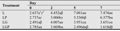
Extend shelf-life. Also shelf-life of fresh cut honeydew melon with functional film extend 5~6 days compared to none film treatment that was easily decay due to
accumulation of carbon dioxide and ethylene which was double level verse control. Also normal and ambient temperature treatment of melon was short shelf-life compared to under low temperature (Table 1).
4. Discussion
We found that combining phage and Gluconobacter asaii
treatments is an effective method for reducing L.
monocytogenes contamination on fresh-cut honeydew melon. Because of previous successes with respect to lytic bacteriophages on honeydew melon (Leverentz 2003) and
Gluconobacter asaii on apples (Leverentz 2006), we hoped to see an added effect to the combination. This was, however, the first test of Gluconobacter asaii on honeydew melon as a biocontrol agent. The recovery of Gluconobacter asaii from honeydew melon pieces stored 10°C was increased over 7 days. This result is similar to those of our previous experiments with Listeria and Salmonella growing on fresh-cut apples. The more growth of Gluconobacter asaii at 10°C with L. monocytogenes compared to Gluconobacter asaii on fresh-cut apples stored at 25°C may be due to the greater cold tolerance of Gluconobacter asaii (Leverentz 2006). Phage was known as effective biocontrol agent about reducing L. monocytogenes and Salmonella on fresh-cut fruits through our previous reports (Leverentz 2004, Leverentz 2003, and Leverentz 2001). Phage recovered well when co-inoculated with L. monocytogenes but no phage was recovered by 7 days
when combined with Gluconobacter asaii. There are several
possibilies to explain the low recovery of phage at 7 days. One of the most reasonable is the low concentration of L. monocytogenes remaining and the phage could not “find” the necessary host. The combining of phage and Gluconobacter asaii was the most effective against controlling L. monocytogenes populations on honeydew melon pieces. In addition, for maximum effectiveness in controlling populations of L. monocytogenes throughout the entire storage period of 7 days at 10°C, there seems to be synergy effect between phage and Gluconobacter asaii. In past experiments we had observed that phage decreased in biocontrol activity over time (Leverentz 2003) and that the
Gluconobacter asaii was increased in biocontrol activity over time effective later (Leverentz 2006). We hypothesized that
the combination would more effectively control L.
Appendices
Fig. 1. Populations of G. asaii alone or in the presence of L. monocytogenes alone or combined with phage when wedges were stored at 10°C over 7 days
Fig. 2. Populations of phage in the presence L. monocytogenes alone or combined with G. asaii when wedges were stored at 10°C over 7 days
Fig. 3. Effect of G. asaii (G) and phage (P) on the recovery of L. monocytogenes (L, 104) from honeydew wedges stored at 10°C over 7 days.
Table 1. Influence of combined treatment in reducing L. monocytogenes from honeydew wedges when stored at 10°C for 7 days.
Treatment Day
0 2 5 7
L 2.637a1γ2 4.452aβ 7.061aα 7.474aα
LP 2.737aγ 3.006bγ 5.536bβ 6.377bα LG 2.491aβ 4.007aα 3.951cα 3.651cα LGP 2.785aα 3.069bα 2.496dαβ 1.618dβ
1 Treatment means within Day with different a, b, c, d letters are different at
the 0.05 significance level.
2 Day means within Treatment with different α, β, γ letters are different at the
0.05 significance level.
Table 2. Index of shelf-life on fresh-cut honeydew melon after treatment by functional film over 2 weeks.
Treatment Index of shelf-life over 2 weeks (days)
0 2 4 6 8 10 12 14
Control 51 5 4 3 2 1
Low-temp.(LT) 5 5 4 4 4 4 3 3
Functional film(FF) 5 5 5 4 4 4 4 3
LT+FF 5 5 5 5 5 5 4 4
1Index of shelf-life.: 5-very good, 4-good, 3-normal, 2-bad, 1-very bad.
Acknowledgements
This study was conducted with support from the National Institute of Horticultural and Herbal Science of Republic of Korea (Research project for establishment of postharvest technology to maintain the freshness Project No. PJ009362) and AVRDC nutrition team in Taiwan (Project No. 10000241-01)
References
[1] Adams, M. H. (1959). Bacteriophages. Interscience publishers, New York, N. Y.
[2] Barrangou, R., S. S. Yoon, F. Breidt, H. P. Fleming, and T. R. Klaenhammer. (2002). Characterization of six Leuconostoc fallax bacteriophages isolated from an industrial sauerkraut fermentation. Appl. Environ. Microbiol. 68:5422-5458.
[3] Bergh, O., K. Y. Borsheim, G. Bratbak, and M. Heldal. (1989). High abundance of viruses found in aquatic environments. Microbes Infect. 4:413-423
[4] Beuchat, L. 2002. Ecological factors influencing survival and growth of human pathogens on raw fruits and vegetables. Microbes infect. 4:413-423.
[5] Cho, M. A., Y. P. Hong, J. W. Choi, and D. A. Jung. (2012). Effect of enhancing shelf-life according to 1-MCP and MAP treatment on fresh Jujube during cold storage. Horticultural Environment and Biotechnology. 30(1):136-137.
[6] Choi, J. W., Y. P. Hong, M. A. Cho, and J. Y. Lee. (2013). Effect of low temperature and MA treatment on oriental melon after harvest. Horticultural Environment and Biotechnology. 31(1):75-76.
[7] Conway, W. S., B. Leveentz, R. A. Saftner, W. J. Janisiewicz, C. E. Sams, and E. Leblanc. (2000). Survival and growth of Listeria monocytogenes on fresh-cut apple slices and its interaction with Glomerella cingulata and Penicillium expansum. Plant Dis. 84:177-181.
[8] Deppenmeier, U., M. Hoffmeister, and C. Prust. (2002). Biochemistry and biotechnological applications of Gluconobacter strains. Appl. Microbiol. Biotechnol. 60:233-242.
[9] Doyle M P (1988) Effect of environmental and processing conditions on Listeria monocytogenes. Food Technology 42. 169-171.
[11] Gupta, A., V. K. Singh, G. N. Qazi, and A. Kumar. 2001. Gluconobacter oxydans:Its biotechonogical applications. Journal of Molecular Microbiology and Technology 3:445-456.
[12] Hong, Y. P., J. H. Lee., and J. W. Choi. (2014). Change of quality according to several film treatment on fresh ginseng during cold storage. Horticultural Environment and Biotechnology. 32(1):217-218.
[13] Leverentz, B., W. S. Conway, Z. Alavidze, W. J. Janisiewicz, Y. Fuchs, M. J. Camp, E. Chighladze, and A. Sulakvelidze. 2001. Examination of bacteriophage as a biocontrol method for Salmonella on fresh-cut fruit – a model study. J. Food Prot. 64:1116-1121.
[14] Leverentz, B., W. S. Conway. M. J. Camp, W. J. Janisiewicz, T. Abuladze, M. Yang, R. Saftner, and A. Sulakvelidze. 2003. Biocontrol of Listeria monocytogenes on fresh-cut produce by treatment with lytic bacteriophages and a bacteriocin. Appl. Environ. Microbiol. 69:4519-4526.
[15] Leverentz, B., W. S. Conway. W. J. Janisiewicz, and M. J. Camp. 2004. Optimizing concentration and timing of a phage spray application to reduce Listeria monocytogenes on honeydew melon tissue. J. of Food Protection, 67:1682-1686
[16] Lu, Z., f. Breidt, V. Plengvidhya, and H. P. Fleming. 2003. Bacteriophage ecology in commercial sauerkraut fermentations. Appl. Environ. Microbiol. 69:3192.
[17] Lu, Z., F. Breidt, H. P. Fleming, E. Altermann. And T. R. Klaenhammer. 2003. Isolation and characterization of a Lactobacillus plantarum bacteriophage. Phi JL-1, from a cucumber fermentation. Int. J. Food Microbiol. 84:225-235.
[18] Macauley, S., B. McNeil, and L. M. Harvey. 2001. The genus Gluconobacter and its applications in biotechnology. Crit. Rev. Biotechnol. 21:1-25.
[19] SAS Institute Inc. 1999. SAS/STATⓡ User’s Guide, Version 8. SAS Institute, Cary, N. C.
[20] Skovgaard, N., and C.-A. Morgen. 1988. Detection of Listeria spp. In faeces from animals, in feeds, and in raw foods of animal origin. Int. J. Food Microbiol. 6:229-242.
[21] Tompkin, R. B. 2002. Control of Listeria monocytogenes in the food-processing environment. J. Food Prot. 65:709-725.
[22] Van de Venter T (2000) Emerging food-borne diseases: a global responsibility. Food nutrition and Agriculture (FAO) 26, 4-13
[23] Van Keer, C., P. Vanden Abeele, J. Swings, F. Gossele, and J. De Lay. 1981. Acetic acid bacteria as causasl agents of apples and pears. Zbl. Bakt. Hyg., I. Abt. Orig. C:197-204.
[24] Weiss, J., and H. P. R. Seeliger. 1975. Incidence of Listeria monocytogenes in nature. Appl. Environ. Microbiol. 30:29-32.
[25] Welshimer, H. J. 1968. Isolation of Listeria monocytogenes from vegetation. J. Bacteriol. 95:300-320.