R E S E A R C H
Open Access
Differences in the epigenetic and reprogramming
properties of pluripotent and extra-embryonic
stem cells implicate chromatin remodelling as an
important early event in the developing mouse
embryo
Joana Santos
1, C Filipe Pereira
1, Aida Di-Gregorio
2, Thomas Spruce
2, Olivia Alder
3, Tristan Rodriguez
2,
Véronique Azuara
3, Matthias Merkenschlager
1, Amanda G Fisher
1*Abstract
Background:During early mouse development, two extra-embryonic lineages form alongside the future embryo: the trophectoderm (TE) and the primitive endoderm (PrE). Epigenetic changes known to take place during these early stages include changes in DNA methylation and modified histones, as well as dynamic changes in gene expression.
Results:In order to understand the role and extent of chromatin-based changes for lineage commitment within the embryo, we examined the epigenetic profiles of mouse embryonic stem (ES), trophectoderm stem (TS) and extra-embryonic endoderm (XEN) stem cell lines that were derived from the inner cell mass (ICM), TE and PrE, respectively. As an initial indicator of the chromatin state, we assessed the replication timing of a cohort of genes in each cell type, based on data that expressed genes and acetylated chromatin domains, generally, replicate early in S-phase, whereas some silent genes, hypoacetylated or condensed chromatin tend to replicate later. We found that many lineage-specific genes replicate early in ES, TS and XEN cells, which was consistent with a broadly ‘accessible’chromatin that was reported previously for multiple ES cell lines. Close inspection of these profiles revealed differences between ES, TS and XEN cells that were consistent with their differing lineage affiliations and developmental potential. A comparative analysis of modified histones at the promoters of individual genes showed that in TS and ES cells many lineage-specific regulator genes are co-marked with modifications associated with active (H4ac, H3K4me2, H3K9ac) and repressive (H3K27me3) chromatin. However, in XEN cells several of these genes were marked solely by repressive modifications (such as H3K27me3, H4K20me3). Consistent with TS and XEN having a restricted developmental potential, we show that these cells selectively reprogramme somatic cells to induce thede novo expression of genes associated with extraembryonic differentiation.
Conclusions:These data provide evidence that the diversification of defined embryonic and extra-embryonic lineages is accompanied by chromatin remodelling at specific loci. Stem cell lines from the ICM, TE and PrE can each dominantly reprogramme somatic cells but reset gene expression differently, reflecting their separate lineage identities and increasingly restricted developmental potentials.
* Correspondence: [email protected] 1
Lymphocyte Development Group, MRC Clinical Sciences Centre, Imperial College School of Medicine, Hammersmith Campus, Du Cane Road, London W12 0NN, UK
Background
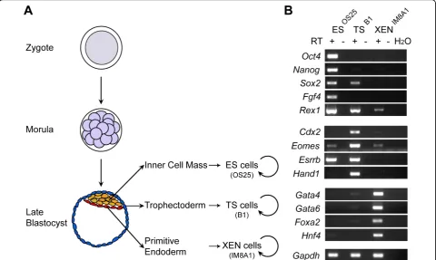
After fertilization, the mouse embryo undergoes a series of sequential cleavage divisions producing an eight-cell embryo, where blastomeres maximize their contact with one another in order to generate a compact sphere of cells. Subsequently, apico-basal polarization and asym-metric divisions generate two distinct cell populations at the 16-cell stage: large peripheral polarized cells and small apolar central cells [1]. The outer, polar cells of the late morula change morphology to form an epithelial monolayer of cells - the trophectoderm (TE), which mediates the implantation and initiation of placentation, while the inner apolar cells become the inner cell mass (ICM) and contain the founder cells of the embryo proper. By the early blastocyst stage (E3.5), these two tissues are morphologically distinct - the outer polarized epithelium, the TE, enclosing the ICM, which is itself heterogeneous [2]. Around the time of implantation, cells within the ICM segregate spatially and morphologi-cally into the epiblast (EPI) and PrE lineages, through the migration of PrE cells to the blastocoelic surface of the ICM. Lineage studies have shown that the cells of the EPI are pluripotent and give rise to all tissues of the fetus plus extra-embryonic mesoderm. TE cells are mul-tipotent differentiating exclusively into the trophoblast lineages that form the majority of the fetal placenta, while the PrE give rise to the visceral and parietal endo-derm layers that will later line the yolk sack. Besides providing growth support and protection within the uterus, the extra-embryonic TE and PrE are sources of signals to the embryonic lineages to promote correct patterning and differentiation [3].
While the molecular mechanisms underlying the gen-eration of the ICM, TE and PrE lineages are not fully understood, several transcription factors that play a role in the development of these three different lineages have been described, including Oct4, Cdx2 and Gata6, which are critical for the development of the ICM, TE and PrE, respectively [4-6]. An appropriate segregation of the ICM and TE has, in addition, been shown to be dependent upon the establishment and maintenance of cell polarity, involving E-cadherin and the Par3/aPKC complex [7-9].
Studies from several laboratories have provided evi-dence of global epigenetic differences between these early lineages that may be important in defining their developmental fate. In particular, a recent study has sug-gested that at the four-cell-stage mouse embryo, blasto-meres with higher levels of histone H3 arginine methylation are more likely to contribute to the pluripo-tent cells of the ICM [10]. Moreover, while the TE (and also the PrE) are hypomethylated both at repetitive and structural gene sequences [11,12] throughout
develop-ment, a striking increase in both DNA and H3K9 methylation levels characterizes the ICM at the blasto-cyst stage [13,14]. In addition, epigenetic asymmetry between embryonic and extra-embryonic tissues is evi-dent during X-inactivation, which is random in embryo-nic but imprinted in the TE and PrE lineages [15,16].
Recently, data from two different sources has provided important insights into how lineage potential is regu-lated at the earliest stages of mammalian development. Studies comparing DNA methylation at gene promoters in embryonic stem (ES) versus trophectoderm stem (TS) cells, germ cells and fibroblasts identified novel factors that act as‘gatekeepers’ for the specification of extra-embryonic tissue [17] and showed that epigenetic repro-gramming, essential for the transmission of pluripo-tency, occurs within the germline prior to fertilization [18]. Another set of reports, in which the chromatin profile of ES cells, somatic stem cells and their differen-tiated progeny were contrasted, provided collective evi-dence that many developmental regulators genes in ES cells are primed for future expression, being marked with histone modifications associated with both active and repressed chromatin [19,20]. In this study we have examined the epigenetic status of other blastocyst-derived lineages required for the successful development of the early mammalian embryo, using stem cells lines isolated from the TE [21] and PrE [22] that self-renew and differentiate into defined extra embryonic tissues (Figure 1A). Our results demonstrate that, following lineage specification to the ICM, TE and PrE, there are predictable changes in the temporal replication and chromatin structure of lineage-determining genes, as reflected in the stem cell lines analysed here. We also show that extra-embryonic endoderm (XEN) and TS cells, like ES cells, can dominantly reprogramme somatic cells (human lymphocytes), but that they initiate discrete and different lineage-specific gene expression pro-grammes. Taken together, these results suggest that dynamic changes in chromatin organization occur within the developing blastocyst and that these epige-netic changes are important for cell specification and conveying lineage identity.
Results
ES (OS25), TS (B1) and XEN (IM8A1) cells selectively express genes characteristic of either the ICM, trophectoderm or primitive endoderm
Reverse transcription polymerase chain reaction (RT-PCR) was used to assess the relative abundance of dif-ferent mRNA transcripts in ES (OS25, [23]), TS (B1, [21]) and XEN (IM8A1, [22]) cell lines. Consistent with previous reports, ES cells expressed Oct4, Nanog,Sox2,
Fgf4,Rex1[24] and Esrrb[25], TS cells expressedCdx2,
Santoset al.Epigenetics Chromatin2010,3:1
http://www.epigeneticsandchromatin.com/content/3/1/1
Eomes,Esrrb andHand1[21], and XEN cells selectively expressed Gata4, Gata6,Foxa2 and Hnf4 [22] (Figure 1B and Additional file 1).Rex1transcripts were detected in all three cell types but were most abundant in ES cells; Sox2transcripts were detected in both ES and TS cells;Eomes transcripts were detected in all three cell types but were most abundant in TS cells. These data show that each of the stem cell lines displays a different profile of gene expression, in line with previous studies [21,22,24] and with their different origins. At the level of specific genes, however, there is considerable overlap in expression between the cell lines.
ES, TS and XEN cell lines have similar but distinct replication timing profiles
In order to directly compare the epigenetic profiles of extra-embryonic stem cell lines with those of pluripotent cell lines, we initially assessed the replication timing of a panel of developmental genes in OS25, B1 and IM8A1 cell lines. Genes include those that encode transcription factors regulating the specification of germ layers in the embryo [19], as well as those encoding transcription fac-tors that are important for the biology of early
embryonic ICM, TE, PrE and EPI lineages. Replication was assessed using a previously established assay [26,27] in which asynchronous cells are pulse-labelled with 5-bromo-2-deoxyuridine (BrdU), fractionated according to cell-cycle stage (see Additional file 2, part A) and the relative abundance of newly synthesized locus-specific DNA is compared between successive cell cycle frac-tions using quantitative PCR. Although the exact rela-tionship between chromatin structure and replication timing is not fully understood, early replication is a characteristic of‘accessible’and highly acetylated chro-matin while late replication is a feature of heterochro-matic domains and some repressed genes [28]. Consistent with this,a-globina constitutively early repli-cating gene, was detected in S1 fractions isolated from ES, TS and XEN cells (Additional file 2, part B top panel), whileAmylase 2.1, a late replicating control, was detected in S3 and peaked in the S4 fractions in all three cell types (Additional file 2, part B middle panel) [19]. Detection of similar levels of BrdU-labelled Gbe
DNA in cell cycle fractions that were ‘spiked’ with a
constant amount of Drosophila BrdU-labelled DNA
(Additional file 2, part B lower panel), confirmed an equivalent recovery of immuno-precipitated DNA in all analyses shown. The replication times of candidate genes were determined from at least two independent experiments, scored according to a peak abundance of
locus-specific DNA (in G1/S1 [early], S2 [middle-early],
S2 and S3[middle], S3 [middle-late] or S4/G2 [late]) and the results were colour-coded to facilitate comparison (see Figure 2, as previously described [19,27]).
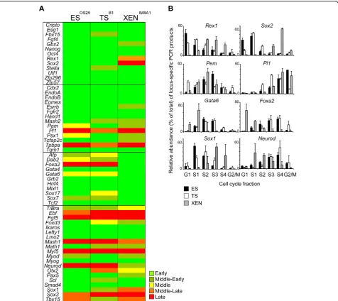
Most of the genes analysed replicated in early (or mid-dle-early) S-phase in all three stem cell lines (44, 44, 45 out of 58 genes in ES, TS and XEN cells, respectively, Figure 2A). These included a subset of ICM-associated genes (Oct4, Nanog andFgf4) expressed by ES cells, as well as genes associated with TE (Cdx2and Hand1) and PrE (Gata4andHnf4). In addition, many genes that are not thought to be expressed at significant levels in any of these cell types, for example Math1, Scland Myog,
Figure 2Embryonic stem (ES), trophectoderm (TS) and extra-embryonic endoderm (XEN) cell populations have distinct replication timing profiles, which reflect their lineage potential. (A) Summary of the replication timing comparison of the selected candidate genes between the three embryo-derived stem cell lines. The replication timing of each gene was defined according to its peak abundance in G1/S1 (early, dark green), S2 (middle-early, light green), S2 and S3 (middle, yellow), S3 (middle-late, orange) or S4/G2 (late, red), determined in at least two independent experiments. Inner cell mass/ES-, TE/TS-, PrE/XEN-relatedlocior genes involved in the specification of somatic cell types are grouped into four different boxes. (B) Histograms comparing the relative abundance oflocus-specific signal forRex1,Sox2,Pem,Pl1,Gata6,Foxa2, Sox1andNeurod lociwithin each cell cycle fraction for ES (black bars), TS (white bars) and XEN (grey bars) cells as assessed by quantitative polymerase chain reaction. Mean and standard deviation of two or more experiments are shown for each cell type analysed.
Santoset al.Epigenetics Chromatin2010,3:1
http://www.epigeneticsandchromatin.com/content/3/1/1
replicate early in all three embryonic stem cell lines. These data suggests that in TS and XEN cells, many developmental regulator genes remain ‘accessible’- as reflected by the prevalence of early replicating loci -similar to that reported previously for ES cells [19]. Overall, the replication timing profiles of ES and TS cells were similar (41/58) or identical (32/58), while XEN cells showed a greater disparity. This is illustrated by a delayed replication of several pluripotency-asso-ciated genes in XEN cells (for example,Rex1andSox2)
and the early replication of PrE-associated genesGata6
and Foxa2(Figure 2B) and is in keeping with the idea
that some tissue-specific genes may replicate earlier when transcriptionally active [29,30]. Similarly,Pemand
Psx1, which encode factors required for extra-embryonic lineages, replicated later in ES cells as compared to TS and XEN cells and the replication ofPl1, a TE-specific factor, was selectively advanced in TS cells (Figure 2B). These results were confirmed by analysing additional independent TS and XEN cell lines (Additional file 1) that were derived from mice carrying floxed Dicer alleles [31]. Comparing TSB1and TSDicerfx/fx or XENIM8A1and
XENDicerfx/fx(Figure 2 and Additional file 1), as well as
numerous different ES cell lines [19,32], confirmed that the replication timing profiles of different embryonic and extra-embryonic cell lines were robustly preserved.
Interestingly, the neural-associated genes Sox1 and
Neurodthat are not expressed by any of the embryonic
stem cell lines, showed clear differences in replication timing between ES, TS and XEN cells (Figure 2B, lower panel).Sox1 replication was advanced in ES cells while
Neurod replicated early in XEN cells. Although
unex-pected, these results suggest underlying changes in the chromatin context of these genes in the stem cell lines. In the case ofNeurod, although the transcription factor is known to function in neuronal development, it has also been shown to have an important role in the devel-opment of specialized cell types arising from the gut endoderm [33]. Despite being derived from the EPI and not from the PrE, gut endoderm cells have morphologi-cal and functional similarities to visceral endoderm cells [34]. The advanced replication ofNeurod in XEN cells might therefore reflect changes in transcriptional com-petence at thelocusthat is associated with an affiliation to the‘endoderm’lineage.
Chromatin profiling of gene promoters in stem cell lines
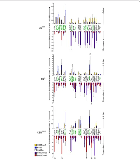
The chromatin profile of important regulator genes was compared between embryo-derived stem cell lines using chromatin immunoprecipitation (ChIP) in order to eval-uate the abundance of specific histone modifications that are associated with either active (H3K4me2, H4ac and H3K9ac) or repressed (H3K27me3 and H4K20me3) chromatin. For these analyses primers were designed to recognize the promoter region (up to 600 kb upstream
the transcriptional start site) of each candidate gene; genes that are known to be abundantly expressed by each cell type were used as positive controls for‘active’ chromatin marks. Pericentric heterochromatin (g -satel-lite repeats) provided controls for H4K20me3 immuno-precipitations, H3K27me3 was validated by analysing known bivalentloci in ES cells [19], and the abundance of modified histones was calculated relative to histone H3.
As anticipated, the promoters of many genes that are overtly expressed in ES cells (shown in bold, Figure 3, upper panel), as well as many bivalent genes (including
Eomes, Fgf5, Foxd3, Mash1, Math1, Sox1 and Tbx15)
were enriched for H3K4me2, and/or H3K9ac and H4ac at their promoters [19,20]. Exceptions included the pro-moters ofTpbpa andPl1, two markers of differentiated trophoblast. In ES cells histone H3K27me3, a modifica-tion catalyzed by polycomb repressor complex 2 (PRC2), was abundant at the promoters of genes that were either not expressed or expressed at low levels, including TS-associated genes (Cdx2, Eomes,Pem, Psx1), PrE-asso-ciated genes (Gata6, Foxa2) and genes that are normally expressed by subsets of differentiated tissue (such as
Mash1, Math1andNeurod) (shown in purple in Figure
3). Some silent late-replicating genes showed only low levels of H3K27 trimethylation (Tpbpaand Pl1) suggest-ing that these genes, in contrast to bivalent genes, are not developmentally‘poised’in ES cells and may, there-fore, require extensive chromatin-remodelling for cor-rect developmental expression. Levels of promoter H4K20me3 (shown in red in Figure 3), a mark asso-ciated with mammalian pericentric heterochromatin [35], were modest in ES cells with the exception ofSox2
(an observation that is likely to reflect the fact that OS25 cells carryingSox2 as a transgene). In TS (B1) and XEN (IM8A1) cell lines, in contrast to ES (OS25), H4K20me3 was detected at the promoters of many genes and was particularly enriched at several silent genes in XEN cells (Sox2, Foxd3, Sox1) (Figure 3, see Additional file 3 and Figure 1A for expression data). Taken as a whole these ChIP analyses suggest that, although the promoters of many development regulator genes are co-marked with histone modifications asso-ciated with active (acetylated, H3K4me2) and repressive (H3K27me3) chromatin in both ES and TS cells, this is not the case in XEN cells. Rather, in XEN cells histone marks that characterize accessible chromatin genes tend to be restricted to genes that are productively expressed at high(Gata6, Foxa2, Pem, Psx1) or moderate levels
(Eomes, Fbx15,Rex1, Tbx15). The exception to this
Figure 3Histone modifications at the promoters of key developmental regulator genes in embryonic stem (ES), trophectoderm stem (TS) and extra-embryonic endoderm (XEN) cells. The abundance of active [histone 3 lysine 4 dimethylation (H3K4me2, yellow bars), histone 4 acetylation (H4ac, blue bars), histone 3 lysine 9 acetylation (H3K9ac, white bars)] and repressive [histone 3 lysine 27 trimethylation (H3K27me3, purple bars), histone 4 lysine 20 trimethylation (H4K20me3, red bars)] histone marks at selectedlociwas assessed in ES, TS and XEN cells by chromatin immunoprecipitation and quantitative polymerase chain reaction. Values are shown as the ratio of modified histone H3 to unmodified histone H3 immunoprecipitations and normalized to an abundantly expressed gene in each cell type;Oct4in ES cells,Cdx2in TS cells andGata6 in XEN cells. Detected transcripts are highlighted in green while overt gene expression is shown in bold green. Primers were designed to the promoter region (100-600 bp upstream the transcriptional start site). Error bars represent the standard deviation of three independent experiments.
Santoset al.Epigenetics Chromatin2010,3:1
http://www.epigeneticsandchromatin.com/content/3/1/1
do not currently know the cause or significance of this single observation, collectively our data suggest that the chromatin structure of many genes is different between the stem cell lines, supporting earlier proposals that epi-genetic reprogramming occurs in embryonic and extra-embryonic lineages during early mouse development [17,18].
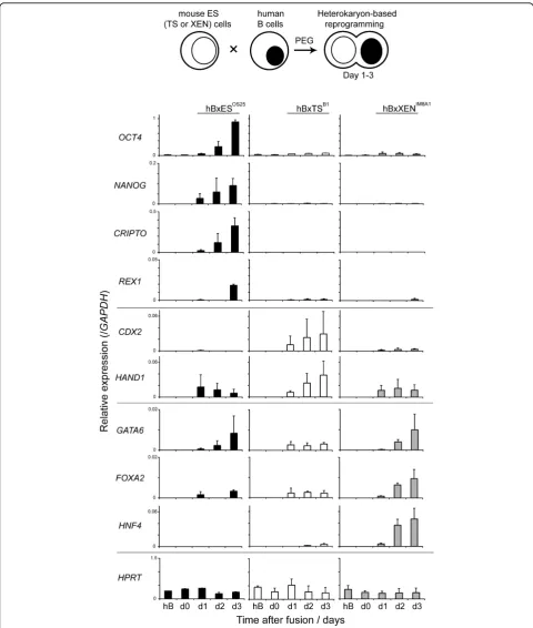
Mouse TS, XEN and ES cell lines dominantly reprogram lymphocytes in interspecies heterokaryons but induce the expression of different lineage-associated genes
Spontaneous and experimental cell fusions between ES cells and cells from a range of somatic tissues result in the nuclei of differentiated cells being reprogrammed to express an ES-specific gene expression pattern (so-called dominant reprogramming) [36,37]. Although the rules of dominance are not fully understood, other stem cell populations, including embryonic germ and embryonic carcinoma cells, can also reprogramme somatic cells towards a pluripotent state in vitro [38,39]. In order to establish whether extra-embryonic stem cell populations have this capacity and, moreover, whether reprogram-ming by these extra-embryonic cells induces thede novo
expression of different cohorts of genes, we tested the ability of TS and XEN cells in order to reprogramme human B cells using a previously established assay sys-tem [40]. Briefly, mouse ssys-tem cells and human B-lym-phocytes were mixed in a 1:1 ratio and inter-species heterokaryons (cells in which parental nuclei share the same cytoplasm but remain spatially separate) were gen-erated by polyethylene glycol (PEG)-mediated cell fusion. Human B cell reprogramming within these het-erokaryons was assessed 1 to 3 days after fusion, by qRT-PCR using primers that were designed (and vali-dated) to specifically amplify human transcripts.
Expres-sion of HPRT served as a positive control in these
analyses and transcripts derived from the human pluri-potency-associated genes (OCT4, NANOG,CRIPTOand
REX1), TE (CDX2 and HAND1) and PrE-associated
genes were examined in detail (GATA6, FOXA2 and
HNF4) (Figure 4). Human B cells (hB) did not express detectable levels of pluripotency-associated transcripts prior to fusion, but following heterokaryon formation
with mouse ES cells (hBxES), expression of OCT4,
NANOG,REX1and CRIPTOwas initiated and increased
up to day 3 (Figure 4, upper panel left). Fusion of hB with mouse TS (B1) or with mouse XEN (IM8A1) cells did not induce the expression of any of the human plur-ipotency-associated genes tested, including CRIPTO. This could be considered surprising as XEN (IM8A1) cells express high levels of mouse Cripto transcripts (several fold more than ES cells) and both TS and XEN cell lines express mouseRex1(Additional file 4). In het-erokaryons formed between hB and TS (B1), expression
of TE-associated genes (CDX2 and HAND1) was
induced, as well as low levels of some PrE-associated
genes (GATA6andFOXA2). Fusions between hBxXEN
cells resulted in a rapid and sustained induction of
GATA6, FOXA2 and HNF4 (Figure 4). In contrast,
mouse lymphocyte-specific transcripts (such asCD19,
CD37 and CD45) were not detected throughout these
experiments (data not shown) which is in line with the dominance of embryonic and extra-embryonic stem cells in reprogramming. These results collectively show that XEN and TS stem cell lines, like ES cells, retain a capacity to dominantly reprogramme somatic cells, but impose a programme of gene expression that is consis-tent with their different lineage affiliations.
Discussion
In this study we show that stem cell lines derived from the ICM, TE and PrE, display distinct epigenetic proper-ties as defined by replication timing, chromatin profiling and reprogramming potential. However, our studies revealed that many genes that are important in deter-mining cellular fate are retained in an‘accessible’ chro-matin state (acetylated and early replicating) in trophoblast-restricted stem cells (TS), being co-marked also by PRC2-mediated H3K27me3. This chromatin configuration, often referred to as ‘bivalent’, is shared with ES cells [19,20] and results in non-productive gene expression [41,42]. It is thought to be important for priming specific cohorts of genes for future develop-mental expression [19,43,44], and may therefore be important for restraining differentiation [41,45,46]. In keeping with this idea, our data show that few develop-mental regulator genes appear to be primed (bivalent) in XEN cells as compared with ES or TS cells, perhaps reflecting their narrower developmental potential. In addition, the delayed replication of several neuronal-associated genes in XEN cells relative to ES (for example
Figure 4Lineage restricted dominant reprogramming of human lymphocytes by embryo-derived stem cells. The reprogramming activity of embryonic stem (ES), trophectoderm stem (TS) and extra-embryonic endoderm (XEN) cells was assessed by heterokaryon formation with human B-lymphocytes (hB). Relative expression levels of human stem cell-specific genes in interspecies heterokaryons were detected by quantitative reverse transcriptase polymerase chain reaction analysis. The transcript levels of ES (OCT4,NANOG,CRIPTO,REX1), TS (CDX2,HAND1) and XEN-specific (GATA6,FOXA2andHNF4)genes were measured 1 to 3 days after fusion and normalized toGAPDHexpression. The constitutively expressed geneHPRTwas included as a control. Values shown are the mean from two independent experiments and error bars indicate standard deviations.
Santoset al.Epigenetics Chromatin2010,3:1
http://www.epigeneticsandchromatin.com/content/3/1/1
conserved role in the development of extra-embryonic endoderm lineages. Consistent with this idea, XEN cells exhibit a strong bias to form parietal endoderm in chi-meras, a tissue which is highly specialized for the synth-esis and secretion of extracellular matrix proteins [22,49].
The similar histone modifications and replication tim-ing profiles between ES and TS cells is consistent with mounting evidence indicating that only relatively few genes are uniquely restricted to the placenta, the vast majority of candidate TS-associated genes being involved in the development of other organs within the
embryo proper [50]. Some genes such as Oct4 and
Nanog, which are downregulated in TS cells (but remain
early-replicating), probably rely on alternative epigenetic mechanisms to suppress transcription in extra-embryo-nic lineages. For example, in TS cellsOct4 andNanog
regulatory domains are hyper (DNA) methylated and hypoacetylated, relative to ES cells [51,52]. Despite the overall similarity between ES and TS, the use of a candi-date-based replication timing assay allowsloci that are subject to chromatin re-modelling events early in mam-malian development to be readily identified. A number of studies have suggested that the generation of the ICM and TE requires the development of cell polarity in the outer cells of the morula, and the linked asymmetric divisions of blastomeres at the eight-cell-stage [53]. The significance of this polarization event is reflected by the identification oflociinvolved in cell polarity and cytos-keleton dynamics among candidates that replicate earlier in TS cells than in ES cells (such as Epb4.1l3,Fez2and
Cdh5, data not shown) in addition to Dab2, which are likely to be functional relevant for the biology of the tro-phoblast lineage.
The reprogramming properties of extra-embryonic stem cells have, to our knowledge, received little atten-tion. Here, experimental heterokaryons were generated to ask whether TS and XEN cells were capable of domi-nant reprogramming human somatic cells and, if so, whether they could impose different lineage-specific gene expression programmes. We demonstrate that TS and XEN cells reprogramme human B-lymphocytes in order to establish TE- or PrE-specific gene expression, respectively, albeit at low levels. As these fusions were performed using cells from different mammalian species, low expression levels may reflect inter-species differ-ences, such as mismatches between mouse factors and
cis acting elements within human genes [54]. Despite this, fusions using ES, TS or XEN cells reprogrammed human lymphocytes differently, the outcome reflecting discrete lineage affiliations. Interestingly, the expression of human transcripts by reprogrammed B-cell nuclei was not identical to that produced by the mouse stem cell eliciting the dominant reprogramming. This
observation mirrors previous reports that fusion with mouse ES cells, results in human B cells expressing a human ES-specific gene expression profile (hSSEA4, hFGF2 and hFGFR1), while hallmark factors of mouse ES cells, such as Lif receptor and Bmp4, are not acti-vated [38]. In this context, it seems likely that repro-grammed hB cells display features of human extra-embryonic-specific gene expression upon heterokaryon formation with mTS or mXEN cells, in agreement with published data [55]. Since extra-embryonic derived human stem cell lines have not been fully characterized, the generation of heterokaryon and hybrid cells using this approach could provide an important tool for study-ing human extra-embryonic lineages.
Conclusions
This report provides a preliminary epigenetic characteri-zation of mouse TE and PrE extra-embryonic lineages using stem cell lines as a model. We provide evidence of qualitative differences in the chromatin profiles between embryo-derived stem cell lines that accurately reflect their different transcriptional, lineage commitment and developmental potentials. These data support previous
in vivo studies of pre-implantation stage embryos
[13,14], showing that dynamic changes in chromatin occur at the earliest stages of mammalian development and are likely to be important for refining cellular potential.
Methods
Cell lines and cell culture
ES cells (OS25) were maintained in an undifferentiated state on 0.1% gelatin (StemCell Technologies, Vancou-ver, Canada)-coated flasks (Fisher Scientific UK Ltd,
Lei-cestershire, UK) in G-MEM-BHK 21 medium
(Invitrogen Ltd, Paisley, UK) supplemented with 10% fetal calf serum (FCS; PAA Laboratories, Gmbh, Pasch-ing, Austria), non-essential amino acids, sodium pyru-vate, sodium bicarbonate, antibiotics, L-glutamine, b -mercaptoethanol (Sigma-Aldrich Co Ltd, Gillingham, UK) and ESGRO-LIF (1000 U/ml) (Chemicon/Millipore, Billerica, USA). TS cell lines (B1 and Dicerfx/fx) were cultured in the presence of 70% mitotically inactivated mouse embryo fibroblast cells-conditioned medium and 30% TS medium to which human recombinant Fgf4 (25 ng/ml) (Sigma-Aldrich) and heparin (1μg/ml) (Sigma-Aldrich) were added. The TS cell medium was RPMI 1640 supplemented with 20% FCS (GlobePharm, Cork, Ireland), sodium pyruvate,b-mercaptoethanol, L-gluta-mine and antibiotics. XEN cell lines (IM8A1 and Dicerfx/fx) were maintained on 0.1% gelatin-coated flasks in RPMI 1640 supplemented with 20% FCS (GlobePharm), sodium pyruvate, L-glutamine,
B-lymphocyte clones were maintained in RPMI med-ium supplemented with 10% FCS (GlobePharm), L-glutamine and antibiotics. All cell lines used in this study were subjected to karyotypic analysis to check chromosome number. XEN cell lines routinely con-tained 40-46 chromosomes consistent with their pre-viously reported aneuploid status [22] while ES and TS cell lines appeared normal.
RT-PCR analysis
RNA extraction from ES, TS, XEN cells and heterokar-yons was performed using RNeasy protect mini kit (Qia-gen, USA) and RNase-free DNase set (Qiagen) for digestion of residual DNA. Total RNA (2.5μg) was then reverse transcribed using the Superscript first-strand synthesis system (Invitrogen) and cDNA of interest amplified in a total reaction volume of 50μL using 500 nM primers, and 1.25 U of HotStarTaq (Qiagen). The PCR cycling conditions were as follows: 95°C for 2 min, 30 cycles 95°C for 30 s, annealing at 60°C or 65°C for 30 s and elongation at 72°C for 2 min, finishing with a step at 72°C for 10 min.
Replication timing assay
BrdU-labelling, ethanol fixation, cell cycle fractionation by flow cytometry and isolation of BrdU-labelled DNA by immunoprecipitation were carried out as previously described [19] with the same BrdU-pulse labelling time for all three stem cell populations (30 min). The abun-dance of newly replicated DNA in each cell-cycle frac-tion was determined by real-time PCR amplificafrac-tion.
Real-Time PCR analysis
Real-Time PCR analysis was carried out on a Opticon™ DNA engine (MJ Research, Inc, MA, USA) under the following cycling conditions: 95°C for 15 min, 40 cycles at 94°C for 15 s, 60°C for 30 s, 72°C for 30 s followed by plate read. PCR reactions were performed in a 30 μL reaction volume containing 2× SYBR Green (Qiagen), 1.5μL of template and 300 nM primers. Each measure-ment was performed in duplicate. For heterokaryon
ana-lysis data were normalised to human GAPDH
expression.
Chromatin immunoprecipitation analysis
Exponentially growing ES, TS and XEN cells were pro-cessed for ChIP analysis as described previously [19]. 140μg chromatin was subjected to immunoprecipitation
with 5 μL anti-H3K9ac (Upstate Biotechnology, NY,
USA), 5 μL anti-H3K4me2 (Upstate), 5 μL anti-H4ac
(Upstate), 5 μL anti-H3K27me3 (Upstate), 5 μL anti-H4K20me3 (Upstate), 2.5μL of a rabbit anti-mouse-IgG antiserum (negative-control) (Dako Inc, CA, USA) and 4 μL of anti-H3 (Abcam, MA, USA). After purification, DNA was resuspended in 80μL TE solution. Quantifica-tion of precipitated DNA was performed using real-time qPCR (quantitative PRC) amplification. Histone’s modi-fication levels were normalized against total H3 detected
and the ratio of modified-H3 to H3 was denoted as rela-tive enrichment. ChIP experiments were performed twice.
Experimental heterokaryons
Heterokaryons were generated by fusing either mouse ES, TS or XEN cells and human B-lymphocytes using 50% polyethylene glycol, pH 7.4 (PEG 1500, Roche, Hertfordshire, UK). Equal numbers of stem cells and B-lymphocytes were mixed, washed twice in phosphate buffered saline at 37°C and 1 mL of PEG at 37°C was added to the pellet of cells over 60 s followed by an incubation at 37°C for 90 s. Cell mixtures were washed with 10 mL of DMEM at 37°C added over 3 min. After centrifugation the pellet was allowed to swell in com-plete medium for 3 min before resuspension. In order to eliminate non-fused hB cells Ouabain (10-5 M) was added to the medium. Proliferating stem cells were eliminated by the addition of 10-5 M Ara-C 6 h after fusion and then removed after 12 h. Fused cells were cultured under conditions promoting the maintenance of undifferentiated mouse stem cells.
Additional file 1: Replication timing and gene expression analysis of additional trophectoderm stem (TS) and extra-embryonic endoderm (XEN) cell lines. (A) Summary of the replication timing comparison of the selected candidate genes in TSDicerfx/fxand XENDicerfx/fxstem cell lines. The replication timing of each gene was defined according to its peak abundance in G1/S1 (early, dark green), S2 (middle-early, light green), S2 and S3 (middle, yellow), S3 (middle-late, orange) or S4/G2 (late, red), determined in at least two independent experiments. Inner cell mass/ES-, trophectoderm/TS-, primitive endoderm/XEN-relatedlocior genes involved in the specification of somatic cell types are grouped into four different boxes. Comparison with data presented in Figure 2A shows that 50/58 and 52/58 showed identical replication times in TS and XEN cells, respectively. Of the remaining genes, 8/8 and 6/6 showed similar replication times (peak abundance in an adjacent cell cycle fraction) in these cell lines. (B) Reverse transcriptase polymerase chain reaction expression analysis of TSDicerfx/fxand XENDicerfx/fxcell lines. RNA was isolated and cDNA analysed using primers forOct4,Nanog,Sox2, Fgf4,Rex1,Cdx2,Eomes,Esrrb,Hand1,Gata4,Gata6,Foxa2,Hnf4and Gapdhas a loading control. +/- indicates presence or absence of reverse transcriptase; H2O, water control.
Click here for file
[ http://www.biomedcentral.com/content/supplementary/1756-8935-3-1-S1.PDF ]
Additional file 2: Replication timing analysis of embryo-derived stem cell lines. (A) Typical cell cycle profiles for each cell population based on propidium iodide (PI) staining. Embryonic stem (ES), trophectoderm stem (TS) and extra-embryonic endoderm (XEN) cells lines were pulse-labelled with BrdU, stained with PI and separated by cell sorting into six cell cycle fractions according to DNA content. The gates used to define G1, S1, S2, S3, S4 and G2/M fractions of the cell cycle are indicated for each cell type. (B) Histograms showing the relative abundance of polymerase chain reaction products for early and late replicating controls (a-globinandAmylase 2.1, respectively) in each cell cycle fraction for ES (black bars), TS (white bars) and XEN (grey bars) cell lines.Drosophila melanogaster Gbeis an internal control for uniform recovery of BrdU-labelled DNA. Mean and standard deviation of two or more experiments are shown for each cell type analysed.
Click here for file
[ http://www.biomedcentral.com/content/supplementary/1756-8935-3-1-S2.PDF ]
Santoset al.Epigenetics Chromatin2010,3:1
http://www.epigeneticsandchromatin.com/content/3/1/1
Additional file 3: Expression analysis of lineage-specific genes in embryonic stem (ES), trophectoderm stem (TS) and extra-embryonic endoderm (XEN) cells. Total RNA was isolated from ES, TS and XEN cells lines, reverse-transcribed and analysed by reverse transcription
polymerase chain reaction using primers for several lineage-specific markers.Gapdhwas used as a loading control. +/-, with or without reverse transcriptase. Control tissues used as positive controls: embryoid bodies differentiated for 2.5 days (Fgf5), embryonic heads E12.5 (Mash1, Math1,NeurodandSox1) and embryonic placenta E12.5 (Pl1andTpbpa). Click here for file
[ http://www.biomedcentral.com/content/supplementary/1756-8935-3-1-S3.PDF ]
Additional File 4: Quantification of lineage-restricted transcription factors in embryo-derived stem cells. The relative expression ofOct4, Nanog, Cripto, Rex1, Sox2, Cdx2, Eomes, Hand1, Gata4, Gata6, FoxA2and Hnf4was assessed by quantitative reverse transcription polymerase chain reaction in embryonic stem (black bars), trophectoderm stem (white bars) and extra-embryonic endoderm (grey) cells. Developmental regulators characteristic of each cell type are highlighted with a blue box. Data was normalised toGapdhexpression. Values shown are the mean from three independent experiments and error bars indicate standard deviations.
Click here for file
[ http://www.biomedcentral.com/content/supplementary/1756-8935-3-1-S4.PDF ]
Abbreviations
ChIP: chromatin immunoprecipitation; EPI: epiblast; ES: embryonic stem; FCS: fetal calf serum; hB: human B cells; ICM: inner cell mass; PBS: phosphate buffered saline; PCR: polymerase chain reaction; PEG: polyethylene glycol; PRC2: polycomb repressor complex 2; PrE: primitive endoderm; qPCR: quantitative PCR; RT: reverse transcription; TE: trophectoderm; TS: trophectoderm stem; XEN: extra-embryonic endoderm.
Acknowledgements
We thank Austin Smith, Neil Brockdorff and Janet Rossant for ES, TS and XEN cell lines, respectively. We are also grateful to Tatyana Nesterova for help and advice with TS cell culture and Eric O’Connor and Eugene Ng for FACS sorting. This work was supported by the Medical Research Council, UK. JS was supported by a doctoral grant from Fundação para a Ciência e a Tecnologia, Portugal.
Author details
1Lymphocyte Development Group, MRC Clinical Sciences Centre, Imperial
College School of Medicine, Hammersmith Campus, Du Cane Road, London W12 0NN, UK.2Molecular Embryology Group, MRC Clinical Sciences Centre, Imperial College School of Medicine, Hammersmith Campus, Du Cane Road, London W12 0NN, UK.3Epigenetics and Development Group, Institute of Reproductive and Developmental Biology, Imperial College School of Medicine, Hammersmith Campus, Du Cane Road, London W12 0NN, UK.
Authors’contributions
JS and CFP contributed equally to this work. JS carried out the experiments with help from OA and analysed the data. CFP carried out the
reprogramming and revision experiments with help from AD, TS and TR. VA, MM and AGF participated on the design of the study. All authors have read and approved the manuscript.
Competing interests
The authors declare that they have no competing interests.
Received: 1 July 2009
Accepted: 12 January 2010 Published: 12 January 2010
References
1. Johnson MH, Ziomek CA:The foundation of two distinct cell lineages within the mouse morula.Cell1981,24:71-80.
2. Chazaud C, amanaka Y, Pawson T, Rossant J:Early lineage segregation between epiblast and primitive endoderm in mouse blastocysts through the Grb2-MAPK pathway.Dev Cell2006,10:615-624.
3. Rossant J, Tam PP:Blastocyst lineage formation, early embryonic asymmetries and axis patterning in the mouse.Development (Cambridge, England)2009,136:701-713.
4. Nichols J, Zevnik B, Anastassiadis K, Niwa H, Klewe-Nebenius D, Chambers I, Scholer H, Smith A:Formation of pluripotent stem cells in the mammalian embryo depends on the POU transcription factor Oct4.Cell 1998,95:379-391.
5. Strumpf D, Mao CA, Yamanaka Y, Ralston A, Chawengsaksophak K, Beck F, Rossant J:Cdx2 is required for correct cell fate specification and differentiation of trophectoderm in the mouse blastocyst.Development 2005,132:2093-2102.
6. Morrisey EE, Tang Z, Sigrist K, Lu MM, Jiang F, Ip HS, Parmacek MS:GATA6 regulates HNF4 and is required for differentiation of visceral endoderm in the mouse embryo.Genes Dev1998,12:3579-3590.
7. Plusa B, Frankenberg S, Chalmers A, Hadjantonakis AK, Moore CA, Papalopulu N, Papaioannou VE, Glover DM, Zernicka-Goetz M:
Downregulation of Par3 and aPKC function directs cells towards the ICM in the preimplantation mouse embryo.J Cell Sci2005,118:505-515. 8. Larue L, Ohsugi M, Hirchenhain J, Kemler R:E-cadherin null mutant
embryos fail to form a trophectoderm epithelium.Proc Natl Acad Sci USA 1994,91:8263-8267.
9. Kan NG, Stemmler MP, Junghans D, Kanzler B, de Vries WN, Dominis M, Kemler R:Gene replacement reveals a specific role for E-cadherin in the formation of a functional trophectoderm.Development2007,134:31-41. 10. Torres-Padilla ME, Parfitt DE, Kouzarides T, Zernicka-Goetz M:Histone
arginine methylation regulates pluripotency in the early mouse embryo.
Nature2007,445:214-218.
11. Chapman V, Forrester L, Sanford J, Hastie N, Rossant J:Cell lineage-specific undermethylation of mouse repetitive DNA.Nature1984,307:284-286. 12. Rossant J, Sanford JP, Chapman VM, Andrews GK:Undermethylation of
structural gene sequences in extraembryonic lineages of the mouse.Dev Biol1986,117:567-573.
13. Santos F, Hendrich B, Reik W, Dean W:Dynamic reprogramming of DNA methylation in the early mouse embryo.Dev Biol2002,241:172-182. 14. Santos F, Zakhartchenko V, Stojkovic M, Peters A, Jenuwein T, Wolf E,
Reik W, Dean W:Epigenetic marking correlates with developmental potential in cloned bovine preimplantation embryos.Curr Biol2003,
13:1116-1121.
15. Okamoto I, Otte AP, Allis CD, Reinberg D, Heard E:Epigenetic dynamics of imprinted X inactivation during early mouse development.Science2004,
303:644-649.
16. Mak W, Nesterova TB, de Napoles M, Appanah R, Yamanaka S, Otte AP, Brockdorff N:Reactivation of the paternal X chromosome in early mouse embryos.Science2004,303:666-669.
17. Ng RK, Dean W, Dawson C, Lucifero D, Madeja Z, Reik W, Hemberger M:
Epigenetic restriction of embryonic cell lineage fate by methylation of Elf5.Nat Cell Biol2008,10:1280-1290.
18. Farthing CR, Ficz G, Ng RK, Chan CF, Andrews S, Dean W, Hemberger M, Reik W:Global mapping of DNA methylation in mouse promoters reveals epigenetic reprogramming of pluripotency genes.PLoS Genet 2008,4:e1000116.
19. Azuara V, Perry P, Sauer S, Spivakov M, Jorgensen HF, John RM, Gouti M, Casanova M, Warnes G, Merkenschlager M, Fisher AG:Chromatin signatures of pluripotent cell lines.Nat Cell Biol2006,8:532-538. 20. Bernstein BE, Mikkelsen TS, Xie X, Kamal M, Huebert DJ, Cuff J, Fry B,
Meissner A, Wernig M, Plath K, Jaenisch R, Wagschal A, Feil R, Schreiber SL, Lander ES:A bivalent chromatin structure marks key developmental genes in embryonic stem cells.Cell2006,125:315-326.
21. Tanaka S, Kunath T, Hadjantonakis AK, Nagy A, Rossant J:Promotion of trophoblast stem cell proliferation by FGF4.Science1998,282:2072-2075. 22. Kunath T, Arnaud D, Uy GD, Okamoto I, Chureau C, Yamanaka Y, Heard E,
Gardner RL, Avner P, Rossant J:Imprinted X-inactivation in extra-embryonic endoderm cell lines from mouse blastocysts.Development 2005,132:1649-1661.
24. Tanaka TS, Kunath T, Kimber WL, Jaradat SA, Stagg CA, Usuda M, Yokota T, Niwa H, Rossant J, Ko MS:Gene expression profiling of embryo-derived stem cells reveals candidate genes associated with pluripotency and lineage specificity.Genome Res2002,12:1921-1928.
25. Zhang X, Zhang J, Wang T, Esteban MA, Pei D:Esrrb activates Oct4 transcription and sustains self-renewal and pluripotency in embryonic stem cells.J Biol Chem2008,283:35825-35833.
26. Hansen RS, Canfield TK, Lamb MM, Gartler SM, Laird CD:Association of fragile X syndrome with delayed replication of the FMR1 gene.Cell1993,
73:1403-1409.
27. Azuara V, Brown KE, Williams RR, Webb N, Dillon N, Festenstein R, Buckle V, Merkenschlager M, Fisher AG:Heritable gene silencing in lymphocytes delays chromatid resolution without affecting the timing of DNA replication.Nat Cell Biol2003,5:668-674.
28. Gilbert DM:Replication timing and transcriptional control: beyond cause and effect.Curr Opin Cell Biol2002,14:377-383.
29. Simon I, Tenzen T, Mostoslavsky R, Fibach E, Lande L, Milot E, Gribnau J, Grosveld F, Fraser P, Cedar H:Developmental regulation of DNA replication timing at the human beta globin locus.Embo J2001,20 :6150-6157.
30. Cimbora DM, Schubeler D, Reik A, Hamilton J, Francastel C, Epner EM, Groudine M:Long-distance control of origin choice and replication timing in the human beta-globin locus are independent of the locus control region.Mol Cell Biol2000,20:5581-5591.
31. Cobb BS, Nesterova TB, Thompson E, Hertweck A, O’Connor E, Godwin J, Wilson CB, Brockdorff N, Fisher AG, Smale ST, Merkenschlager M:T cell lineage choice and differentiation in the absence of the RNase III enzyme Dicer.J Exp Med2005,201:1367-1373.
32. Hiratani I, Ryba T, Itoh M, Yokochi T, Schwaiger M, Chang CW, Lyou Y, Townes TM, Schubeler D, Gilbert DM:Global reorganization of replication domains during embryonic stem cell differentiation.PLoS Biol2008,6: e245.
33. Naya FJ, Huang HP, Qiu Y, Mutoh H, DeMayo FJ, Leiter AB, Tsai MJ:
Diabetes, defective pancreatic morphogenesis, and abnormal enteroendocrine differentiation in BETA2/neuroD-deficient mice.Genes Dev1997,11:2323-2334.
34. Bielinska M, Narita N, Wilson DB:Distinct roles for visceral endoderm during embryonic mouse development.Int J Dev Biol1999,43:183-205. 35. Schotta G, Lachner M, Sarma K, Ebert A, Sengupta R, Reuter G, Reinberg D,
Jenuwein T:A silencing pathway to induce H3-K9 and H4-K20 trimethylation at constitutive heterochromatin.Genes Dev2004,18 :1251-1262.
36. Ying QL, Nichols J, Evans EP, Smith AG:Changing potency by spontaneous fusion.Nature2002,416:545-548.
37. Tada M, Takahama Y, Abe K, Nakatsuji N, Tada T:Nuclear reprogramming of somatic cells by in vitro hybridization with ES cells.Curr Biol2001,
11:1553-1558.
38. Tada M, Tada T, Lefebvre L, Barton SC, Surani MA:Embryonic germ cells induce epigenetic reprogramming of somatic nucleus in hybrid cells.
Embo J1997,16:6510-6520.
39. Miller RA, Ruddle FH:Pluripotent teratocarcinoma-thymus somatic cell hybrids.Cell1976,9:45-55.
40. Pereira CF, Terranova R, Ryan NK, Santos J, Morris KJ, Cui W, Merkenschlager M, Fisher AG:Heterokaryon-based reprogramming of human B lymphocytes for pluripotency requires Oct4 but not Sox2.PLoS Genet2008,4:e1000170.
41. Stock JK, Giadrossi S, Casanova M, Brookes E, Vidal M, Koseki H,
Brockdorff N, Fisher AG, Pombo A:Ring1-mediated ubiquitination of H2A restrains poised RNA polymerase II at bivalent genes in mouse ES cells.
Nat Cell Biol2007,9:1428-1435.
42. Guenther MG, Levine SS, Boyer LA, Jaenisch R, Young RA:A chromatin landmark and transcription initiation at most promoters in human cells.
Cell2007,130:77-88.
43. Lee TI, Jenner RG, Boyer LA, Guenther MG, Levine SS, Kumar RM, Chevalier B, Johnstone SE, Cole MF, Isono K, Koseki H, Fuchikami T, Abe K, Murray HL, Zucker JP, Yuan B, Bell GW, Herbolsheimer E, Hannett NM, Sun K, Odom DT, Otte AP, Volkert TL, Bartel DP, Melton DA, Gifford DK, Jaenisch R, Young RA:Control of developmental regulators by Polycomb in human embryonic stem cells.Cell2006,125:301-313.
44. Boyer LA, Plath K, Zeitlinger J, Brambrink T, Medeiros LA, Lee TI, Levine SS, Wernig M, Tajonar A, Ray MK, Bell GW, Otte AP, Vidal M, Gifford DK,
Young RA, Jaenisch R:Polycomb complexes repress developmental regulators in murine embryonic stem cells.Nature2006,441:349-353. 45. Spivakov M, Fisher AG:Epigenetic signatures of stem-cell identity.Nat Rev
Genet2007,8:263-271.
46. Endoh M, Endo TA, Endoh T, Fujimura Y, Ohara O, Toyoda T, Otte AP, Okano M, Brockdorff N, Vidal M, Koseki H:Polycomb group proteins Ring1A/B are functionally linked to the core transcriptional regulatory circuitry to maintain ES cell identity.Development2008,135:1513-1524. 47. Lehtonen E, Laasonen A, Tienari J:Teratocarcinoma stem cells as a model
for differentiation in the mouse embryo.Int J Dev Biol1989,33:105-115. 48. Yang Q, Bermingham NA, Finegold MJ, Zoghbi HY:Requirement of Math1
for secretory cell lineage commitment in the mouse intestine.Science 2001,294:2155-2158.
49. Hogan BL, Barlow DP, Kurkinen M:Reichert’s membrane as a model for studying the biosynthesis and assembly of basement membrane components.Ciba Found Symp1984,108:60-74.
50. Cross JC, Baczyk D, Dobric N, Hemberger M, Hughes M, Simmons DG, Yamamoto H, Kingdom JC:Genes, development and evolution of the placenta.Placenta2003,24:123-130.
51. Hattori N, Nishino K, Ko YG, Ohgane J, Tanaka S, Shiota K:Epigenetic control of mouse Oct-4 gene expression in embryonic stem cells and trophoblast stem cells.J Biol Chem2004,279:17063-17069.
52. Hattori N, Imao Y, Nishino K, Ohgane J, Yagi S, Tanaka S, Shiota K:
Epigenetic regulation of Nanog gene in embryonic stem and trophoblast stem cells.Genes Cells2007,12:387-396.
53. Johnson MH, McConnell JM:Lineage allocation and cell polarity during mouse embryogenesis.Semin Cell Dev Biol2004,15:583-597.
54. Odom DT, Dowell RD, Jacobsen ES, Gordon W, Danford TW, MacIsaac KD, Rolfe PA, Conboy CM, Gifford DK, Fraenkel E:Tissue-specific transcriptional regulation has diverged significantly between human and mouse.Nat Genet2007,39:730-732.
55. Adewumi O, Aflatoonian B, Ahrlund-Richter L, Amit M, Andrews PW, Beighton G, Bello PA, Benvenisty N, Berry LS, Bevan S, Blum B, Brooking J, Chen KG, Choo AB, Churchill GA, Corbel M, Damjanov I, Draper JS, Dvorak P, Emanuelsson K, Fleck RA, Ford A, Gertow K, Gertsenstein M, Gokhale PJ, Hamilton RS, Hampl A, Healy LE, Hovatta O,et al:
Characterization of human embryonic stem cell lines by the International Stem Cell Initiative.Nat Biotechnol2007,25:803-816.
doi:10.1186/1756-8935-3-1
Cite this article as:Santoset al.:Differences in the epigenetic and reprogramming properties of pluripotent and extra-embryonic stem cells implicate chromatin remodelling as an important early event in the developing mouse embryo.Epigenetics & Chromatin20103:1.
Publish with BioMed Central and every scientist can read your work free of charge
"BioMed Central will be the most significant development for disseminating the results of biomedical researc h in our lifetime."
Sir Paul Nurse, Cancer Research UK
Your research papers will be:
available free of charge to the entire biomedical community
peer reviewed and published immediately upon acceptance
cited in PubMed and archived on PubMed Central
yours — you keep the copyright
Submit your manuscript here:
http://www.biomedcentral.com/info/publishing_adv.asp
BioMedcentral
Santoset al.Epigenetics Chromatin2010,3:1
http://www.epigeneticsandchromatin.com/content/3/1/1