Complete genome sequence of Arcobacter nitrofigilis
type strain (CI
T)
Amrita Pati1, Sabine Gronow3, Alla Lapidus1, Alex Copeland1, Tijana Glavina Del Rio1, Matt Nolan1, Susan Lucas1, Hope Tice1, Jan-Fang Cheng1, Cliff Han1,2, Olga Chertkov1,2, David Bruce1,2, Roxanne Tapia1,2, Lynne Goodwin1,2, Sam Pitluck1, Konstantinos Liolios1, Natalia Ivanova1, Konstantinos Mavromatis1, Amy Chen4, Krishna Palaniappan4, Miriam Land1,5, Loren Hauser1,5, Yun-Juan Chang1,5, Cynthia D. Jeffries1,5, John C. Detter1,2, Manfred Rohde6, Markus Göker3, James Bristow1, Jonathan A. Eisen1,7, Victor Markowitz4, Philip Hugenholtz1, Hans-Peter Klenk3, and Nikos C. Kyrpides1*
1 DOE Joint Genome Institute, Walnut Creek, California, USA
2 Los Alamos National Laboratory, Bioscience Division, Los Alamos, New Mexico, USA 3 DSMZ – German Collection of Microorganisms and Cell Cultures GmbH, Braunschweig,
Germany
4 Biological Data Management and Technology Center, Lawrence Berkeley National Laboratory, Berkeley, California, USA
5 Oak Ridge National Laboratory, Oak Ridge, Tennessee, USA
6 HZI – Helmholtz Centre for Infection Research, Braunschweig, Germany 7 University of California Davis Genome Center, Davis, California, USA
*Corresponding author: Nikos C. Kyrpides
Keywords: symbiotic, Spartina alterniflora Loisel, nitrogen fixation, micro-anaerophilic, mo-tile, Campylobacteraceae, GEBA
Arcobacter nitrofigilis (McClung et al. 1983) Vandamme et al. 1991 is the type species of the genus Arcobacter in the family Campylobacteraceae within the Epsilonproteobacteria. The species was first described in 1983 as Campylobacter nitrofigilis [1] after its detection as a free-living, nitrogen-fixing Campylobacter species associated with Spartina alterniflora Loisel roots [2]. It is of phylogenetic interest because of its lifestyle as a symbiotic organism in a ma-rine environment in contrast to many other Arcobacter species which are associated with warm-blooded animals and tend to be pathogenic. Here we describe the features of this or-ganism, together with the complete genome sequence, and annotation. This is the first com-plete genome sequence of a type stain of the genus Arcobacter. The 3,192,235 bp genome with its 3,154 protein-coding and 70 RNA genes is part of the GenomicEncyclopedia of Bac-teria andArchaea project.
Introduction
Strain CI
T(= DSM 7299 = ATCC 33309 = CCUG
15893) is the type strain of the species
Arcobacter
nitrofigilis
, which is the type species of the genus
Arcobacter
[1]. Strain CI
Twas isolated from roots
of
Spartina alterniflora
Loisel (cordgrass) growing
in salty marshes at the East coast of Canada. It was
the first description of an organism in this kind of
habitat that belonged to the genus
Campylobacter,
as described based on phenotypic and biochemical
traits [2]. The species epithet
nitrofigilis
means
'nitrogen-fixing' and is based on the outstanding
characteristic of this species [3]. The new genus
Arcobacter
, meaning 'bow-shaped rod', was
intro-duced in 1991 and its separation from the genus
Campylobacter
was based on DNA and
DNA-rRNA hybridization [1]. Up to now, the genus
Ar-cobacter
comprises nine species, some of which
are associated with warm-blooded animals
whe-reas others are found in marine environments.
Within the
Campylobacteraceae
several
whole-genome sequences have already been deciphered:
the only member of the genus
Arcobacter
, as well
as genomes from seven species of the genus
Cam-pylobacter
, and
Sulfurospirillum deleyianum
[5].
Only few additional strains belonging to the
spe-cies
A. nitrofigilis
are known in the literature, with
F2176 and F2173 [6] being the closest related
ones (99% sequence identity). The type strains of
the other species of the genus
Arcobacter
share
93.8-94.6% 16S rRNA sequence identity with
strain CI
T, whereas the type strains from other
genera in the family
Campylobacteraceae
share
less than 89% sequence identity with strain CI
T[7]. There are plenty of phylotypes (uncultured
bacteria) known from marine environments such
as the ridges flanking crustal fluids in oceanic
crust (AY704399, clone FD118-51B-02, 98.6%),
sea water from Ishigaki port in Japan
(AB262370/-71, 96.4%), a mangrove of the
Dan-shui river estuary of northern Taiwan (DQ234254,
95.8%) [8], costal water in the Bohai Bay, China,
(FJ155005, 95.8%), in Black Sea shelf sediments in
Romania (AJ271655, 95.8%), or from activated
sludge in New Zealand (EU104146, 95.8%).
Envi-ronmental screens and marine metagenome
libra-ries do not contain more than a handful of
se-quences with >93% 16S rRNA gene sequence
identity indicating a sparse representation of
closely related strains in the habitats analyzed
(status March 2010). Here we present a summary
classification and a set of features for
A. nitrofigilis
strain CI
T, together with the description of the
complete genome sequencing and annotation.
Classification and features
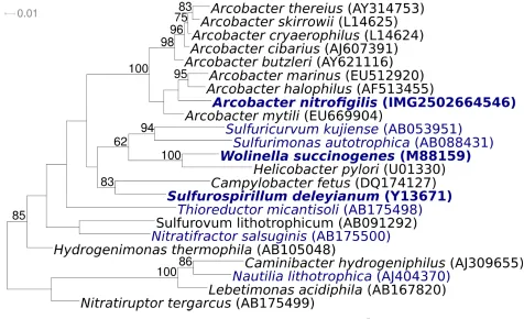
Figure 1 shows the phylogenetic neighborhood of
A. nitrofigilis
strain CI
Tin a 16S rRNA based tree.
The four 16S rRNA gene sequences in the genome
differ from each other by up to two nucleotides,
and differ by up to three nucleotides from the
pre-viously published 16S rRNA sequence (L14627)
generated from CCUG 15893, which contains nine
ambiguous base calls.
Figure 1. Phylogenetic tree highlighting the position of A. nitrofigilis strain CIT relative to the type strains of the
A. nitrofigilis
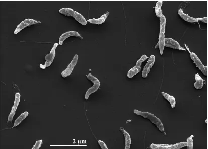
cells are Gram-negative, bow-shaped
or curved rods of 1–3 µm length and 0.2–0.9 µm
width (Figure 2 and Table 1). Motility is based on
a single, polar flagellum and results in rapid
corkscrew motion. Older cultures also show
coc-coid cells [2]. The habitat of all known
A.
nitrofigi-lis
isolates is either the roots or the sediment
around the roots of
S. alterniflora
Loisel growing
in salt marshes [3]. Although no pathogenic
asso-ciation has been described so far,
A. nitrofigilis
was among five
Arcobacter
species that were
iso-lated from food samples such as meat and
shell-fish varieties [27]. The optimum growth
tempera-ture of
A. nitrofigilis
is 30°C, the temperature
range is from 10–37°C [28]. Neither spores nor
granules are present but a brown pigment is
formed from tryptophan [2]. All strains of the
spe-cies show positive reactions for nitrogenase,
cata-lase and oxidase. Growth occurs under
microae-rophilic conditions with oxygen as terminal
elec-tron acceptor, under anaerobic conditions
fuma-rate or aspartate are necessary, the presence of
nitrate is detrimental [2]. Hydrogen is not
neces-sary for growth [1]. Nitrate is reduced to nitrite
and sulfide is produced from cysteine [3]. Strain
CI
Ttested positive for urease, other strains of the
species do not [3]. The metabolism of
A. nitrofigilis
is chemoorganotrophic; organic acids and amino
acids are used as carbon sources but
carbohy-drates are neither oxidized nor fermented [2]. All
strains of the species are halotolerant. They
re-quire a minimum of 0.5% NaCl for growth and can
tolerate up to 7% NaCl [28].
A. nitrofigilis
is
sus-ceptible to cephalothin and nalidixic acid but
isre-sistant to vancomycin [3]. The G+C content of the
DNA was determined by thermal denaturation to
be 28.0% [3] which is slightly below the 28.4%
found in the genome.
Figure 2. Scanning electron micrograph of A. nitrofigilis strain CIT
Genome sequencing and annotation
Genome project history
This organism was selected for sequencing on the
basis of its phylogenetic position [29], and is part
of the
Genomic
Encyclopedia of
Bacteria and
Arc-haea
project [30]. The genome project is
depo-sited in the Genomes OnLine Database [15] and
Table 1. Classification and general features of A. nitrofigilis strain CIT according to the MIGS recommendations [17]
MIGS ID Property Term Evidence code
Current classification
Domain Bacteria TAS [18]
Phylum ‘Proteobacteria’ TAS [19] Class Epsilonproteobacteria TAS [20,21] Order Campylobacterales TAS [20,22] Family Campylobacteraceae TAS [23] Genus Arcobacter TAS [1] Species Arcobacter nitrofigilis TAS [1]
Type strain CI TAS [3]
Gram stain negative TAS [2]
Cell shape bow-shaped rods TAS [2]
Motility motile TAS [2]
Sporulation non-sporulating TAS [2]
Temperature range mesophile, 10-37°C TAS [2]
Optimum temperature 30°C TAS [24]
Salinity halotolerant up to 7% NaCl TAS [2]
MIGS-22 Oxygen requirement microaerophilic TAS [2]
Carbon source organic and amino acids TAS [1]
Energy source chemoorganotroph TAS [3]
MIGS-6 Habitat marine TAS [2]
MIGS-15 Biotic relationship symbiotic TAS [2]
MIGS-14 Pathogenicity none NAS
Biosafety level 1 TAS [25]
Isolation roots of the marshplant Spartina alterniflora TAS [2] MIGS-4 Geographic location Conrads Beach (Dartmouth), Nova Scotia (Canada) TAS [2] MIGS-5 Sample collection time about or before 1980 TAS [2] MIGS-4.1
MIGS-4.2
Latitude Longitude
44.65
-63.60 NAS
MIGS-4.3 Depth unknown
MIGS-4.4 Altitude sea level
Evidence codes - IDA: Inferred from Direct Assay (first time in publication); TAS: Traceable Author Statement (i.e., a direct report exists in the literature); NAS: Non-traceable Author Statement (i.e., not directly observed for the living, isolated sample, but based on a generally accepted property for the species, or anecdotal evidence). These evidence codes are from of the Gene Ontology project [26]. If the evidence code is IDA, then the proper-ty was directly observed by one of the authors or an expert mentioned in the acknowledgements.
Chemotaxonomy
The major respiratory quinones are menaquinone
6 and a second atypical menaquinone 6 that has
not yet been clearly identified [1]. The major fatty
acids in whole cells of
A. nitrofigilis
are
hexadece-noic (C
16:0), cis-9-hexadecenoic (cis-C
16:1ϖ7c) and
cis-9-octadecenoic acid (cis-C
18:1ϖ7c) [24]
Growth conditions and DNA isolation
A. nitrofigilis
strain CI
T, DSM 7299, was grown on
DSMZ medium 429 (Columbia agar including 5%
horse blood) [31] at 28°C. DNA was isolated from
1-1.5 g of cell paste using Qiagen Genomic 500
DNA Kit (Qiagen, Hilden, Germany) with lysis
modification st/LALMP according to Wu
et al
.
[30].
Genome sequencing and assembly
of 24 kb were generated for this genome. All general
aspects of library construction and sequencing can
be found a
was based on 138 Mb 454 standard and 454 paired
end data (498,215 reads). Newbler (Roch, version
2.0.0PostRelease10/28/2008) parameters are
-consed -a 50 -l 350 -g -m -ml 20. The initial Newbler
assembly contained 42 contigs in 3 scaffolds. It was
converted into a phrap assembly by making fake
reads from the consensus and collecting the read
pairs in the 454 paired end library. Illumina
se-quencing data was assembled with Velvet [32], and
the consensus sequences were shredded into 1.5 kb
overlapped fake reads and assembled together with
the 454 data. Th
package was used for sequence assembly and
quali-ty assessment in the following finishing process.
Af-ter the shotgun stage, reads were assembled with
parallel phrap (High Performance Software, LLC).
Possible mis-assemblies were corrected with
PCR fragments with subcloning or transposon
bombing [33]. Gaps between contigs were closed by
editing in Consed, by PCR and by Bubble PCR primer
walks (J-F.Cheng, unpublished). A total of 480
addi-tional Sanger reactions were necessary to close gaps
and to raise the quality of the finished sequence.
Il-lumina reads were also used to improve the final
consensus quality using an in-house developed tool
(the Polisher). The error rate of the completed
ge-nome sequence is less than 1 in 100,000.
Table 2. Genome sequencing project information
MIGS ID Property Term
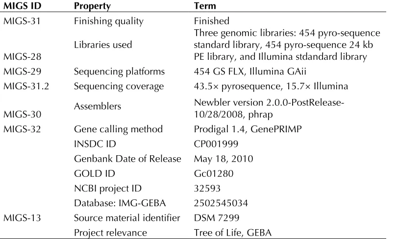
MIGS-31 Finishing quality Finished
MIGS-28 Libraries used
Three genomic libraries: 454 pyro-sequence standard library, 454 pyro-sequence 24 kb PE library, and Illumina stdandard library MIGS-29 Sequencing platforms 454 GS FLX, Illumina GAii
MIGS-31.2 Sequencing coverage 43.5× pyrosequence, 15.7× Illumina
MIGS-30 Assemblers Newbler version 2.0.0-PostRelease-10/28/2008, phrap MIGS-32 Gene calling method Prodigal 1.4, GenePRIMP
INSDC ID CP001999
Genbank Date of Release May 18, 2010
GOLD ID Gc01280
NCBI project ID 32593 Database: IMG-GEBA 2502545034 MIGS-13 Source material identifier DSM 7299
Project relevance Tree of Life, GEBA
Genome annotation
Genes were identified using
of the Oak Ridge National Laboratory genome
an-notation pipeline, followed by a round of manual
curation using the JGI
The predicted CDSs were translated and used to
search the National Center for Biotechnology
In-formation (NCBI) nonredundant database,
Uni-Prot, TIGR-Fam, Pfam, PRIAM, KEGG, COG, and
In-terPro databases. Additional gene prediction
anal-ysis and functional annotation was performed
within the Integrated Microbial Genomes - Expert
Review (IMG-ER) platform [36].
Genome properties
Table 3. Genome Statistics
Attribute Value % of Total
Genome size (bp) 3,192,235 100.00%
DNA coding region (bp) 3,009,967 94.29%
DNA G+C content (bp) 905,345 28.36%
Number of replicons 1
Extrachromosomal elements 0
Total genes 3,224 100.00%
RNA genes 70 2.17%
rRNA operons 4
Protein-coding genes 3,154 97.83%
Pseudo genes 70 2.17%
Genes with function prediction 2,324 72.08%
Genes in paralog clusters 454 14.08%
Genes assigned to COGs 2,363 73.29%
Genes assigned Pfam domains 2,480 76.92%
Genes with signal peptides 597 18.52%
Genes with transmembrane helices 838 25.99%
CRISPR repeats 1
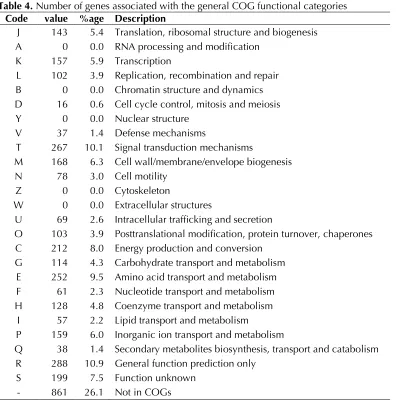
Table 4. Number of genes associated with the general COG functional categories Code value %age Description
J 143 5.4 Translation, ribosomal structure and biogenesis A 0 0.0 RNA processing and modification
K 157 5.9 Transcription
L 102 3.9 Replication, recombination and repair B 0 0.0 Chromatin structure and dynamics D 16 0.6 Cell cycle control, mitosis and meiosis
Y 0 0.0 Nuclear structure V 37 1.4 Defense mechanisms
T 267 10.1 Signal transduction mechanisms
M 168 6.3 Cell wall/membrane/envelope biogenesis N 78 3.0 Cell motility
Z 0 0.0 Cytoskeleton
W 0 0.0 Extracellular structures
U 69 2.6 Intracellular trafficking and secretion
O 103 3.9 Posttranslational modification, protein turnover, chaperones C 212 8.0 Energy production and conversion
G 114 4.3 Carbohydrate transport and metabolism E 252 9.5 Amino acid transport and metabolism F 61 2.3 Nucleotide transport and metabolism H 128 4.8 Coenzyme transport and metabolism
I 57 2.2 Lipid transport and metabolism
P 159 6.0 Inorganic ion transport and metabolism
Q 38 1.4 Secondary metabolites biosynthesis, transport and catabolism R 288 10.9 General function prediction only
S 199 7.5 Function unknown - 861 26.1 Not in COGs
Acknowledgements
We would like to gratefully acknowledge the help of Sabine Welnitz for growing the A. nitrofigilis cells, and Susanne Schneider for DNA extraction and quality analysis (both at DSMZ). This work was performed un-der the auspices of the US Department of Energy Office of Science, Biological and Environmental Research Pro-gram, and by the University of California, Lawrence Berkeley National Laboratory under contract No.
DE-AC02-05CH11231, Lawrence Livermore National La-boratory under Contract No. DE-AC52-07NA27344, Los Alamos National Laboratory under contract No. DE-AC02-06NA25396, and UT-Battelle Oak Ridge National Laboratory under contract DE-AC05-00OR22725, as well as German Research Foundation (DFG) INST 599/1-2.
References
1. Vandamme P, Falsen E, Rossau R, Hoste B, Segers P, Tytgat RDE, Ley J. Revision of Campylobacter, Helicobacter, and Wolinella taxonomy: emenda-tion of generic descripemenda-tions and proposal of Arco-bacter gen. nov. Int J Syst Bacteriol 1991; 41 :88-103 2. McClung CR, Patriquin DG. Isolation of a
nitro-gen-fixing Campylobacter species from the roots of Spartina alterniflora Loisel. Can J Microbiol
1980; 26:881-886
3. McClung CR, Patriquin DG, Davis RE. Campylo-bacter nitrofigilis sp. nov. a nitrogen-fixing bacte-rium associated with roots of Spartina alterniflora Loisel. Int J Syst Bacteriol 1983; 33:605-612.
ge-nome sequence and analysis of the Epsilonpro-teobacterium Arcobacter butzleri.PLoS ONE
2007; 2:e1358
5. Sikorski J, Lapidus A, Copeland A, Glavina Del Rio T, Nolan M, Lucas S, Chen F, Tice H, Cheng JF, Saunders E, et al. Complete genome of Sulfu-rospirillum deleyianum type strain (5175T). Stand
Genomic Sci 2010; 2:149-157.
6. Figueras MJ, Collado L, Guarro J. A new 16S rRNA-RFLP method for the discrimination of the accepted species of Arcobacter.Diagn Microbiol Infect Dis 2008; 62:11-15
7. Chun J, Lee JH, Jung Y, Kim M, Kim S, Kim BK,
Lim YW. EzTaxon: a web-based tool for the iden-tification of prokaryotes based on 16S ribosomal RNA gene sequences. Int J Syst Evol Microbiol 2007; 57:2259-2261
8. Liao PC, Huang BH, Huang S. Microbial commu-nity composition of the Danshui river estuary of northern Taiwan and the practicality of the phy-logenetic method in microbial barcoding. Microb Ecol 2007; 54:497-507
9. Castresana J. Selection of conserved blocks from multiple alignments for their use in phylogenetic analysis. Mol Biol Evol 2000; 17:540-552
10. Lee C, Grasso C, Sharlow MF. Multiple sequence alignment using partial order graphs. Bioinformat-ics 2002; 18:452-464
11. Stamatakis A, Hoover P, Rougemont J. A Rapid Bootstrap Algorithm for the RAxML Web Servers. Syst Biol 2008; 57:758-771
12. Felsenstein J. Evolutionary trees from DNA se-quences: a maximum likelihood approach. J Mol Evol 1981; 17:368-376
13. Yarza P, Richter M, Peplies J, Euzeby JP, Amann R, Schleifer KH, Ludwig W, Glöckner FO, Rossel-lo-Mora R. The All-Species Living Tree project: A 16S rRNA-based phylogenetic tree of all se-quenced type strains. Syst Appl Microbiol 2008;
31:241-250
14. Pattengale ND, Alipour M, Bininda-Emonds ORP, Moret BME, Stamatakis A. How Many Bootstrap
Replicates Are Necessary? Lect Notes Comput Sci
2009; 5541:184-200
15. Liolios K, Chen IM, Mavromatis K, Tavernarakis N, Hugenholtz P, Markowitz VM, Kyrpides NC. The Genomes On Line Database (GOLD) in 2009: status of genomic and metagenomic projects and their associated metadata. Nucleic Acids Res 2010; 38:D346-D354
16. Baar C, Eppinger M, Raddatz G, Simon J, Lanz C, Klimmek O, Nandakumar R, Gross R, Rosinus A, Keller H, et al. Complete genome sequence and analysis of Wolinella succinogenes.Proc Natl Acad Sci USA 2003; 100:11690-11695
17. Field D, Garrity G, Gray T, Morrison N, Selengut J, Sterk P, Tatusova T, Thompson N, Allen MJ, Anguiuoli SV, et al. Towards a richer description of our complete collection of genomes and meta-genomes: the “Minimum Information about a Ge-nome Sequence” (MIGS) specification. Nat Bio-technol 2008; 26:541-547
18. Woese CR, Kandler O, Wheelis ML. Towards a natural system of organisms: proposal for the do-mains Archaea, Bacteria, and Eucarya. Proc Natl Acad Sci USA 1990; 87:4576-4579
19. Garrity GM, Holt JG. The Road Map to the Ma-nual. In: Garrity GM, Boone DR, Castenholz RW (eds), Bergey's Manual of Systematic Bacteriology, Second Edition, Volume 1, Springer, New York, 2001, p. 119-169.
20. Validation List No. 107. List of new names and new combinations previously effectively, but not validly, published. Int J Syst Evol Microbiol 2006;
56:1-6
21. Garrity GM, Bell JA, Lilburn T. Class V. Epsilon-proteobacteria class. nov. In: Garrity GM, Brenner DJ, Krieg NR, Staley JT (eds), Bergey's Manual of Systematic Bacteriology, Second Edition, Volume 2, Part C, Springer, New York, 2005, p. 1145. 22. Garrity GM, Bell JA, Lilburn T. Order I.
Campylo-bacterales ord. nov. In: Garrity GM, Brenner DJ, Krieg NR, Staley JT (eds), Bergey's Manual of Sys-tematic Bacteriology, Second Edition, Volume 2, Part C, Springer, New York, 2005, p. 1145. 23. Vandamme P, De Ley J. Proposal for a new
fami-ly, Campylobacteraceae. Int J Syst Bacteriol 1991;
24. Donachie SP, Bowman JP, On SL, Alam M. Arco-bacter halophilus sp. nov., the first obligate halo-phile in the genus Arcobacter.Int J Syst Evol Mi-crobiol 2005; 55:1271-1277
25. Classification of. Bacteria and Archaea in risk groups. www.baua.de TRBA 466.
26. Ashburner M, Ball CA, Blake JA, Botstein D, But-ler H, Cherry JM, Davis AP, Dolinski K, Dwight SS, Eppig JT, et al. Gene ontology: tool for the un-ification of biology. Nat Genet 2000; 25:25-29
27. Collado L, Guarro J, Figueras MJ. Prevalence of Arcobacter in meat and shellfish. J Food Prot 2009; 72:1102-1106
28. Tenover FC, Fennell CL. The genera Campylobac-ter and Helicobacter. In: The Prokaryotes. Vol. IV, 2nd ed. 1992, Springer-Verlag, New York.
29. Klenk HP, Göker M. En route to a genome-based taxonomy of Archaea and Bacteria? Syst Appl Mi-crobiol 2010; 33:175-182
30. Wu D, Hugenholtz P, Mavromatis K, Pukall R, Dalin E, Ivanova N, Kunin V, Goodwin L, Wu M, Tindall BJ, et al. A phylogeny-driven genomic en-cyclopedia of Bacteria and Archaea. Nature 2009; 462:1056-1060
31. List of growth media used at DSMZ:
32. Zerbino DR, Birney E. Velvet: algorithms for de novo short read assembly using de Bruijn graphs. Genome Res 2008; 18:821-829
33. Sims D, Brettin T, Detter JC, Han C, Lapidus A, Copeland A, Glavina Del Rio T, Nolan M, Chen F, Lucas S, et al. Complete genome of Kytococcus sedentarius type strain (541T). Stand Genomic Sci
2009; 1:12-20
34. Hyatt D, Chen GL, Locascio PF, Land ML, Lari-mer FW, Hauser LJ. Prodigal Prokaryotic Dynam-ic Programming Genefinding Algorithm. BMC Bioinformatics 2010; 11:119
35. Pati A, Ivanova N, Mikhailova N, Ovchinikova G, Hooper SD, Lykidis A, Kyrpides NC. GenePRIMP: A Gene Prediction Improvement Pipeline for mi-crobial genomes. Nat Methods 2010; 7:455-457
![Table 1. Classification and general features of A. nitrofigilis strain CIT according to the MIGS recommendations [17] MIGS ID Property Term Evidence code](https://thumb-us.123doks.com/thumbv2/123dok_us/674319.2066142/4.612.68.558.71.497/classification-general-features-nitrofigilis-according-recommendations-property-evidence.webp)