214 *Corresponding author: Dr Seyed Mohammad Jazayeri
MD, PhD-Associate Professor, Clinical Virologist, Hepa-titis B Lab-Dept Virology, School of Public Health, Tehran University of Medical Sciences, Tehran, Iran
Zip Code: 14155-6446
Evolution of hepatitis B virus surface gene and protein among Iranian
chronic carriers from different provinces
Fatemeh Ramezani1, Seyed Moayed Alavian2, Ahmadreza Sadeghi1, Abolfazl Khedive1, Leila
Ghalichi3, Mehdi Norouzi1, Hadi Karimzadeh1, Reza Malekzadeh4, Ghodrat Montazeri4, Azim
Nejatizadeh5, Masood Ziaee6, Farshid Abedi7, Behrooz Ataei8, Majid Yaran8, Babak Sayad9, Mohamad
Hosein Somi10, Gholamreza Sarizadeh11, Ismail Sanei-Moghaddam12, Fariborz Mansour-Ghanaei13,
Houshang Rafatpanah14, Hossein Keyvani15, Ebrahim Kalantari16, Mehdi Saberfiroozi17, Reza Rezaee18,
Maryam Daram1, Mostafa Mahabadi1, Zahra Goodarzi1, Vahdat Poortahmasebi1, Babak Geravand18,
Azam Khamseh1, Masoud Mahmoodi1, Seyed Mohammad Jazayeri1*.
1Hepatitis B Molecular Laboratory-Department of Virology-School of Public Health-Tehran Uni-versity of Medical Sciences, Tehran, Iran.
2Middle East Liver Diseases Center (MELD Centers), Tehran, Iran.
3Mental health research center, Iran University of Medical Sciences, Tehran, Iran.
4Digestive Disease Research Center, Tehran University of Medical Sciences, Tehran, Iran.
5Research Center for Molecular Medicine, Hormozgan University of Medical Sciences, Bandar Abbas, Iran.
6Hepatitis Research Center, Department of Internal Medicine, Faculty of Medicine, Birjand University of Medical Sciences, Birjand, Iran.
7Department of Infectious Disease, Mashhad University of Medical Sciences, Iran.
8Infectious Diseases and Tropical Medicine Research Center, Isfahan University of Medical Sciences, Isfahan, Iran.
9Kermanshah Liver Diseases and Hepatitis Research Center, Kermanshah, Iran.
10Liver and Gastrointestinal Disease Research Center, Tabriz University of Medical Sciences, Tabriz, Iran.
11Educational Region of Khozestan Blood Transfusion Organization, Ahvaz, Iran.
12Department of Gastroenterology, Zahedan University of Medical Sciences, Zahedan, Iran.
13Gastrointestinal & Liver Diseases Research Center, Guilan University of Medical Sciences, Rasht, Iran.
14Immunology Research Center, Mashhad University of Medical Sciences, Mashhad, Iran.
15Department of Virology, School of Medicine, Iran University of Medical Sciences, Tehran, Iran.
16Gholhack medical laboratory Tehran, Iran.
17Gastroenterohepatology Research Center, Shiraz University of Medical Sciences, Shiraz, Iran.
18Ministry of Health and Medical Education, Deputy of Curative Affairs, Budget Administration, Tehran, Iran.
Received: February 2014, Accepted: August 2014
ORIGINAL
AR
TICLE
INTRODUCTION
Hepatitis B virus (HBV) is a hepatotropic, non-cy-topathic DNA virus which has been estimated to in-fect one third of global population, of whom 350 to 400 million suffering from chronic infection. This chronicity estate is responsible for major complica-tions of HBV infection including cirrhosis and hepa-tocellular carcinoma.
The HBV infection prevalence in Iran has been es-timated at 2.14% to 2.6% (1-3). This prevalence in males is about 25% higher than females (2.55% vs. 2.03% rates, respectively) (1).
According a systematic review by Alavian et al, about 1.5 million people are suffering from HBV in-fection in Iran, of whom15% to 40% (1) are at risk of developing cirrhosis and/or hepatocellular carcino-ma (HCC) without intervention (4, 5). They also esti-mated that 225,000 to 600,000 Iranian chronic HBV carriers are at risk of serious health problems related to HBV infection and need immediate attention (1). Other patients are HBsAg positive carriers that may disseminate infection to healthy people vertically or horizontally. Moreover, 51% to 56% of Iranian cir-rhotic patients are HBsAg positive (6, 7). The most common routes of transmission reported in national surveys are perinatal transmission and intravenous drug abuse (8). The geographic distribution of HBV
infection in Iran showed heterogeneous patterns with the highest prevalence rates (more that 3% of the population) in northeastern region of the country while central and western regions showed the lowest prevalence rates (1% to 2%) (1).
Different epidemiological studies with alternative
methodologies have been taken place to find out the
risk factors and the routes of transmission of hepati-tis B virus in Iran. Factors predicting HBV infection included family history of hepatitis B infection, his-tory of receiving blood transfusion, hospitalization, unsafe sex, male gender, and living in city area (3, 8-10).
A national-wide Iranian study had undertaken for HBV genotyping between 2006 and 2011 by Iranian Hepatitis Network and Hepatitis B laboratory at Tehran University of Medical sciences, 360 sam-ples from different regions and ethnic groups. An extensive molecular analysis based on surface genes and proteins of isolates carried out as a whole which published in recent years. However, the details of differences of molecular analysis between provinc-es have not published yet. Therefore, the aim of this study was to summarize the data extracted from de-mographic, biochemical, serological/molecular epi-demiology analysis among different ethnic groups of Iranian HBV chronic carriers.
ABSTRACT
Background and Objectives: Iranian chronic HBV carrier’s population has shown a unique pattern of genotype D distri-bution all around the country. The aim of this study was to explore more details of evolutionary history of carriers based on structural surface proteins from different provinces.
Materials and Methods: Sera obtained from 360 isolates from 12 Different regions of country were used for amplification and sequencing of surface proteins. A detailed mutational analysis was undertaken.
Results: The total ratio for Missense/Silent nucleotide substitutions was 0.96. Sistan and Kermanshah showed the lowest rate of evolution between provinces (P = 0.055). On the other hand, Khorasan Razavi and Khoozestan contained the highest ratio (P = 0.055). The rest of regions were laid between these two extremes. Azarbayjan and Guilan showed the highest proportion of immune epitope distribution (91.3% and 96%, respectively). Conversely, Sistan and Tehran harbored the least percentage (66.6% and 68.8%, respectively). Kermanshah province contained only 5.2%, whereas Isfahan had 54.5% of B cell epitope distribution. In terms of T helper epitopes, all provinces showed a somehow homogeneity: 22.58% (Fars) to 46.6% (Khuz-estan). On the other hand, distribution of substitutions within the CTL epitopes showed a wide range of variation between 6.6% (Khuzestan) and 63% (Kermanshah).
Conclusion: Further to low selection pressure found in Iranian population, the variations between different regions designate random genetic drift within the surface proteins. These finding would have some applications in terms of specific antiviral regimen, design of more efficient vaccine and public health issues.
http://ijm.tums.ac.ir MATERIALS AND METHODS
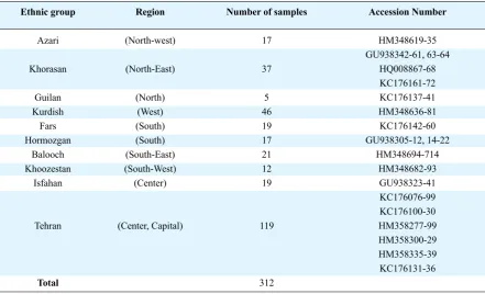
360 HBsAg-positive chronic carriers who were referred to the Iranian Hepatitis Network (2004-2010) were enrolled in a cross-sectional study. Dif-ferent regions based on population and geographical zones were chosen to cover the whole ethnic parts of country (Table 1). The capital city, Tehran, with a population around 13 million was chosen as a versa-tile multi-cultural and multi-ethnic region without a
definite ethnic background. All patients were known
chronic carriers. They had no evident of
co-infec-tion with other hepatitis virus, human immunodefi -ciency virus (HIV), and they were treatment-naive. The diagnosis of chronic liver disease was made by clinical, biochemical, radiological and endoscopic criteria. 5 ml aliquots of whole blood samples were withdrawn from each participant. Serum was, asep-tically, separated. All sera were referred to Hepatitis B Laboratory, School of public Health for serological and molecular analysis. The methodology was
216 IRAN. J. MICROBIOL. Volume 7 Number 4 (August 2015) 214-220
tical for all specimens. The HB viral DNA was ex-tracted from 200 µl of sera using Qiagen Mini Blood Kit (Qiagen, Hilden, Germany) according to manu-facturer’s instruction. DNA was eluted using 100 µl of elution buffer, stored at -20°C. Polymerase chain reaction (PCR) was carried out in 100 µl of a mixture containing 5 µl of the extracted DNA using standard
methods. The complete surface gene was amplified
using S1, S2, S6 and S7 primers which included the region of surface gene specifying HBV genotypes/ subtype (amino acid positions 122-160). Direct sequencing of surface gene was carried out (Genet-ic Analyzer ABI- 3130 DNA Sequencer, Fostercity, CA, USA) using 2 pmol of appropriate primers: S6C and S7D for surface gene. The electropherograms were examined visually using Chromas program. Sequences of surface gene were aligned using the BioEdit Package version 7.0.9. 312 sequences were submitted to Genbank, under individual accession number (Table 1).
Ethnic group
Azari
Khorasan
Guilan Kurdish
Fars Hormozgan
Balooch Khoozestan
Isfahan
Tehran
Total
Region
(North-west)
(North-East)
(North) (West) (South) (South) (South-East) (South-West) (Center)
(Center, Capital)
Number of samples
17
37
5 46 19 17 21 12 19
119
312
Accession Number
HM348619-35 GU938342-61, 63-64
HQ008867-68 KC176161-72 KC176137-41 HM348636-81 KC176142-60 GU938305-12, 14-22
HM348694-714 HM348682-93
GU938323-41 KC176076-99 KC176100-30 HM358277-99 HM358300-29 HM358335-39 KC176131-36
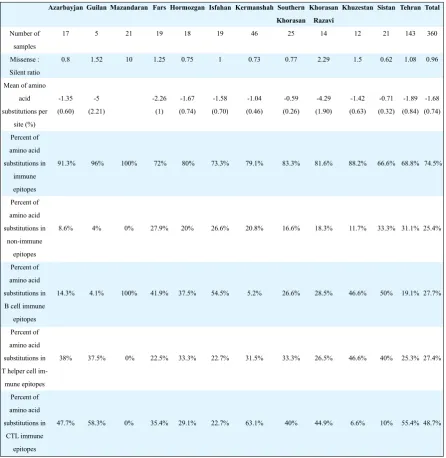
In addition, it was possible to identify the level of S proteins evolution between isolates by measuring the mutation frequency (Missense/Silent, dN/dS) of in-dividual sequences. The average mutation frequency of all sequences was 0.96 according to the number of mutations per site. The total number of amino acid substitutions was 542, of which 404 (74.5%) were dis-tributed in known immune epitopes of surface protein (Table 2). The average amino acid mutation frequency was 1.68 mutations per sample. There was a probabil-ity of 0.74% for substitution per amino acid position.
Comparison of evolution history of HBsAg be-tween provinces. Previous results as a whole were presented elsewhere (12). The results obtained from the comparison between amino acid substitutions and demographic, biochemistry and type of substitutions
among provinces showed no significant associations
between the number of amino acid mutations with age, gender and the ALT levels of chronic carriers. On the other hand, strong correlations were found be-tween the HBeAg status of the patients and the
occur-rence of amino acid substitutions; a significant pro -portion of such changes occurred in anti-HB epositive individuals. Moreover, substantial relationships were found between the occurrence of mutations within and outside of surface protein immune epitope residues, a majority were occurred in anti-HBe positive patients. Also, comparison between the distribution of amino acid mutations within the B, Th and CTL epitopes among provinces showed considerable variations.
Provinces comparison. In terms of evolutionary history of HBV among different population (Table 2), due to great divergence between results obtained for all provinces, a precise molecular analysis was carried out on individual patients in each region, instead of individual provinces. Results showed that
ignoring small, non-significant differences, almost all
locations showed a homogenous pattern (results not shown). The exception was non-synonymous/ synon-ymous (dN/dS) ratio. Sistan and Kermanshah showed the least ration of evolution between provinces (0.62 and 0.73, respectively, P = 0.055). On the other hand, Khorasan Razavi and Khoozestan contained the high-est ratio (2.29 and 1.5, respectively, P = 0.055). The rest of regions were laid between these two extremes. The distribution of amino acid changes showed that of 542 mutated residues, 404 (74.5%) were occurred
in immune epitopes (Table 2), a novel finding that has
For the purpose of mutational analysis, we used a genotype D sequence isolated from New Guin-ea reported by Okomoto et al. (accession number AB033559). Our previous search for the best com-parable sequence has led to the selection of this
iso-late as the best fit reference sequence for comparison
alignment. The differences between this isolate and Iranian were only one and six mismatches in terms of amino acid and nucleotide, respectively. The dis-tributions of amino acid changes were determined according to known immune epitope residues of HBsAg (11). For the purpose of evolutionary history study of isolates, previously we used Silent/Missense (dS/dN) ratio. For a more clear analysis we used Mis-sense/Silent (dN/dS) ratio in this study.
RESULTS
Basic characteristics. 360HBsAg-positive chron-ic patients were enrolled in this study; all were native residents between different regions of Iran. All were chronic carriers, HBV DNA positive and treat-ment-naive. 247(68.61%) were male and 113 (31.38%) were female with a mean age of 36.34 ±12.42 (mean±SD). The mean ALT and viral load level were 81.95 ± 64.85 IU/L (mean±SD) and 15000 copy/mL, respectively (results not shown). 20.5%(n=74) and 68.8% (n=248) were HBeAg and anti-HBe positive, respectively. Also 9.72% (n=35) and 0.83% (n=3) were negative and positive for both markers, respectively.
Genotyping results. The results of the first large
phylogenetic tree (contained all the studied-sequenc-es) revealed that Iranian HBV isolates were of gen-otype D, supported by 95% bootstrap value (1,000 replicates, results not shown). Phylogenetic analy-sis of 360 complete surface genes could distinguish
five sub-genotypes, D (100%); sub-genotypes were
D1:97.7% (n = 352), D2: 0.55% (n = 2), D3: 0.27% (n = 1), D5: 0.83% (n = 3) and D8: 0.55% (n = 2). Finally, all isolates belonged to subtype ayw2 (100%) (Results not shown).
http://ijm.tums.ac.ir Total 360 0.96 -1.68 (0.74) 74.5% 25.4% 27.7% 27.4% 48.7%
218 IRAN. J. MICROBIOL. Volume 7 Number 4 (August 2015) 214-220 not been reported for treatment/vaccine naive chronic HBV patients so far. Azarbayjan and Guilan showed the highest proportion of immune epitope distribution (91.3% and 96%, respectively). Conversely, Sistan and Tehran harbored the least percentage (66.6% and 68.8%, respectively).
112 (27.7%), 111 (27.4%) and 197 (48.7%) of mu-tations which clustered within immune epitopes were localized to B, T helper and CTL epitopes within the surface protein, respectively. Distribution of amino
acid substitution within the B cell epitope (residue 100-160) showed that Kermanshah province con-tained only 5.2%, whereas Isfahan had 54.5% of such distribution. In terms of T helper epitopes, all provinc-es showed a somehow homogeneity; 22.58% (Fars) to 46.6% (Khuzestan). On the other hand, distribution of substitutions within the CTL epitopes showed a wide range of variation between 6.6% (Khuzestan) and 63% (Kermanshah). For more molecular details as a whole, readers refer to the previous report (12).
Table 2. Details of mutational analysis from different provinces
Tehran 143 1.08 -1.89 (0.84) 68.8% 31.1% 19.1% 25.3% 55.4% Sistan 21 0.62 -0.71 (0.32) 66.6% 33.3% 50% 40% 10% Khuzestan 12 1.5 -1.42 (0.63) 88.2% 11.7% 46.6% 46.6% 6.6% Khorasan Razavi 14 2.29 -4.29 (1.90) 81.6% 18.3% 28.5% 26.5% 44.9% Southern Khorasan 25 0.77 -0.59 (0.26) 83.3% 16.6% 26.6% 33.3% 40% Kermanshah 46 0.73 -1.04 (0.46) 79.1% 20.8% 5.2% 31.5% 63.1% Isfahan 19 1 -1.58 (0.70) 73.3% 26.6% 54.5% 22.7% 22.7% Hormozgan 18 0.75 -1.67 (0.74) 80% 20% 37.5% 33.3% 29.1% Fars 19 1.25 -2.26 (1) 72% 27.9% 41.9% 22.5% 35.4% Mazandaran 21 10 100% 0% 100% 0% 0% Guilan 5 1.52 -5 (2.21) 96% 4% 4.1% 37.5% 58.3% Azarbayjan 17 0.8 -1.35 (0.60) 91.3% 8.6% 14.3% 38% 47.7% Number of samples Missense : Silent ratio Mean of amino
acid substitutions per site (%) Percent of amino acid substitutions in immune epitopes Percent of amino acid substitutions in non-immune epitopes Percent of amino acid substitutions in B cell immune
DISCUSSION
According to CDC classification for the worldwide
prevalence of HBV, Iran has been located as interme-diate area with a mean prevalence of 2.14. This article represents the molecular data based on comparison between 360 chronicin active patients from 12 differ-ent parts of Iran. The proportion of amino acid muta-tion in present reports showed that as high as 61.6% of chronic HBV patients obtain at least one amin oac-id mutation within the structural surface protein. This
finding is much higher than other reports obtained
from genotypes B, C and D worldwide. The impact of these genotypic alterations on the pathogenesis of HBV in chronic carriers phenotypically deserves fur-ther in vitro investigations.
Our previous reports on these samples indicated that a majority of such changes occurred in anti-HBe pos-itive cases (13-15). Transition from HBeAg positivity to anti-HBe positivity is somehow a prolonged pro-cess, during which a number of amino acid changes are proportional to the interaction between virus and host immune status. Not surprising, it seems that in the presence of host immune surveillance, the occur-rence of such mutations is inevitable (14).
Comparison of evolutionary changes within surface proteins between different provinces showed that Kho-rasan Razavi contained the most versatile features of mutation. On the other hand South Khorasan (North-East) and Sistan-Baloochestan Provinces showed the less variable features (along with Kermanshah and Hormozgan). We already discussed that the low evo-lutionary history found in Sistan Baloochestan and South Khorasan (14, 16).We assume that eastern bor-der of Iran which include these provinces contains a different history of HBV evolution compared to other parts of country. We already hypothesized that the rea-son for low prevalence of HBV liver disease compli-cations in these two regions, might be related to slow rate of substitutions (regardless of being synonymous or non-synonymous) during the course of chronicity (14, 16). Obviously, we cannot exclude the same hy-pothesis for other regions who contained Missense/ Silent (dN/dS)ratio <1 (see Table 2), however, details of molecular analysis in these parts and the nature of amino acid substitutions were distinguished Eastern part from other regions with somehow the same ratio. One of interesting features in the present study which make it different from other published data on chron-ic HBV carriers is the unique pattern of amino acid
distribution in CTL epitopes (47.8%) and on the other hand, a low prevalence of “a” determinant variation in the surface proteins from 61.6% carriers. Previous in-vestigation showed a prevalence of 10.8% and 11.2% of HBsAg variation in chronic carriers, mainly located within the “a” determinant (17-19). All reported sur-veys from Iran showed that the only HBV genotype is D. Is there any correlation between this unique pattern of genotype and the number and the nature of distribu-tion of amino acid changes? To answer this quesdistribu-tion, other similar investigations from other parts of worlds which contain the genotype D predominant is need-ed to avoid bias on recombination between genotype D and other genotypes. The best candidate would be the sequences from countries of Caucasian origin like Turkey, Pakistan and India with prevalence of geno-type D being: 87%, 62% and 67%, respectively (20).
We did not find similar results on comparison between
available surface proteins in international databases. Therefore, we assume that Iranian genetic background (perhaps types of HLA) might be responsible for a high number of amino acid changes in surface protein despite being chronic carriers. We already hypothe-sized that differences in distribution of HLA antigens, or other immune genes between diverse geographical areas probably contributed to the selection of amino acid variation (21).
In keeping with circulation of HBV amongst Ira-nian ethnic group sit seems that after seroconversion to anti-HBe, no further selection pressure exists for substitutions in surface proteins among other geno-type D-infected ethnic groups (other non-Iranian Cau-casians). The same situation is true for Iranian ethnic group, as the total (dN/dS) ratio was 0.96, indicating a natural negative selection pressure. However, we be-lieve that in the long term of progression of chronicity and as time goes by, random genetic drift is responsi-ble for such high number (and also the distribution of amino acids). In fact, once a mutation has arisen in a
population what determine the outcome ( to be fixed
or to be lost), is not always down to how much bet-ter or worse it is (natural selection) compared to those variations already present in the population; instead it may simply be down to chance. As random genet-ic drift observed most strongly in small populations, substitutions of amino acid in various regions of coun-try with versatile features of B, Th and CTL epitopes distribution (see Table 2) designate this unique
muta-tional profile. Does this phenomenon related to either
http://ijm.tums.ac.ir types) factors need to be explored in the future,
ap-plying longitudinal, cohort studies including patients from different phases of chronicity (before and after HBeAg seroconversion).
In conclusion, the significance of unique pictures of
mutational distribution despite low evolutionary rate in Iranian chronic carriers came up from the present study would have some applications in terms of
specif-ic antiviral regimen, design of more effspecif-icient vaccine
and public health issues in the next future.
REFERENCES
1. Alavian SM, Ahmadzad-Asl M, Hajariazdeh B, Kabir A, Bagheri Lankarani K. Hepatitis B Virus Infec-tion in Iran: A Systematic Review. Hepatitis Monthly
2008;8: 14.
2. Alavian SM, Fallahian F, Bagheri Lankarani K. The changing epidemiology of viral hepatitis B in Iran. J Gastrointestin Liver Dis 2007;16(4): 403-406.
3. Merat S, Rezvan H, Nouraie M, Jamali A, Assari S, Abolghasemi H, et al. The prevalence of hepatitis B surface antigen and anti-hepatitis B core antibody in Iran: a population-based study. Arch Iran Med
2009;12: 225-231.
4. McMahon BJ. Epidemiology and natural history of hepatitis B. Semin Liver Dis 2005;25 Suppl 1: 3-8. 5. McMahon BJ. Natural history of chronic hepatitis B -
clinical implications. Medscape J Med 2008;10: 91. 6. Bagheri Lankarani K, Saberi Firoozi M, Nabipoor I,
Fattahi F, Sarafraz YazdiM, Malek Zadeh R, et al. Re-assesment of the role of hepatitis B and C viruses in postnecrotic cirrhosis and chronic Hepatitis in south-ern Iran. Iran J Med Sci 1999;24: 117-121.
7. Shamszad M. Hepatitis B related cirrhosis and hepato-cellular carcinoma in Iran. J Iran Med Council 1982;8: 228.
8. Merat S, Malekzadeh R, Rezvan H, Khatibian M. Hep-atitis B in Iran. Arch Iran Med 2000;3: 192-201. 9. Vahid T, Alavian SM, Kabir A, Kafaee J, Yektaparast
B.Hepatitis B Prevalence and Risk Factors in Blood Donorsin Ghazvin. Hep Mon 2005;5: 117-122.
10. Alavian SM, Mostajabi P, Malekzadeh R, Azimi K, Vosoogh H, Sarrafi M, et al. Evaluation of Hepatitis B Transmission Risk Factors in Tehran Blood Donors.
Govaresh 2004;9(169-75): 169.
11. Alavian SM, Carman WF and Jazayeri SM.HBsAg
variants: Diagnostic-escape and diagnostic dilemma.
J Clin Virol 2013; 57: 201-208.
12. Khedive A, Norouzi M, Ramezani F, Karimzadeh H, Alavian SM, Malekzadeh R, et al. Hepatitis B virus surface protein mutations clustered mainly in CTL im-mune epitopes in chronic carriers: results of an Iranian nationwide study. J Viral Hepat 2013;20: 494-501. 13. Norouzi M, Ghorashi SA, Ataei B, Yaran M,
Malekza-deh R, Alavian SM, et al. Hepatitis B Virus Surface Antigen Variants Clustered Within Immune Epitopes in Chronic Hepatitis B Carriers from Hormozgan Province, South of Iran. Iran JBasic Med Sci 2010; 13: 213-224.
14. Ghaziasadi A, Ziaee M, Norouzi M, Malekzadeh R, Alavian SM, Saberfar E, et al. The Prevalence of Hep-atitis B Virus Surface Antigen (HBsAg) Variations and Correlation with the Clinical and Serologic Pic-tures in Chronic Carriers from Khorasan Province, North-East of Iran. Acta Med Iran 2012;50: 265-272. 15. Sayad B, Anvari FA, Alavian SM, Norouzi M,
Ham-zelooie M, Shirvani M, et al. Correlation of Hepatitis B surface antigen mutations with clinical status of the chronically infected patients from Kermanshah, West of Iran. Minerva Gastroenterol Dietol 2012;58: 9-18. 16. Khedive A, Sanei-Moghadam I, Alavian SM,
Saber-far E, Norouzi M, Judaki MA, et al. Hepatitis B Vi-rus Surface Antigen (HBsAg) Mutations Are Rare but Clustered in Immune Epitopes in Chronic Carriers from Sistan-Balouchestan Province, Iran. Arch Iran Med 2013;16: 385-389.
17. Avellon A and Echevarria JM. Frequency of hepatitis B virus 'a' determinant variants in unselected Spanish chronic carriers. J Med Virol 2006;78: 24-36.
18. Guptan RC, Thakur V, Sarin SK, Banerjee K, Khandekar P. Frequency and clinical profile of precore and surface hepatitis B mutants in Asian-Indian pa-tients with chronic liver disease. Am J Gastroenterol
1996;91: 1312-1317.
19. Yamamoto K, Horikita M, Tsuda F, Itoh K, Akahane Y, Yotsumoto S, et al. Naturally occurring escape mu-tants of hepatitis B virus with various mutations in the S gene in carriers seropositive for antibody to hepatitis B surface antigen. J Virol 1994;68: 2671-2676. 20. Jazayeri SM, Alavian SM, Carman WF. Hepatitis B
virus: origin and evolution. J Viral Hepat 2010;17: 229-235.
21. JazayeriSM, Basuni AA, Sran N, Gish R, Cooksley G, Locarnini S, et al. HBV core sequence: definition of genotype-specific variability and correlation with geo -graphical origin. J Viral Hepat 2004;11: 488-501.