The repair of full‑thickness articular
cartilage defect using intra‑articular
administration of
N
‑acetyl‑
d
‑glucosamine in the
rabbit knee: randomized controlled trial
Nai‑Jen Chang
1, Yi‑Ting Lin
2, Chih‑Chan Lin
3, Hsueh‑Chun Wang
2, Horng‑Chaung Hsu
4and Ming‑Long Yeh
2,5*Abstract
Background: Although various alterative models of therapy are used for cartilage repair, no definite conclusion has been reached. Glucosamine (GlcN) is widely used as a nutritional supplement. However, the clinical‑ evidence‑based outcome of GlcN administration remains controversial. N‑acetyl‑d‑glucosamine (GlcNAc), a derivative
of GlcN, shows chondroprotective activity and mediates the activation of articular chondrocytes. Therefore, we investigated the effect of intra‑articular administration of GlcNAc in rabbits’ knee joints with experimental full‑thickness articular cartilage (FTAC) defects.
Methods: Twelve male adult New Zealand white rabbits, providing 24 knees, were used in this study. FTAC defects were created in the high‑weight‑bearing area of the medial femoral condyles of bilateral knees. All rabbits were randomly allocated to analysis at postsurgical week 4 or postsurgical week 12. In the week 4 group, rabbits’ knees (six per group) were intra‑articularly injected with normal saline or with GlcNAc twice per week for 3 weeks, beginning 1 week postoperatively. In the week 12 group, the rabbits’ knees (six in each group) were intra‑articularly injected with normal saline or with GlcNAc twice per week for 4 weeks, beginning 1 week postoperatively. Rabbits were sacrificed at 4 or 12 weeks after surgery for macroscopic, histological and radio‑ logical examinations of the knee joints.
Results: All rabbits had no systemic or local adverse effects. The saline and GlcNAc groups showed visible differences in healing of the FTAC defect at the end of testing. At week 4, the GlcNAc group had a higher level of collagen type II (COL II) and showed up‑regulated production of transforming growth factor (TGF)‑β2 and TGF‑β3, suggest‑ ing the involvement of endogenous growth factors. At week 12, the GlcNAc group displayed formation of hyaline‑like cartilage regeneration with mature chondrocytes (SOX9+), robust glycosaminoglycan (GAG) content, and positive COL II content in both the adjacent cartilage and reparative sites. However, the saline group demonstrated mainly fibrocartilage scar tissue, indicating COL I expression. Furthermore, the GlcNAc group had significantly higher bone volume per tissue volume and higher trabecular thickness than the saline group.
Conclusions: Intra‑articular GlcNAc may promote the repair of experimental FTAC defects in the rabbit knee joint model.
Keywords: Glucosamine, Intra‑articular injection, Tissue engineering, Cartilage
Open Access
© 2015 Chang et al. This article is distributed under the terms of the Creative Commons Attribution 4.0 International License (http:// creativecommons.org/licenses/by/4.0/), which permits unrestricted use, distribution, and reproduction in any medium, provided you give appropriate credit to the original author(s) and the source, provide a link to the Creative Commons license, and indicate if changes were made. The Creative Commons Public Domain Dedication waiver (http://creativecommons.org/publicdomain/ zero/1.0/) applies to the data made available in this article, unless otherwise stated.
RESEARCH
Background
The repair of articular cartilage defects is particularly challenging because of cartilage’s hypocellularity and insufficient nutrient supply, and the inability of bone marrow stem cells or resident chondroprogenitor cells to form hyaline cartilage. Current clinical ther-apeutic treatments for cartilage repair include intra-articular therapy, e.g., non-steroidal anti-inflammatory drugs, corticosteroids, dextrose prolotherapy, hyaluronic acid (HA) or platelet-rich plasma [1, 2], microfracture, autologous chondrocyte implantation, and osteochondral transplantation techniques [3, 4]. However, these treatment options, par-ticularly for large cartilage defects, present not only the formation of fibrocartilage scar tissue but also poor integration of hyaline cartilage in the host [5, 6]. Hence, regarding their effectiveness for large cartilage lesions, there is a lack of both clinical consensus and reproducible evidence that currently available treatment significantly changes the progression of osteoarthritis, based on prospective, randomized studies [7–9]. Thus far, no defined conclusion for cartilage repair has been reported.
Glucosamine (GlcN) is one of the most widely utilized dietary supplements. It is also believed to stimulate the metabolism of chondrocytes, thus providing beneficial effects on articular health. However, the clinical outcomes of GlcN administration orally remain controversial due to the various formulations and purity of GlcN [10–13]. GlcN intake as a nutritional supplement is commercially available in one of three forms: GlcN hydro-chloride, GlcN sulfate, and N-acetyl-d-glucosamine (GlcNAc). Since a molecule of pure GlcN is essentially unstable, it should be salt-stabilized (e.g., chloride or sulfate) [14]. In particular, GlcNAc, classified as a monosaccharide derived from glucose, has a relatively stable structure.
and effects on glucose transport, glucose transporter expression, and synthesis of sul-fated GAG and HA [27]. A previous study investigated oral GlcNAc and GlcN admin-istration on plasma total free amino acid (PFAA) concentrations in dogs [28]. GlcNAc group has the higher Glu, Gly, and Ala concentrations after 1 h administration compared with GlcN group. The three kind of non essential amino acid are the main components of type II collagen in cartilage.
Therefore, we investigated whether intra-articular injection of GlcNAc could provide a safe niche for the repair of full-thickness articular cartilage (FTAC) defects in rabbit knee joints.
Methods Reagent
GlcNAc (molecular formula: C8H15NO6) was purchased from Sigma (SI-A3286, MO,
USA). It was dissolved in 0.9 % saline solution, and sterile filtered The sterilized solution was stored at 4 °C.
Surgical procedure
All surgical procedures and animal ethics were approved by the Animal Care and Use Committee of National Cheng Kung University. Twelve 4–5-month-old male New Zea-land white rabbits (Livestock Research Institute, Taiwan) weighing 2.5–3 kg were used in this experiment, providing 24 knees. Before surgery, anesthesia was induced with Zoletil 50 (25 mg/kg) (Virbac, France) and maintained with a mixture of 2 % isoflurane (Pan-ion & BF Biotech Inc., Taiwan) and oxygen/nitrous oxide (1/0.4 L/min). Both knees of each rabbit were shaved and disinfected with 10 % ethanol-iodine solution. Longitudinal incisions were made along the parapatellar and capsular ligaments. The medial femo-ral condyle was exposed by latefemo-ral patellar dislocation. A critical FTAC defect, 3 mm in diameter and 3 mm deep, was created with an electric drill in the high-weight-bearing region of the medial femoral condyle, simultaneously irrigated with saline for cooling, followed by patellar relocation and joint closure. After surgery, the rabbits were permit-ted to move freely in their cages. Appetite, body weight, skin wound healing and func-tional activities were monitored.
Intra‑articular injection
Each animal was given either saline or GlcNAc. At 7 days after surgery, in the week 4 group, rabbits’ knees (six knees per group) were injected with 0.3 ml of normal ster-ile saline (0.9 % NaCl) or intra-articularly with GlcNAc solution twice per week start-ing 1 week post-surgery for a period of 3 weeks. In the week 12 group, rabbits’ knees (six knees per group) were injected with normal saline or intra-articularly with GlcNAc twice per week starting 1 week post-surgery for a period of 4 weeks. The single dose of GlcNAc was 80 mg/0.3 ml per joint. In addition, the recommended frequency of intra-articular injection for optimal therapeutic efficacy is twice per week [18].
Macroscopic evaluation
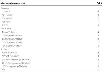
neo-tissue coverage, color and surface condition were assessed independently by two investigators. The maximum possible total score was 12 points.
Micro‑CT evaluation
Micro-computed tomography (CT) can provide quantitative and qualitative measure-ments to identify the healing of bone architecture. A high-resolution microtomograph 1076 scanner (SkyScan, Kontich, Belgium) was employed to scan the distal femur FTAC defect in the saline and GlcNAc groups at 4 and 12 weeks after surgery. The voltage and beam current of the X-ray source were set at 50 kV and 160 μA, respectively. The pixel size was set at 18 μm in resolution. The samples were scanned with 360° rotation with 1° of rotation interval.
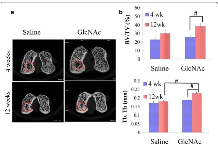
The SkyScan software package, including SkyScan CT-Analyzer v.1.8, and CT-Volume v.2.0, were used to reconstruct the image data and visualize the representation of the newly formed bone. From the data set, a cylindrical region of interest (ROI) 3 mm in diameter within the repaired site was selected for analysis. This ROI corresponds to the original defect region. The bone volume per tissue volume (BV/TV) and trabecular thickness (Tb.Th) were obtained to evaluate the bone volume density and to measure the thickness of trabecular structures, respectively.
Histological and immunohistochemistry processing
All histological sections were performed by the Department of Pathology at Chi-Mei Medical Center. The femur ends were dissected, fixed in 10 % neutral buffered formal-dehyde solution, decalcified and embedded in paraffin blocks. From each sample, the tissue was sliced by a microtome into 4 μm thick sections for histological staining. Hematoxylin and eosin (H&E) staining was performed to allow general observations; Table 1 A modified Wayne’s grading scale scoring system for gross appearance
Macroscopic appearance Points
Coverage
>75 % fill 4
50–75 % fill 3
25–50 % fill 2
<25 % fill 1
0 % fill 0
Tissue color
Normal/whitish 4
>25 % yellow/reddish 3
>50 % yellow/reddish 2
>75 % yellow/reddish 1
100 % yellow/reddish 0
Surface
Normal, smooth 4
Smooth but raised 3
25–50 % irregularity/fibrillation 2
50–75 % irregularity/fibrillation 1
>75 % irregularity/fibrillation 0
Masson’s trichrome stain was used for total collagen and alignment; Alcian blue staining was performed for GAG synthesis. Furthermore, immunohistochemistry analysis was conducted to confirm the expression of collagen type I (COL I, fibrocartilage), collagen type II (COL II, hyaline cartilage), endogenous growth factors (i.e., 2 and TGFβ-3) as well as SOX9 (marker of mature chondrocytes) for the repair of FTAC defects. All staining protocols followed the manufacturers’ recommended guidelines. Briefly, the sections were treated with endogenous peroxidase and incubated overnight with a mon-oclonal primary antibody diluted 1:100: COL I (Bioworld Technology), COL II (Bioss), TGFβ-2 or TGFβ-3 (Spring Bioscience), or SOX9 (Bioss). The rabbit/mouse HRP-DAB polymer detection kit (BioSB) was used as the secondary antibody at room temperature for 30 min. Then, DAB (3,3′ diaminobenzidine) staining in brown represented positive immunoreactivity. The sections were counterstained with hematoxylin (BioSB) and then recorded using a light microscope (Olympus IX71, Tokyo, Japan) and a digital CCD camera (Olympus DP72, Tokyo, Japan).
Statistical analysis
The software package SPSS v17.0 was used for the statistical analysis. All of the data (six knees per group at week 4 or 12) are expressed as the mean ± standard error of the mean. Owing to the concern of repeated measurements from bilateral knees on the same individual [30], a linear model using generalized estimating equations (GEE) was employed for statistical comparison at each time point. P-values <0.05 were defined as statistically significant. The statistical power of this study was 0.9 as determined by the mean, variation, and sample size.
Results
Macroscopic observations and scoring in the FTAC model
The gross appearance of FTAC defects was assessed at both 4 and 12 weeks after surgery (Fig. 1a). No infection, synovitis or osteophyte formation was observed at either time point. At week 4, the defect areas in both the saline and GlcNAc groups were visibly concave although new tissue had developed inwards from the outer area of the defect edges. At week 12, the GlcNAc group had formed a transparent neo-cartilage-like tissue. Meanwhile, the saline group showed incomplete filling, with hybrid whitish/yellow tis-sues and an obvious fissure in the reparative site (Fig. 1a).
The total scores in both saline (6.00 ± 0.40) and GlcNAc groups (7.33 ± 0.76) were comparable at week 4 after surgery. However, at week 12 after surgery, the total score for the GlcNAc group (8.33 ± 0.47) was superior to that for the saline group (5.36 ± 0.37).
In addition, the GlcNAc group showed a significant increase in total scores from 4 to 12 weeks (p < 0.01) (Fig. 1b).
Subchondral bone formation using Micro‑CT evaluation
In addition, BV/TV and Tb.Th were used as measures of subchondral bone regenera-tion. With respect to BV/TV, the values in the GlcNAc group (38.3 ± 3.5) were markedly higher than the saline group (30.0 ± 5.0), particularly at week 12 (Fig. 2b). The GlcNAc group had a 1.5-fold greater increase in BV/TV compared to the saline group from week 4 to week 12 (Fig. 2b). Moreover, the GlcNAc group showed a significant increase in BV/ TV from week 4 (26.0 ± 2.6) to week 12 (38.3 ± 3.5) (p = 0.002) (Fig. 2b). With respect to Tb.Th, at 12 weeks, the values in the GlcNAc group (0.23 ± 0.01) were significantly higher than those in the saline group (0.19 ± 0.01) (p < 0.001). In addition, the GlcNAc
group showed a significant increase in Tb.Th over time (p < 0.001) (Fig. 2b).
Fig. 1 a Representative images of the gross appearances at 4 and 12 weeks after operation. Circles indicate the repaired osteochondral defect area. b Quantitative scores of the gross appearance. #p < 0.01
Histological findings
At 4 weeks after operation
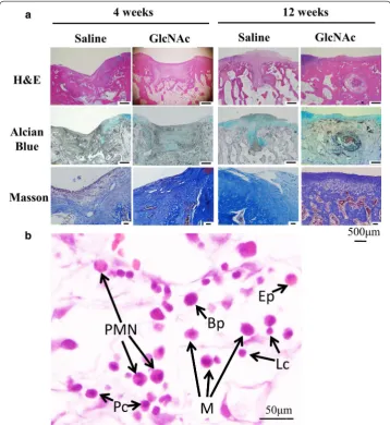
In the saline group, the defect regions were filled with hyperplasic blood vessels, con-tained disoriented fibrous tissue together with fibroblast-like cells, and showed a shortage of GAG synthesis (Fig. 3a). Furthermore, representative inflammatory cells, including plasma cells, lymphocytes and polymorphonuclear neutrophils (PMNs) were found in the reparative region (Fig. 3b).
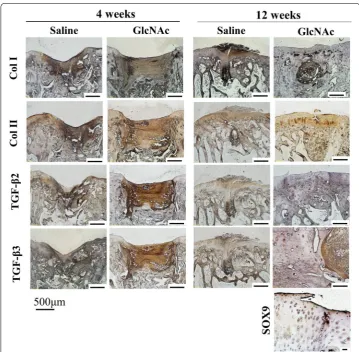
However, in the GlcNAc group, the migration of undifferentiated chondroblasts together with increasing GAG synthesis had begun in the reparative regions at week 4. Furthermore, the GlcNAc group showed newly generated collagen matrix, coupled with underlying osteoid at the defect edges (Fig. 3a). Moreover, the GlcNAc group displayed higher levels of COL I and COL II than the saline group (Fig. 4). Surprisingly, the two
decisive endogenous growth factors (i.e., TGF-β2 and TGF-β3) were highly expressed compared to the saline group (Fig. 4).
At 12 weeks after operation
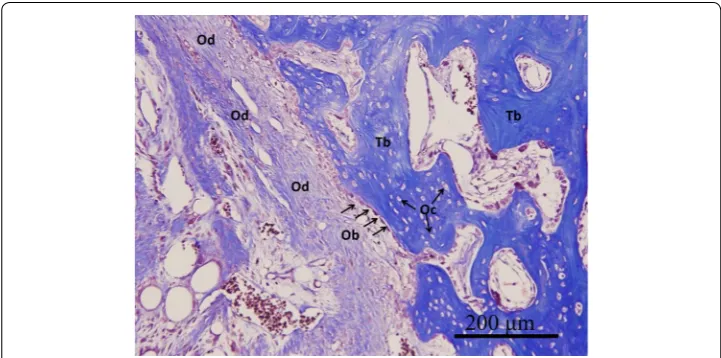
Compared to the saline group, the GlcNAc group showed obvious histological changes in both hyaline cartilage regeneration and subchondral bone formation (Fig. 3a). The saline group still showed irregular surfaces, disorganized collagen, and GAG depletion in the adjacent cartilage (Fig. 3a). In contrast, the GlcNAc group showed potential posi-tive outcomes including the regeneration of hyaline-like cartilage corresponding with chondrocytes in lacunae (SOX9+), rich GAG content, reconstructed COL II content (hyaline cartilage) as well as low levels of COL I (fibrocartilage) in the adjacent cartilage and reparative site (Figs. 3a, 5). Furthermore, in the GlcNAc group, the trabecular bone was embedded with osteocytes, along with osteoid matrix surrounding osteoblasts, indi-cating sound bone remodeling (Fig. 5).
Discussion
The fate of healing articular cartilage is fundamentally dependent on better healing in the early stages and mechanically subchondral bone regeneration [31]. In this study, the GlcNAc group showed higher levels of tissue calcification, indicating beneficial trends for higher BV/TV and Tb.Th.
The TGF-β superfamily plays significant roles in enhancing chondrocyte growth, chondrocyte proliferation and osteochondrogenic differentiation [25]. TGF-β2 can stim-ulate chondrocyte proliferation and cartilage regeneration [32, 33]; TGF-β3 may act as a chemotactic molecule (i.e., biological cue) that can recruit bone marrow stem cells, induce the recruited stem cells toward chondrogenesis, and enhance the compressive properties of neocartilage [34]. Based on the current data, we thus suggest that direct manipulation of in situ microenvironments (i.e., GlcNAc injection) provides a better mix of endogenous growth factors and cytokines, especially in the early stage, for cartilage repair. In this study, GlcNAc showed no locally or systemically adverse reactions, con-sistent with reported outcomes. Previous studies suggest that GlcNAc has an excellent safety profile in humans. In addition, it has been reported that GlcNAc has additional potential advantages over GlcN as a potential therapeutic anti-inflammatory agent. GlcN initiates phosphorylation by glucokinase and competes with glucose for binding to glucokinase in the cell [35], thereby giving rise to GlcN-induced insulin resistance [36]. In contrast, GlcNAc has much lower affinity toward glucokinase than do either glucose or GlcN; thus, it does not respond significantly to glucose metabolism [37].
Based on this study, one possible major reparative mechanism may function through changes to the in situ microenvironments in the reparative site. Transport of nutrients and a subset of signal molecules can be supplied from the synovial membrane to syno-vial fluid [24]; as an alternative source, nutritional support also diffuses from subchon-dral bone [38].
Conclusions
This study demonstrated that intra-articular injection of GlcNAc is safe and has the poten-tial to improve the repair of FTAC in the rabbit knee joint model. Promising outcomes include the improvement of damaged articular surface, formation of hyaline-like cartilage, rich GAG synthesis, and subchondral bone regeneration. This strategy warrants further investigation to support an effective transition from animal models to human patients.
Abbreviations
HA: hyaluronic acid; GlcN: glucosamine; GlcNAc: N‑acetyl‑d‑glucosamine; PFAA: plasma total free amino acid; FTAC: full‑
thickness articular cartilage; H&E: hematoxylin and eosin; COL I: collagen type I; COL II: collagen type II; TGF‑β: transform‑ ing growth factor‑β; GAG: glycosaminoglycan; CT: computed tomography; BV/TV: bone volume per tissue volume; Tb.Th: trabecular thickness.
Authors’ contributions
NJC, YIL, HCW, and MLY conceived and designed the study; NJC, YIL, CCL, HCW performed the experiments; YIL, CCL, and HCW analyzed the data; and NJC, HCH and MLY jointly prepared the manuscript. All authors read and approved the final manuscript.
Author details
1 Department of Sports Medicine, Kaohsiung Medical University, Kaohsiung, Taiwan. 2 Department of Biomedical Engi‑ neering, National Cheng Kung University, 1 University Rd., Tainan 701, Taiwan. 3 Laboratory Animal Center, Department of Medical Research, Chi‑Mei Medical Center, Tainan, Taiwan. 4 Department of Orthopedics, China Medical University Hospital, Taichung, Taiwan. 5 Medical Device Innovation Center, National Cheng Kung University, Tainan 701, Taiwan.
Acknowledgements
This work was supported by the Ministry of Science and Technology, Taiwan (Grant 101‑2221‑E‑006‑062‑MY3) and the Kaohsiung Medical University Research Foundation (KMU‑Q104003). We would like to thank Dr. Liang, Peir‑In, Depart‑ ment of Pathology, Kaohsiung Medical University Hospital, for help with histological judgement.
Competing interests
The authors declare that they have no conflict of interests related to this work.
Received: 10 July 2015 Accepted: 6 November 2015
References
1. Laudy AB, Bakker EW, Rekers M, Moen MH. Efficacy of platelet‑rich plasma injections in osteoarthritis of the knee: a systematic review and meta‑analysis. Br J Sports Med. 2015;49(10):657–72. doi:10.1136/bjsports‑2014‑094036. 2. McAlindon TE, Bannuru RR, Sullivan MC, Arden NK, Berenbaum F, Bierma‑Zeinstra SM, et al. OARSI guidelines for the
non‑surgical management of knee osteoarthritis. Osteoarthr Cartil OARS Osteoarthr Res Soc. 2014;22(3):363–88. doi:10.1016/j.joca.2014.01.003.
3. Lynch TS, Patel RM, Benedick A, Amin NH, Jones MH, Miniaci A. Systematic review of autogenous osteochon‑ dral transplant outcomes. Arthrosc J Arthrosc Relat Surg Off Publ Arthrosc Assoc N Am Int Arthrosc Assoc. 2015;31(4):746–54. doi:10.1016/j.arthro.2014.11.018.
4. Oussedik S, Tsitskaris K, Parker D. Treatment of articular cartilage lesions of the knee by microfracture or autologous chondrocyte implantation: a systematic review. Arthrosc J Arthrosc Relat Surg Off Publ Arthrosc Assoc N Am Int Arthrosc Assoc. 2015;31(4):732–44. doi:10.1016/j.arthro.2014.11.023.
5. Mano JF, Reis RL. Osteochondral defects: present situation and tissue engineering approaches. J Tissue Eng Regen Med. 2007;1(4):261–73. doi:10.1002/term.37.
6. Goyal D, Keyhani S, Goyal A, Lee EH, Hui JH, Vaziri AS. Evidence‑based status of osteochondral cylinder transfer techniques: a systematic review of level I and II studies. Arthrosc J Arthrosc Relat Surg Off Publ Arthrosc Assoc N Am Int Arthrosc Assoc. 2014;30(4):497–505. doi:10.1016/j.arthro.2013.12.023.
7. Saw KY, Anz A, Siew‑Yoke Jee C, Merican S, Ching‑Soong Ng R, Roohi SA, et al. Articular cartilage regeneration with autologous peripheral blood stem cells versus hyaluronic acid: a randomized controlled trial. Arthrosc J Arthrosc Relat Surg Off Publ Arthrosc Assoc N Am Int Arthrosc Assoc. 2013;29(4):684–94. doi:10.1016/j.arthro.2012.12.008. 8. Horas U, Pelinkovic D, Herr G, Aigner T, Schnettler R. Autologous chondrocyte implantation and osteochondral
cylinder transplantation in cartilage repair of the knee joint. A prospective, comparative trial. J Bone Joint Surg Am Vol. 2003;85A(2):185–92.
9. Saris DB, Vanlauwe J, Victor J, Haspl M, Bohnsack M, Fortems Y, et al. Characterized chondrocyte implantation results in better structural repair when treating symptomatic cartilage defects of the knee in a randomized controlled trial versus microfracture. Am J Sports Med. 2008;36(2):235–46. doi:10.1177/0363546507311095.
11. Pavelka K, Gatterova J, Olejarova M, Machacek S, Giacovelli G, Rovati LC. Glucosamine sulfate use and delay of progression of knee Osteoarthritis—A 3‑year, randomized, placebo‑controlled, double‑blind study. Arch Intern Med. 2002;162(18):2113–23. doi:10.1001/archinte.162.18.2113.
12. Clegg DO, Reda DJ, Harris CL, Klein MA, Jr OD, Hooper MM et al. Glucosamine, chondroitin sulfate, and the two in combination for painful knee osteoarthritis. N Engl J Med. 2006;354(8):795–808. doi: 10.1056/Nejmoa052771. 13. Sawitzke AD, Shi H, Finco MF, Dunlop DD, Harris CL, Singer NG, et al. Clinical efficacy and safety of glucosamine,
chondroitin sulphate, their combination, celecoxib or placebo taken to treat osteoarthritis of the knee: 2‑year results from GAIT. Ann Rheum Dis. 2010;69(8):1459–64. doi:10.1136/ard.2009.120469.
14. Miller KL, Clegg DO. Glucosamine and chondroitin sulfate. Rheumatic Dis Clin N Am. 2011;37(1):103–18. doi:10.1016/j.rdc.2010.11.007.
15. De Mattei M, Pellati A, Pasello M, de Terlizzi F, Massari L, Gemmati D, et al. High doses of glucosamine‑HCl have detrimental effects on bovine articular cartilage explants cultured in vitro. Osteoarthr Cartil. 2002;10(10):816–25. doi:10.1053/joca.2002.0834.
16. Fenton JI, Chlebek‑Brown KA, Peters TL, Caron JP, Orth MW. The effects of glucosamine derivatives on equine articu‑ lar cartilage degradation in explant culture. Osteoarthr Cartil. 2000;8(6):444–51. doi:10.1053/joca.2000.0320. 17. Derfoul A, Miyoshi AD, Freeman DE, Tuan RS. Glucosamine promotes chondrogenic phenotype in both chondro‑
cytes and mesenchymal stem cells and inhibits MMP‑13 expression and matrix degradation. Osteoarthr Cartil/OARS, Osteoarthr Res Soc. 2007;15(6):646–55. doi:10.1016/j.joca.2007.01.014.
18. Shikhman AR, Amiel D, D’Lima D, Hwang SB, Hu C, Xu A, et al. Chondroprotective activity of N‑acetylglucosamine in rabbits with experimental osteoarthritis. Ann Rheum Dis. 2005;64(1):89–94. doi:10.1136/ard.2003.019406.
19. Shikhman AR, Kuhn K, Alaaeddine N, Lotz M. N‑acetylglucosamine prevents IL‑1 beta‑mediated activation of human chondrocytes. J Immunol. 2001;166(8):5155–60.
20. d’Abusco AS, Corsi A, Grillo MG, Cicione C, Calamia V, Panzini G, et al. Effects of intra‑articular administration of glucosamine and a peptidyl‑glucosamine derivative in a rabbit model of experimental osteoarthritis: a pilot study. Rheumatol Int. 2008;28(5):437–43. doi:10.1007/s00296‑007‑0463‑x.
21. Sato H, Takahashi T, Ide H, Fukushima T, Tabata M, Sekine F, et al. Antioxidant activity of synovial‑fluid, hyaluronic‑ acid, and 2 subcomponents of hyaluronic‑acid—synovial‑fluid scavenging effect is enhanced in rheumatoid‑arthri‑ tis patients. Arthritis Rheum. 1988;31(1):63–71. doi:10.1002/art.1780310110.
22. Rubin BRTJ, Kongtawelert P, Pertusi RM, Forman MD, Gracy RW. Oral polymeric N‑acetyl‑d‑glucosamine and osteoar‑ thritis. J Am Osteopath Assoc. 2001;101(6):339–44.
23. Tamai Y, Miyatake K, Okamoto Y, Takamori Y, Sakamoto K, Minami S. Enhanced healing of cartilaginous injuries by
N‑acetyl‑d‑glucosamine and glucuronic acid. Carbohydr Polym. 2003;54(54):251–62.
24. Laverty S, Sandy JD, Celeste C, Vachon P, Marier JF, Plaas AHK. Synovial fluid levels and serum pharmacokinetics in a large animal model following treatment with oral glucosamine at clinically relevant doses. Arthritis Rheum. 2005;52(1):181–91. doi:10.1002/Art.20762.
25. Setnikar I, Giacchetti C, Zanolo G. Pharmacokinetics of glucosamine in the dog and in man. Arzneimittel‑Forschung/ Drug Res. 1986;36–1(4):729–35.
26. Gaulden EC, Keating WC. The effect of intravenous N‑acetyl‑d‑glucosamine on the blood and urine sugar concen‑
trations of normal Subjects. Metab Clin Exp. 1964;13:466–72.
27. Shikhman AR, Brinson DC, Valbracht J, Lotz MK. Differential metabolic effects of glucosamine and N‑acetylglucosa‑ mine in human articular chondrocytes. Osteoarthr Cartil/OARS Osteoarthr Res Soc. 2009;17(8):1022–8. doi:10.1016/j. joca.2009.03.004.
28. Azuma K, Osaki T, Tsuka T, Imagawa T, Okamoto Y, Takamori Y, et al. Effects of oral glucosamine hydrochloride admin‑ istration on plasma free amino acid concentrations in dogs. Mar Drugs. 2011;9(5):712–8. doi:10.3390/md9050712. 29. Wayne JS, McDowell CL, Shields KJ, Tuan RS. In vivo response of polylactic acid‑alginate scaffolds and bone marrow‑
derived cells for cartilage tissue engineering. Tissue Eng. 2005;11(5–6):953–63. doi:10.1089/ten.2005.11.953. 30. Ranstam J. Repeated measurements, bilateral observations and pseudoreplicates, why does it matter? Osteoarthr
Cartil/OARS Osteoarthr Res Soc. 2012;20(6):473–5. doi:10.1016/j.joca.2012.02.011.
31. Schlichting K, Schell H, Kleemann RU, Schill A, Weiler A, Duda GN, et al. Influence of scaffold stiffness on subchondral bone and subsequent cartilage regeneration in an ovine model of osteochondral defect healing. Am J Sports Med. 2008;36(12):2379–91. doi:10.1177/0363546508322899.
32. Chang NJ, Lam CF, Lin CC, Chen WL, Li CF, Lin YT, et al. Transplantation of autologous endothelial progenitor cells in porous PLGA scaffolds create a microenvironment for the regeneration of hyaline cartilage in rabbits. Osteoarthr Cartil/OARS Osteoarthr Res Soc. 2013;21(10):1613–22. doi:10.1016/j.joca.2013.07.016.
33. Das R, Timur UT, Edip S, Haak E, Wruck C, Weinans H, et al. TGF‑beta2 is involved in the preservation of the chondro‑ cyte phenotype under hypoxic conditions. Ann Anat. 2015;198:1–10. doi:10.1016/j.aanat.2014.11.003.
34. Hara ES, Ono M, Pham HT, Sonoyama W, Kubota S, Takigawa M, et al. Fluocinolone acetonide is a potent synergistic factor of TGF‑beta3‑associated chondrogenesis of bone marrow‑derived mesenchymal stem cells for articular surface regeneration. J Bone Miner Res. 2015;. doi:10.1002/jbmr.2502.
35. Van Schaftigen E. Glucosamine‑sensitive and ‑insensitive detritiation of [2‑3H]glucose in isolated rat hepatocytes: a study of the contributions of glucokinase and glucose‑6‑phosphatase. Biochem J. 1995;308(Pt 1):23–9.
36. Ciaraldi TP, Carter L, Nikoulina S, Mudaliar S, McClain DA, Henry RR. Glucosamine regulation of glucose metabolism in cultured human skeletal muscle cells: divergent effects on glucose transport/phosphorylation and glyco‑ gen synthase in non‑diabetic and type 2 diabetic subjects. Endocrinology. 1999;140(9):3971–80. doi:10.1210/ endo.140.9.6974.
37. Miwa I, Mita Y, Murata T, Okuda J, Sugiura M, Hamada Y, et al. Utility of 3‑O‑methyl‑N‑acetyl‑d‑glucosamine, an
N‑acetylglucosamine kinase inhibitor, for accurate assay of glucokinase in pancreatic islets and liver. Enzyme Protein. 1994;48(3):135–42.