Bhoi et al. Page 1 of 115
MICROBUBBLES: ROLE IN THERAPEUTICS
Biswaranjan Ray1 and Shubhrasweta Bhoi*1
Department of Pharmacology, Gayatri College of Pharmacy, Sambalpur, Odisha, India.
Received date: 24 August 2019 Revised date: 14 September 2019 Accepted date: 04 October 2019
INTRODUCTION
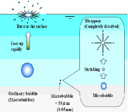
Microbubbles are an extremely small bubble, usually only a few micrometers in diameter that can be uniformly suspended in a liquid such as blood.
Microbubbles first catapulted to prominence in the mid-'90s, when the FDA cleared them for use in imaging applications. They quickly became a cheap alternative to expensive scans – all that's needed is a shot and a portable ultrasound machine. Plus, the technique is fast; microbubbles can be imaged in minutes, while CTs and MRIs take hours.
Since then, researchers have demonstrated that little bubbles have another use: delivering pharmaceutical payloads inside the body. Bubbles coated with specific molecules will selectively bind to certain cellular receptors. Once they're attached, a strong burst of ultrasound is all that's needed to pop the bubbles and free what's inside. Delivering drugs directly to a tumor this way would allow for smaller doses – no more bombarding the whole body with radioactive material – thereby radically reducing side effects. Gene therapy, which treats genetic diseases by using viruses to deliver DNA, could also be safer if the material were administered via microbubble. Both techniques are being tested on animals.
The most impressive accomplishment on the
microbubble's resume, however, may be its ability to get drugs across the blood-brain barrier. Small molecules like alcohol can make it into the gray matter, but anything larger is typically kept out – a trait that protects us but has severely limited the treatment options for
neurological diseases like Alzheimer's.[1] Over the past few years, scientists at UC Davis have been working to show that microbubbles can be "pushed" to the barrier and then exploded, opening up small pores through which drugs can pass. The result may eventually give doctors a much larger palette of drugs for brain diseases.[2]
Fig. 1: Basic difference between an ordinary bubble and a microbubble.
Fundamental properties of microbubble 1. Increase in interior gas pressure2. Increase in ion concentration around the gas-water interface
Generation of free-radicals Ions
Generation of Nano-bubble
Review Article www.wjahr.com
ISSN: 2457-0400
Volume: 3. Issue: 5.
Page N. 110-115
Year: 2019
WORLD JOURNAL OF ADVANCE
HEALTHCARE RESEARCH
*Corresponding author: Shubhrasweta Bhoi
Department of Pharmacology, Gayatri College of Pharmacy, Sambalpur, Odisha, India.
ABSTRACT
Microbubbles are very small-encapsulated gas bubbles (diameters of micrometers) that can be used in diagnostic and therapeutic applications. Upon exposure to sufficiently intense ultrasound, microbubbles will cavitate, rupture, disappear, release gas content, etc. Such characteristics of the microbubbles can be used to enhance diagnostic tests, dissolve blood clots, and deliver drugs or genes for therapy.
Preparation of Microbubbles
The mentioned application depends strongly on the physical and chemical properties of the Ultrasound Contrast Agents (UCA), which, amongst others depends on the method of preparation. With the traditional UCA production methods like sonication, it is difficult to have such particle properties. In this project, UCA will be prepared by emulsification. Different techniques such as
membrane-, micro channel- and micro sieve
emulsification will be examined and compared for dedicated UCA production.[3]
Figure 2: Shows a schematic diagram of a cross flow membrane emulsification process.
How Microbubbles work?
Microbubbles work by resonating in an ultrasound beam, rapidly contracting and expanding in response to the pressure changes of the sound wave. By a fortunate coincidence, they vibrate particularly strongly at the high frequencies used for diagnostic ultrasound imaging. This makes them several thousand times more reflective than normal body tissues. In this way they enhance both grey scale images and flow mediated Doppler signals. As well as being useful in it self, the resonance that microbubbles produce has several special properties that can be exploited to improve diagnoses. Just as with a musical instrument, multiple harmonic signals or overtones are produced, Ultrasound scanners can be tuned to “listen” to these harmonics, producing strong preferential imaging of the microbubbles in an image. The selective
excitation produced can also destroy microbubbles relatively easily, an effect that can be useful both in imaging and in emerging therapeutic applications.
Fig. 3: Shows the working of Microbubbles and the effect of ultrasound on it.
Microbubble Contrast Agents: A New Era in Ultrasound
Contrast agents are widely used in imaging, but until recently they had little place in ultrasonography. This has changed with the introduction of Microbubbles small (typically 3 μm in diameter) gas filled bubbles that are usually injected intravenously. Injecting a gas into the circulation may seem potentially hazardous, but extensive clinical experience has shown that the tiny volume of air or gas given (under 200 μl) is not dangerous, and the safety of microbubbles compares well to that of conventional agents in radiography and magnetic resonance imaging. Although microbubbles were originally designed simply to improve conventional ultrasound scanning, recent discoveries have opened up powerful emerging applications.[4]
Treatment with microbubbles
Microbubbles can aid drug delivery in themselves (by acting as “cavitation nuclei”) and as agents to carry drugs for site-specific treatment. Their most exciting application is in the emerging area of gene therapy, where delivery of genetic material to a chosen site is difficult.[5,6]
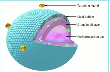
Gas filled microspheres may be designed so that their interior is loaded with drug and gas. A stabilizing material, here a lipid, surrounds the perfluorocarbon bubble. Drugs may be incorporated by themselves or, if insoluble in water, in an oil layer.
The microsphere may be targeted to specific tissue by incorporating protein ligands on the surface.
Ultrasound can potentiate drug delivery by creating transient non-lethal perforations in cell membranes to aid ingress of large molecules and particles into the cells (“sonoporating”). In general this requires high acoustic power, substantially beyond that permitted for imaging, but the power needed is greatly reduced when microbubbles are present. This is because microbubbles lower the amount of energy necessary for cavitation, the process in which extreme oscillations induced by ultrasound pulses lead to microbubble collapse. The potential of this in gene therapyhas already been shown. Cavitation of microbubbles in capillary beds also increases capillary permeability, which improves local access of the released therapeutic agent.[7]
Microbubbles as Drug Delivery Vehicles
Microbubbles can be formulated to carry therapeutic agents. Some albumin based microbubbles and those with shells of charged lipids take up genetic material directly. Hydrophilic compounds can be encased within lipid membranes or polymeric shells that stabilise the microbubbles. The circulation of these loaded microbubbles can be followed with ultrasound, and when they reach the target area they can be disrupted, releasing their therapeutic payload to the surrounding tissue (fig 5). A recent study showed that transfection of a reporter gene in a mouse heart model was increased 10-fold using microbubbles loaded with an adenovirus gene vector.[8,9]
Fig. 5: Gene delivery using ultrasound and microbubbles. The presence of gas in the gene- filled microbubble allows ultrasound energy to "pop" the bubble. An energetic wave is then created which allows the genetic material to enter surrounding cells.
Delivery to a specific site can be aided by incorporating ligands into the membrane of the microbubbles that target receptors on cell membranes. For example, incorporation of a surface ligand that binds to the GPIIB/IIIA receptors on activated platelets allows microbubbles to bind to a thrombus and deliver thrombolytic agents.[10,11]
Mechanisms for Microbubble Delivery
Ultrasound-mediated microbubbles destruction has been proposed as an innovative method for noninvasive delivering of drugs and genes to different tissues. Microbubbles are used to carry a drug or gene until a specific area of interest is reached, and then ultrasound is used to burst the microbubbles, causing site-specific delivery of the bioactive materials.[12] Furthermore, the ability of albumin-coated microbubbles to adhere to vascular regions with glycocalix damage or endothelial dysfunction is another possible mechanism to deliver drugs even in the absence of ultrasound.[13] The recent advances in gene therapy and molecular biology have improved the interest in methods of noninvasive delivery of therapeutic agents. Besides the well-known application of microbubbles as contrast agents for diagnostic ultrasound, microbubbles have also been demonstrated an effective technique for targeted delivery of drugs and genes.
Drugs can be incorporated into the microbubbles in a number of different ways, including binding of the drug to the microbubble shell and attachment of site-specific ligands. As perfluorocarbon-filled microbubbles are sufficiently stable for circulating in the vasculature as blood pool agents, they act as carriers of these agents until the site of interest is reached. Ultrasound applied over the skin surface can then be used to burst the microbubbles at this site, causing localized release of the drug. This technique then permits using lower concentrations of drugs systemically and concentration of the drug only where it is needed. This improved therapeutic index may be extremely advantageous in cases of drugs with hazardous systemic side effects, like cytotoxic agents. Albumin-encapsulated microbubbles have also demonstrated to adhere to the vessel walls in the setting of endothelial dysfunction.This also may be a method of targeting delivery with microbubbles but without the application of ultrasound.
Mechanisms for Target Drug Delivery Using Microbubbles
Different drugs and genes can be incorporated into the ultrasound contrast agents
Perfluorocarbon-filled albumin microbubbles avidly bind proteins and synthetic oligonucleotides.[14]
Microbubbles directly take up genetic material, such as plasmids and adenovirus.
Phospholipid-coated microbubbles have a high affinity for chemotherapeutic drugs.
Specific ligands for endothelial cell adhesion molecules, such as P-selectin and leukocyte intercellular adhesion molecule 1 (ICAM-1), can be attached to both lipid- and albumin-encapsulated microbubbles, which increases their deposition to activated endothelium.[15,16,17]
The mechanisms by which ultrasound facilitates the delivery of drugs and genes results from a complex interplay among the therapeutic agent, the microbubble characteristics, the target tissue, and the nature of ultrasound energy.
The presence of microbubbles in the insonified field reduces the peak negative pressure needed to enhance drug delivery with ultrasound. This occurs because the microbubbles act as nuclei for cavitations, decreasing the threshold of ultrasound energy necessary to cause this phenomenon.[18,19]
The results of optical and acoustical studies have suggested the following mechanisms for microbubble destruction by ultrasound.
Gradual diffusion of gas at low acoustic power,
Formation of a shell defect with diffusion of gas,
Immediate expulsion of the microbubble shell at high acoustic power,
Dispersion of the microbubble into several smaller bubbles.
Cavitation of the bubbles is characterized by rapid destruction of contrast agents due to a hydrodynamic instability excited during large amplitude oscillations, and is directly dependent on the transmission pressure.
The formation of pores in the membranes of cells as a result of ultrasound-induced microbubble cavitation has been proposed as a mechanism for facilitating the drug deposition.
Another important therapeutic property of microbubbles is their increased adherence to damaged vascular endothelium. Albumin-coated microbubbles do not adhere to normally functioning endothelium, but their adherence does occur to activated endothelial cells or to extra-cellular matrix of the disrupted vascular wall, and this interaction could be a marker of endothelial integrity. Because of this characteristic, the delivery of drugs or genes bound to albumin-coated microbubbles could be selectively concentrated at the site of vascular injury in the presence or absence of ultrasound application.[20,21,22]
Targeted Molecular Imaging and Therapy
Benefits of microbubble target delivery system include:
Real-time imaging,
Relatively short and efficient imaging protocols,
Non-invasive with minima patient discomfort and low operating costs.
Two possible approaches for targeted imaging are
Passive targeting
Active targeting
Passive targeting relies on the materials of the shell and the size of the microbubble (Fig 6).
Active targeting relies on adhesive ligands, as markers of inflammation and thrombus, among other factors.
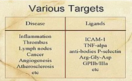
Microbubbles today have different characteristics that allow them to be used as targeting materials, including various sizes and the types of gases, and shell materials. Under development are microbubble agents, liposomal agents, and perflurocarbon emulsion nanoparticles. Disease and ligand targets are shown in (Figure 7).
Fig. 6: Targeted imaging using passive and active approaches.
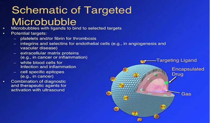
Fig. 8: Schematic diag. of a Microbubble used for targeted imaging.
The microbubble is comprised of an outer shell, which can contain target ligands, and gases within the microbubble. Drugs can be encapsulated within the microbubbles, which can be targeted for drug release or enhancement of drug effects using Ultrasound. Ultrasound alone enhances drug delivery and efficiency. Thrombolysis can be enhanced by the combination of Ultrasound and thrombolytic agents. Tissue penetration can be enhanced by combining Ultrasound and transdermal drug delivery. Hall and colleagues demonstrated nanoparticles that adhere to fibrin and the nanoparticles can be visualized by Ultrasound. Another example is a microbubble with GPIIb/IIIa receptor that adheres to thrombus, which had been demonstrated in an animal model. Lanza et al had shown that using nanoparticle accumulation and US imaging, it is possible to obtain enhanced imaging of balloon-injured arteries, whereas there was no enhancement in the control animals.[23,24,25]
CONCLUSION
Firstly, specific acoustic and biological properties make microbubbles a promising tool as a vehicle for drug and gene delivery. In vivo studies have been performed and have showed the expression of genes delivered by microbubbles in combination with ultrasound. Although results were positive, more in vivo animal studies are needed to investigate possible future applications in humans. An interesting problem is the precise interaction of microbubbles with living cells. Although several options, like transient cell membrane holes, endocytosis, phagocytosis and fusion of microbubble shell components with the cell membrane have been proposed, the exact mechanism remains to be elucidated. Recent advances in live-cell imaging techniques (e.g. multidimensional digital imaging microscopy) offer excellent opportunities to study this process at the (sub) cellular level in real-time, thereby creating the possibility to visualize the interaction of fluorescent labeled
microbubbles and myocardial or endothelial cells under high ultrasound pressure.
Secondly, the use of targeted microbubbles has been a great step forward. Microbubbles have been targeted to receptors of leukocytes and blood clots, and may in the future probably be used in diagnostic imaging of thrombo-embolic or inflammatory processes. Targeted microbubbles create various challenging therapeutic options, not only in cardiovascular disease but also in treatment of inflammatory and malignant diseases.[26,27] These microbubbles can be used as a vehicle for drugs or genes and local delivery can be achieved by destruction of microbubbles with ultrasound. The same mechanism could be used in treatment of thrombo-embolic processes. Enhancement of thrombolysis with the use of microbubbles and ultrasound looks promising in vitro and in vivo. The development of microbubbles targeted to GP-IIbIIIa receptors could be a step forward in treatment of vascular thrombi. Attachment of microbubbles loaded with thrombolytic agents and administration of ultrasound may in the future well be a therapeutic option.
In summary, over the past years, contrast agents in echocardiography have rapidly evolved from a diagnostic adjuvant to a possible therapeutic agent. In the coming years, this promising technique needs further development to make it available for clinical application.
FINAL REFERENCES
1. Albrecht T, Urbank A, Mahler M, Bauer A, Dore CJ, Blomley MJ, et al. Prolongation and optimization of Doppler enhancement with a microbubble US contrast agent by using continuous infusion: preliminary experience. Radiology, 1998; 207: 339-347
transcranial Doppler signal in humans. Stroke, 1993; 24: 1903-1909.
3. Cosgrove D. Why do we need contrast agents for ultrasound? Clin Radiol, 1996; 51(suppl 1): 1-4. 4. Nanda NC, Carstensen C. Echo-enhancing agents:
safety. In: Nanda NC, Schlief R, Goldberg BB, eds. Advances in echo imaging using contrast enhancers. Dordrecht: Kluwer, 1997; 115-131.
5. Russell SJ. Science, medicine, and the future: gene therapy. BMJ, 1997; 315: 1289-1292.
6. Apfel RE, Holland CK. Gauging the likelihood of cavitation from short-pulse, low-duty cycle diagnostic ultrasound. Ultrasound Med Biol., 1991; 17: 179-185.
7. Porter TR, Iverson PL, Li S, Xia F. Interaction of
diagnostic ultrasound with synthetic
oligonucleotide-labeledperfluorocarbon-exposed sonicated dextrose albumin microbubbles. J Ultrason Med, 1996; 15: 577-584.
8. Shohet RV, Chen S, Zhou Y-T, Wang Z, Meidell RS, Unger RH, et al. Echocardiographic destruction of albumin microbubbles directs gene delivery to the myocardium. Circulation, 2000; 101: 2554-2556. 9. Price RJ, Skyba DM, Kaul S, Skalak TC. Delivery
of colloidal particles and red blood cells to tissue through microvessel ruptures created by targeted
microbubble destruction with ultrasound.
Circulation, 1998; 98: 1264-1267.
10. Wu Y, Unger EC, McCreery TP, Sweitzer RH, Shen D, Wu G, et al. Binding and lysing of blood clots using MRX-408. Invest Radiol, 1998; 33: 880-885. 11. Miller MW. Gene transfection and drug delivery.
Ultrasound Med Biol., 2000; 26(suppl 1): S59-S62. 12. Skyba DM, Price RJ, Linka AZ, Skalak TC, Kaul S:
Direct in vivo visualization of intravascular destruction of microbubbles by ultrasound and its local effects on tissue. Circulation, 1998; 98: 290-293.
13. Main ML, Grayburn PA: Clinical applications of transpulmonary contrast echocardiography.Am Heart J, 1999; 137: 144-153.
14. Wei K, Skyba DM, Firschke C, Jayaweera AR, Lindner JR, Kaul S: Interactions between microbubbles and ultrasound: in vitro and in vivo observations. J Am Coll Cardiol, 1997; 29: 1081-1088.
15. Unger EC, McCreery TP, Sweitzer RH, Caldwell VE, Wu Y: Acoustically active lipospheres containing paclitaxel: a new therapeutic ultrasound contrast agent. Invest Radiol, 1998; 33: 886-892. 16. Taniyama Y, Tachibana K, Hiraoka K, Namba T,
Yamasaki K, Hashiya N, et al.: Local delivery of plasmid DNA into rat carotid artery using ultrasound. Circulation, 2002; 105: 1233-1239. 17. Chen S, Shohet RV, Bekeredjian R, Frenkel P,
Grayburn PA: Optimization of ultrasound
parameters for cardiac gene delivery of adenoviral or plasmid deoxyribonucleic acid by ultrasound-targetedmicrobubbledestruction. J Am Coll Cardiol, 2003; 42: 301-308.
18. Mukherjee D, Wong J, Griffin B, Ellis SG, Porter T, Sen S, et al.: Ten-fold augmentation of endothelial uptake of vascular endothelial growth factor with ultrasound after systemic administration. J Am Coll Cardiol, 2000; 35: 1678-1686.
19. Villanueva FS, Jankowski RJ, Manaugh C, Wagner WR: Albumin microbubble adherence to human coronary endothelium: implications for assessment of endothelial function using myocardial contrast echocardiography. J Am Coll Cardiol, 1997; 30: 689-693.
20. Unger EC, McCreery TP, Sweitzer RH, Caldwell VE, Wu Y: Acoustically active lipospheres containing paclitaxel: a new therapeutic ultrasound contrast agent. Invest Radiol, 1998; 33: 886-892. 21. Fritz TA, Unger EC, Sutherland G, Sahn D: Phase I
clinical trials of MRX-115. A new ultrasound contrast agent. Invest Radiol, 1997; 32: 735-740. 22. Lindner JR, Song J, Christiansen J, Klibanov AL,
Xu F, Ley K: Ultrasound assessment of
inflammation and renal tissue injury with microbubbles targeted to P-selectin. Circulation, 2001; 104: 2107-2112.
23. Weller GE, Lu E, Csikari MM, Klibanov AL, Fischer D, Wagner WR, et al.: Ultrasound imaging of acute cardiac transplant rejection with microbubbles targeted to intercellular adhesion molecule-1. Circulation, 2003; 108: 218-224. 24. Chomas JE, Dayton P, Allen J, Morgan K, Ferrara
KW: Mechanisms of contrast agent destruction. IEEE. Trans Ultrason Ferroelectr Freq Control, 2001; 48: 232-248.
25. Chomas JE, Dayton P, May D, Ferrara K: Threshold of fragmentation for ultrasonic contrast agents. J Biomed Opt., 2001; 6: 141-150.
26. Porter TR, Hiser WL, Kricsfeld D, Deligonui U, Xie F, Iversen P, et al.: Inhibition of carotid artery
neointimal formation with intravenous
microbubbles. Ultrasound Med Biol., 2001; 27: 259-265.