Extraction Socket Preservation with or without Membranes,
Soft Tissue Influence on Post Extraction Alveolar Ridge
Preservation: a Systematic Review
Ricardo Faria-Almeida
1, Inesa Astramskaite-Januseviciene
2, Algirdas Puisys
3, Francisco Correia
11Department of Oral Surgery and Oral Medicine, Porto University of Dental Medicine, Portugal.
2Department of Oral and Maxillofacial Surgery, Faculty of Odontology, Medical Academy, Lithuanian University of Health Sciences, Kaunas, Lithuania.
3VIC clinic, private practice, Vilnius, Lithuania.
Corresponding Author: Ricardo Faria-Almeida
Department of Oral Surgery and Oral Medicine Porto University of Dental Medicine
Rua Dr. Manuel Pereira da Silva, 4200-393 Porto Portugal
Phone: +351937230675 Fax: +3515193259
E-mail: [email protected]
ABSTRACT
Objectives: The purpose of this systematic review was to assess quantitatively and qualitatively the influence of two different factors: membranes and soft tissue graft influence for the extraction socket preservation.
Material and Methods: A wide-ranging electronic search was performed in six databases up to 30 of November 2018 in order to identify all the clinical and randomized clinical trials performed in humans published with no data restriction. The inclusion criteria were extraction socket preservation with and without membranes or a soft tissue graft in a intact socket with at least six months of follow-up, have more than 12 patients or treat more than 12 sites per group and evaluated at least one of the primary outcomes measures (radiographic measures histological assessment, clinical measures).
Results: From an initial search of 1524 studies only 6 papers fulfil the inclusion and exclusion criterions. All the six selected papers, presented a wide heterogeneity of treatments used, evaluated variables and observation period that made impossible to recommend any specific techniques and/or material to achieve better results. The limited data found suggest that the used of membrane reveals to achieve better results. It wasn’t possible to observe in any clinical trial that compares the used of soft tissue graft.
Conclusions: New trials need to be performed in order to identify what specific techniques and/or materials are better to decrease the reabsorption of the socket after tooth extraction. Clinical trials designed to understand when/how the soft tissues grafts influence at the socket preservation is needed.
Keywords: alveolar bone loss; bone remodeling; tooth socket; systematic review.
Accepted for publication: 5 September 2019 To cite this article:
Faria-Almeida R, Astramskaite-Januseviciene I, Puisys A, Correia F.
Extraction Socket Preservation with or without Membranes, Soft Tissue Influence on Post Extraction Alveolar Ridge Preservation: a Systematic Review
J Oral Maxillofac Res 2019;10(3):e5
JOURNAL OF ORAL & MAXILLOFACIAL RESEARCH Faria-Almeida et al.
INTRODUCTION
One of the goals of the periodontal treatment is to maintain the teeth in good conditions to provide health, function and aesthetics to the patients [1]. However, sometimes as a result of caries, periodontal disease, endodontic lesions and others, tooth extraction is inevitable.
There are several options to give to the patients as an alternative to replace the extracted teeth, from
removal to fixed dental prosthesis, from implant to adhesive fixed prosthesis, each of them with good
results in terms of survival rates with long term follow-ups according to the literature, when well indicated [2,3]. It is clear that all of these options give to the patients an alternative to the teeth lost, but they don´t solve the biological and dimensional changes that occur after tooth extraction.
Tooth extraction begins a sequence of biological changes, with intense resorption of the alveolar bone,
invagination of the mucosa, just in the first weeks
after [4-8]. The quantity and extent of bone process changes are dependent on several factors, which in all of the situations leads to alveolar ridge resorption, in the three-dimensional space [9,10].
In the last years, the goal was not only to solve the existing problem but mainly to avoid the problem or at least to reduce the amount of hard and soft
tissue reabsorption over the time. Several different techniques and approaches offer clinicians different
possibilities to reach this goal.
From the alveolar ridge preservation [11,12], to immediate implants [13-15] and also to the socket
shield technique to immediate implant placement [ 16-18] the clinician have now several different options with a different type of indications as well as different level of scientific background.
A wide variety of alveolar ridge preservation treatment modalities have been described in the last
20 years, including socket grafting with a biomaterial
alone [19], overbuilding of the facial bone wall [20],
occluding the access to the socket by interposing a
barrier element [12], or a combination of some of them [21], with or without using soft tissue grafts to allow primary intention healing.
There are already in the literature several systematics reviews [22-26] that were designed to give the clinicians the state of art of these techniques to
allow them to make the best clinical decision.
However, there is limited information based on randomized clinical trials or clinical trials to address the real advantages of alveolar ridge preservation techniques.
Unfortunately, is not clear yet, which technique and
materials are the most suitable for socket preservation:
autogenous [27], allogenic [19,28] or alloplastic [29]. Furthermore, is still unclear the need of a bone graft, a membrane or only soft tissue graft, as well as the
influence of soft tissue impact to socket preservation.
Therefore, preservation of alveolar ridge may lead to less reabsorption, but the quantity of regenerated bone, as well as the density, might be variable [30]. Hence, a systematic review of the current literature was conducted in order to analyse the outcome of
socket preservation with and without membrane and determine the influence of soft tissues for ridge
preservation.
MATERIAL AND METHODS
This systematic review attempted to assess
quantitatively and qualitatively the influence of two different factors i.e. membranes and soft tissue graft influence, for the extraction socket preservation.
Protocol and registration
The review was registered in an international prospective register for systematic reviews “PROSPERO”.
This systematic review was performed according to
the guidance of the Cochrane hand book [31] and the writing obeyed to the PRISMA Statement (Preferred Reporting Items for Systematic Review and Meta-Analysis) [32].
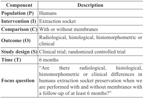
Focus question
The research questions of this systematic review were “Are there radiological, histological,
histomorphometric or clinical differences in humans extraction socket preservation when we are performed
with and without membranes with a follow-up of at least 6 months?” and “Are there radiological,
histological, histomorphometric or clinical differences in humans extraction socket preservation when a soft
tissue graft is performed with a follow-up of at least 6 months?”
The focus questions development according to the PICOST criterions for this systematic review is described in Table 1 and 2.
Information sources
using the following electronic databases: LILACS; PubMed; SciELO; ScienceDirect; Scopus; Web of ScienceTM.
Search
Electronic searches for clinical trials and randomized clinical trials published with no data restriction and written in English, Portuguese, Spanish, Italian, German, Lithuanian or French that presented at
least one of the keyword combinations: “socket
preservation”, “ridge preservation” was performed. In PubMed a MeSH terms explore was performed
too “((“Tooth Socket”[MeSH]) AND “Preservation,
Biological”[Mesh]) OR “Alveolar Ridge Augmentation”[MeSH]))”.
An additional manual search among periodical
journals and the grey literature of relevant papers was
also performed.
Selection of studies
The paper selection based on titles, abstracts and full text was performed by CF and FAR.
In all of this steps attention was payed for the
inclusion/exclusion characteristics in order to
locate all the paper included in this systematic review.
Disagreements were solved by reviewers discussion
prior to final inclusion or exclusion. Kappa test of the agreement was performed and confirmed the high
agreement between researches.
Types of publications
This systematic review included only human clinical trials or randomized clinical trials. Letters, editorials, PhD theses, and abstracts were excluded.
Types of studies
This systematic review included clinical trials or randomized clinical trials performed in human with at least of six months of follow-up, with more than 12 patients or treat more than 12 sites per group.
Types of participants/population
Human patients that had been performed an extraction
socket preservation with or without membranes or a soft tissue graft in an intact socket.
Inclusion and exclusion criteria
The studies selection were based on the following inclusion and exclusion criteria:
Inclusion criteria:
• Humans;
• Clinical trial or randomized clinical trial;
• Minimum of 12 patients or 12 sites per group;
• Group with and without membrane;
• Use of membrane or soft tissue graft;
• Report of at least one primary outcome. Exclusion criteria:
• Animal studies;
• Sockets that weren’t intact.
Sequential search strategy
The duplicated titles were excluded prior to the two investigators (CF and FAR) carefully screened all studies based on titles and abstracts considering the inclusion and exclusion criteria.
For the study eligibility, the full texts were accessed
and confirmed that they have the characteristics to be
included in this systematic review. The authors of the papers were contacted when data was missing or to clarify ambiguous reports.
Table 1. Focus question 1 development according to the PICOST
Component Description
Population (P) Humans
Intervention (I) Extraction socket
Comparison (C)With or without membranes
Outcome (O) Radiological, histological, histomorphometric or clinical Study design (S)Clinical trial; randomized controlled trial
Time (T) 6 months
Focus question
“Are there radiological, histological, histomorphometric or clinical differences in humans extraction socket preservation when we are performed with and without membranes with a follow-up of at least 6 months?”
Table 2. Focus question 2 development according to the PICOST
Component Description
Population (P) Humans
Intervention (I) Extraction socket
Comparison (C)Soft tissue graft
Outcome (O) Radiological, histological, histomorphometric or clinical Study design (S)Clinical trial; randomized controlled trial
Time (T) 6 months
Focus question
JOURNAL OF ORAL & MAXILLOFACIAL RESEARCH Faria-Almeida et al.
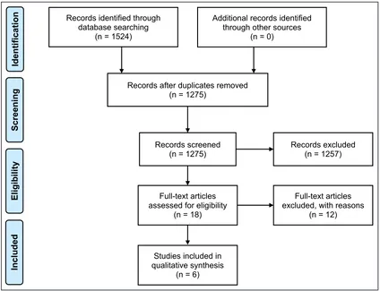
The flow chart representing the literature search is
shown in Figure 1.
Data extraction
After the selection of the papers to be included data regarding the methods, participants, intervention,
outcomes and the risk of bias were extracted.
Data items
Primary outcome measures:
• Radiographic measures: clinical trials that radiographically evaluated the ridge dimensions
previous to the socket preservation and
at follow-up.
• Histological/histomorphometric assessment: clinical trials that assess bone quantity and/or quality and/or residual graft material at follow-up. • Clinical measures: clinical trials that in a clinical
way evaluating the ridge dimensions and\or
gingival recession and\or keratinized tissue width previous to the socket preservation and
at follow-up.
Secondary outcome measures:
• Clinical outcome: clinical findings reported using
any biomaterial.
• Clinical complications: e.g. membrane exposure, graft failure, infections.
Risk of bias within studies
The present systematic review was written in accordance with the PRISMA statement guidelines of transparent reporting for systematic review and meta-analyses [32]. The risk of bias assessment
was carried out based on the guidance of the
Cochrane handbook [31] for systematic review interventions.
The risk of bias was ranked on a three-level scale:
1. Low risk of bias: possible risk of bias which is unlikely to alter report outcomes.
2. Moderated risk of bias: possible risk of bias which
may alter report outcomes.
3. High risk of bias: possible risk of bias which seriously weakens the confidence in reported
outcomes.
Statistical analysis
Based on the large variation types of the intervention included, as well as the studies design, a meta-analysis was not possible to be performed.
Figure 1. Literature search and procedure for the choice of the papers.
Records identified through database searching
(n = 1524)
Scr
eeni
ng
Incl
uded
Elig
ib
ility
Ident
ifi
cat
ion Additional records identified through other sources
(n = 0)
Records after duplicates removed (n = 1275)
Records screened
(n = 1275) Records excluded (n = 1257)
Full-text articles assessed for eligibility
(n = 18)
Full-text articles excluded, with reasons
(n = 12)
Studies included in qualitative synthesis
RESULTS Study selection
The initial search identified a total of 1524 articles,
249 were excluded subsequently because of being duplicate. Following the screening of article titles,
242 potentially relevant articles where identified for
abstract reading were the inclusion and exclusion criteria were applied. Independent screening of abstracts resulted in 18 possibly included articles. After read and analysed the 18 full-text articles a total of six articles met the inclusion criteria and were selected for this systematic review.
The κ values for the inter-reviewer agreement of
the two researchers (FC and FAR) for potentially relevant articles were > 0.9 (titles and abstracts) and > 0.9 (full-text articles), indicating an “almost perfect agreement” between the 2 reviewers.
Figure 1 displays the search strategy and the screening process leading to the selection of the included six trials.
Exclusion of studies
The most frequent reasons for excluding the twelve studies after full-text assessment was as follows: 1. Report guided bone regeneration and not socket
preservation [33-36].
2. The number of sites per group, less than 12 [ 37-39].
3. Follow-up, less than 6 months [40-42].
4. Type of study wasn’t a clinical trials or randomized clinical trials [43].
5. Doesn’t report information about the use of a membrane or a soft tissue graft against a control group [44].
Quality assessment
The quality assessment (Table 3) revealed that two
articles met a low risk of bias [45,46] and four [12,21,47,48] met a high risk of bias.
Types of studies
Three [21,45,48] were randomized control trials, one [12] was split-mouth randomized control trial and two [46,47] were designed as a clinical trial.
The studies that compose this systematic review were
conducted in seven different countries: two at USA
[21,46] and at Italy [45,48]; one at Serbia [12] and Korea [47].
Only one of the included papers of Nam at al. [47] report received funding source from a Ministry of Knowledge and Economy of Seoul, Korea and National Research Foundation of Seoul, Korea.
The studies research were performed at Universities in four of the trials [12,46-48], one of the trials [45] was performed in two private practices private centres and one [21] doesn’t report where it was performed.
Characteristics of outcome measures
The included papers present a wide heterogeneity of treatments used, evaluated variables and observation period.
Related to the clinical variables used, three trials evaluated the dimensional changes based on cone-beam computed tomography (CBCT) analysis [21,45,48] (Table 4), one trial based on periapical X-ray [47] (Table 5); one trial based in clinical variables [12] (Table 6); and four trials gives some information about the
histological/ histomorphometric analysis [21,46-48] (Table 7).
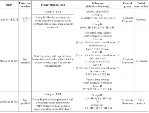
Cone-beam computed tomography analysis
Table 4 shows the selected papers [21,45,48] that present results based on the CBCT analysis.
Table 3. Risk of bias at the selected studies
Study publicationYear of Adequate sequence generation?
Allocation
concealment? All outcomesBlinding?
Incomplete outcome data
addressd?
Risk of bias
Lekovic et al. [12] 1998 Low Low Low Low Low
Iasella et al. [21] 2003 High Low Unclear Unclear High
Meloni et al. [45] 2015 Low Low Low Unclear Low
Perelman-Karmon et al. [46] 2012 Low Low Unclear Unclear Low
Nam et al. [47] 2010 Unclear Unclear Unclear High High
Barone et al. [48] 2008 High Low Low Unclear High
JOURNAL OF ORAL & MAXILLOFACIAL RESEARCH Faria-Almeida et al.
Table 4. Study’s that reported cone-beam computed tomography analysis
Study Extraction location Preservation method (initial vs follow-up)Difference Control group observationPeriod
Iasella et al. [21] 1, 2, 3, 4, 5, 6
Group A - EXT Alveolar ridge width: Group A:
17.28 (SD 1.2)/15.68 (SD 1.13) vs
Group B:
16.91 (SD 1.34)/15.44 (SD 1.33)
12 patients,
12 sockets 6 months Group B: RP with a mineralized
freeze-dried bone allograft, 500 to 1,000 μm particle size, and a collagen
membrane
Meloni et al. [45] specifiedNot
Socket grafting with deproteinized bovine bone and sealed with epithelial
connective tissue graft or porcine collagen matrix
Horizontal bone volume (with collagen vs control):
Level A
(1 mm below the most coronal aspect of the bone crest):
8.24/7.7 vs 8.39/7.72. Level B
(3 mm below the most coronal aspect of the bone crest)
8.18/7.35 vs 8.53/7.62. Level C
(5 mm below the most coronal aspect of the bone crest)
8.21/7.99 vs 8.27/7.96
15 patients,
15 sockets 17 months
Vertical bone volume (with collagen vs control):
Level D
17.28/16.58 vs 16.91/15.44
Barone et al. [48] specifiedNot
Group A - EXT Group RP:
10.6 (SD 1)/8.1 (SD 1.4) vs
Group EXT: 10.8 (SD 0.8)/6.3 (SD 0.8)
20 patients,
20 sockets months7 to 9 Group B: preservation procedure with
corticocancellous porcine bone (MP3, Osteobiol®) and collagen membrane (Evolution, Osteobiol®)
EXT = extraction alone; RP = ridge preservation.
Table 5. Study’s that reported periapical X-ray analysis
References Extraction location Preservation method (initial vs follow-up)Difference Control group observationPeriod
Nam et al. [47] Various
Group A - bovine bone mineral:
OSC-B (Nano - Intelligent Bioengineering, Seoul, Korea) vs
Synthetic oligopeptide - Ossegen-X15 (Nano Intelligent) vs
collagen membrane - Bio-Gide®
Vertical buccal: 15.8/16 vs 12.7/11.8
42/15 6 - 8 months Vertical lingual:
13.9/13.8 vs 11.8/12.7 Width: 9.1/9 vs 7.9/7.7
Table 6. Study’s that reported clinical analysis
References Extraction location Preservation method (experimental vs control, mm)Difference Control group observationPeriod
Lekovic et al. [12] premolar teethAnterior and
Control group: blood clot External vertical measurement:3.19/2.81 vs 3.31/1.81
16 6 months Test group: sockets grafted with
bovine bone mineral and covered with bioabsorbable membrane made of glycolide
and lactide polymers
Internal vertical measurement: 6.88/1.06 vs 6.94/3 Horizontal measurement:
Iasella et al. [21] and Barone et al. [48], used a collagen membrane in their test groups comparing to the control group which was for both the extraction alone. In the Iasella et al. [21] study, the filling
material was a mineralized freeze-dried bone allograft and in the Barone et al. [48] study, a corticocancellous porcine bone.
After an observation period of 6 months, Iasella et al. [21] concluded that ridge preservation using FDBA and a collagen membrane presented better results in terms of ridge height and width dimensions when compared to extraction alone. In the study of Barone et al. [48], after 7 to 9 months of healing period it was concluded that the ridge-preservation approach using porcine bone in combination with collagen membrane
significantly limited the resorption of hard tissue ridge
after tooth extraction compared to extraction alone. Similar outcomes were found in a study of Meloni et al. [45], where after 17 months of the observation period, when two groups were compared, which have
been grafting the sockets with deproteinized bovine
bone sealed with epithelial connective tissue graft or a porcine collagen matrix. The authors concluded that the use of porcine collagen matrix allowed
simplification of treatment because no palatal donor
site was involved.
When a CBCT analysis was used comparing different
types of treatment for the alveolar preservation, all results are better, independently of the material used,
when compared to extraction socket alone. Anyhow
there was a great variability of dimension changes between all of the studies analysed.
Periapical X-ray analysis
Data of Table 5 included only study of Nam et al. [47]
and evaluated the results after 6 - 8 months. Authors
used different protocols, with the same purpose, to
evaluate the dimensional changes in the alveolar ridge after tooth extraction.
Nam et al. [47], compared Group A - bovine bone mineral (BBM), OSC-B (Nano -Intelligent Bioengineering, Seoul, Korea) vs Group B - synthetic oligopeptide - Ossegen-X15® (Nano Intelligent) and
collagen membrane - Bio-Gide® (Geistlich Pharma®,
Wolhusen, Switzerland). The ridge preservation was made in 42 patients, and they concluded that the ridge-preservation approach using synthetic oligopeptide coated bone mineral with collagen membrane
effectively prevented the resorption of hard tissue with
higher bone-to-graft contact, and the oligopeptide-coated bone may be a choice for ridge-preservation procedures while assuring new bone formation.
With limited information in the periapical X-ray analysis, it seems that the use of an alveolar preservation technique reduced the bone changes after tooth extraction.
Clinical analysis
Table 6 presents the data of Lekovic et al. [12]
study, which evaluate the clinical effectiveness of
the bioabsorbable membrane made of glycolide and lactide polymers in preserving alveolar ridges following tooth extraction.
Sixteen patients requiring extractions of two anterior teeth or bicuspids participated in the study (split-mouth design). Following the elevation of buccal and
lingual full-thickness flaps and extraction of teeth,
experimental sites were covered with bioabsorbable membranes; control sites did not receive any membrane.
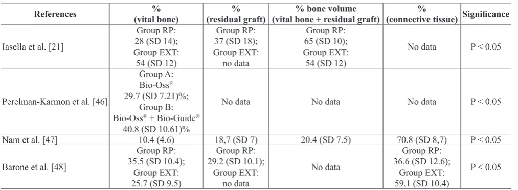
Table 7. Study’s that reported histological and histomorphometric analysis
References (vital bone)% (residual graft)% (vital bone + residual graft)% bone volume (connective tissue) Significance%
Iasella et al. [21]
Group RP: 28 (SD 14); Group EXT: 54 (SD 12)
Group RP: 37 (SD 18); Group EXT:
no data
Group RP: 65 (SD 10); Group EXT: 54 (SD 12)
No data P < 0.05
Perelman-Karmon et al. [46]
Group A: Bio-Oss® 29.7 (SD 7.21)%;
Group B: Bio-Oss® + Bio-Guide®
40.8 (SD 10.61)%
No data No data No data P < 0.05
Nam et al. [47] 10.4 (4.6) 18,7 (SD 7) 20.4 (SD 7.5) 70.8 (SD 8,7) P < 0.05
Barone et al. [48]
Group RP: 35.5 (SD 10.4);
Group EXT: 25.7 (SD 9.5)
Group RP: 29.2 (SD 10.1);
Group EXT: no data
No data
Group RP: 36.6 (SD 12.6);
Group EXT: 59.1 (SD 10.4)
P < 0.05
JOURNAL OF ORAL & MAXILLOFACIAL RESEARCH Faria-Almeida et al.
Flaps were advanced in order to achieve primary closure of the surgical wound.
Three different alveolar bone volume parameters
were chosen: external vertical measurement and internal vertical measurements to rate the vertical
changes of the post extraction socket and horizontal
measurement. The results showed that the higher external vertical volume change from the baseline to the 6 months follow-up was found in the control group (-0.38 [SD 0.22] vs -1.5 [SD 0.26]). However, the internal vertical (-5.81 [SD 0.29] vs -3.94 [SD 0.35] and horizontal measurements (-1.31 [SD 0.24]
vs -4.56 [SD 0.33]) presented with significantly less loss of alveolar bone height, more internal socket bone fill, and less horizontal reabsorption of the
alveolar bone ridge during the follow-up in the control group.
Like in the previous studies, clinical analysis revealed
that the alveolar preservation technique could be an advantage in reducing the alveolar bone dimensions after tooth extraction.
Histological and histomorphometric analysis
From the six papers included in Table 7, four trials [21,46-48] reported information about the histological variables: percentage of vital bone (four studies [21,46-48]); percentage of residual graft (three studies [21,47,48]), percentage of bone volume (two studies [21,47]), and percentage of connective tissue (two studies [47,48]).
Barone et al. in 2008 [48] presented the histologic
analysis showing a significantly higher percentage of
trabecular bone and total mineralized tissue in ridge-preservation sites compared to extraction-alone sites 7 months after tooth removal.
Nam et al. [47], presented the results of the histomorphometric analysis: percentage of residual graft, percentage of new bone, percentage of connective tissue and contact between particle and percentage of new bone, comparing the two groups. Results of these two trials [47,48] confirmed
advantage of the preservation technique comparing to the control.
In 2012, Perelman-Karmon et al. [46], compared extraction sites augmented with BBM with and without resorbable membrane coverage. BBM particles were grafted in fresh human extraction
sockets of 23 patients; in 12 of these patients, a
guided tissue regeneration method with collagen membrane was applied. After 9 months cylindric hard tissue sample were obtained for histomorphometric evaluation. Percent bone area fractions of the crestal, middle and apical sections from each
sample were calculated using the point-counting technique. Changes in values were compared. In sites augmented with BBM, the mean BAF ranged from 22.8% (coronal) to 36.3% (apical) compared to sites augmented with BBM and collagen membrane (35.2% [coronal] to 47% [apical]). Comparison between the
different depths and the two groups showed a distinct
increase in BAF from coronal to apical regions (P < 0.001). This pattern was observed in both groups (P
< 0.001) and was significantly higher in the group
augmented with BBM and collagen membrane (P < 0.05). In the immediate post extraction phase, BBM as a grafted biomaterial preserved the
socket volume and enabled newly formed bone
for future implant site preparation. The amount of the osseous fraction increased with collagen membrane.
In a study of Iasella et al. [21], the quantity of bone observed on histomorphometric analysis was
slightly greater (statistically significant difference) in
preservation sites, although these sites included both vital and non-vital bone but when analysed only the amount of vital bone this parameter was higher in control group. The most predictable maintenance of ridge width, height, and the position was achieved when a ridge preservation procedure was employed. The two other authors [46,48] showed that vital
bone was higher in sockets that were preserved with
a membrane. However, in one study [48] the natural healing was chosen as a control group and in the other [46] the grafting with a bovine bone. Therefore, even
though the statistically significant differences were
achieved in various studies, the results are heterogenic between the studies.
The results presented for the different papers must be analysed with caution because they used different materials in the preservation techniques and different time of analysis. They also included different types
of defects, so it is impossible to compare the results
between the different studies.
DISCUSSION
This systematic review was focused on evaluating the
effect of different alveolar preservation techniques
performed immediately after tooth extraction, in particular, to assess quantitatively and qualitatively
the influence of two different factors (membranes and
soft tissue graft).
The research questions of this systematic review were “Are there radiological, histological,
with and without membranes with a follow-up of at least 6 months” and “Are there radiological,
histological, histomorphometric or clinical differences in humans extraction socket preservation when a soft
tissue graft is performed with a follow-up of at least 6 months”
For this purpose, it was made a wide-ranging electronic search for all related articles up to 30 of November 2018 in the following electronic databases: LILACS, PubMed, SciELO, ScienceDirect, Scopus and Web of Science, with no data restriction.
Based on a clinical purpose, the included papers were only clinical trials and randomized clinical trials,
using keywords combinations already presented. It
was also made an additional manual search among
periodical journals and grey literature of relevant
papers.
No animal studies were included. Only intact
sockets were included following the same definition propose by Hämmerle et al. [49] “ridge preservation = preserving the ridge volume within the envelope existing at the time of extraction”. The included studies should have at least 12 patients or treated 12 sites per group and when
the studies tried to analyse the membrane effect
a control group without membrane need to be present.
Based on the inclusion and exclusion criteria there
was a lack of information, as reported previously, to take strength conclusions.
There was a great variability of the techniques and materials used, analysed variables, location of the extraction sites and also the time of evaluation.
Treatment combinations
A. Control group (blood clot) vs test group (collagen
membrane + filling material): two studies (Iasella
et al. [21] and Barone et al. [48]).
B. Control group (blood clot) vs test group (BBM + bioabsorbable membrane made of glycolide
and lactide polymers): one study (Lekovic et al.
[12]).
C. Control group (deproteinized bovine bone + epithelial connective tissue graft) vs test group (deproteinized bovine bone + collagen matrix): one study (Meloni et al. [45]).
D. Control group (Bio-Oss® [Geistlich Pharma®,
Wolhusen, Swiss]) vs test group (Bio-Oss® +
collagen membrane): one study (Perelman-Karmon et al. [46]).
E. Control group (deproteinized bovine bone) vs test group (collagen membrane) - one study (Nam et al. [47]).
Analysed variables
From the six included studies, three evaluated the results based on the CBCT analysis [21,45,48], one on the periapical X-ray [47], one on the clinical variables [12] and four on the histological and histomorphometric analysis [21,46-48].
Extraction sites
Most of the papers referred that the tooth extractions were made in the anterior zone of the maxilla, but in
some of them, there was a lack of information [45,48].
Time of evaluation
Most of the papers evaluate the results at 6 months after tooth extraction, but there are some that the evaluation period was later, at 9 and 17 months [45].
The risk of bias was high in four of six papers, and it
was low in two of them.
As one of the focus questions was related to the advantage or not by using a membrane in alveolar preservation techniques, there were only two studies that compare the use of membrane against blood clot (Iasella et al. [21] and Barone et al. [48]) which presented better results for the test (membrane) group. Both studies made a CBCT and histomorphometric analysis.
There was another study (Perelman-Karmon et al. [46]) which compare the use of BBM (Bio-Oss®)
in the control group versus BBM (Bio-Oss®) and
collagen membrane in the test group. They made a histological and histomorphometric analysis and
confirm the advantages of the use of a membrane. The
conclusion was that the BBM, as a grafted biomaterial
in fresh extraction sockets, preserved its volume and
enabled newly formed bone for future implant site preparation and the application of collagen membrane increased the osseous fraction.
The other focus question was about the advantage of using the soft tissue graft. For this particular question, there was one selected study by Meloni et al. [45] using CBCT analysis as control method. Meloni et al. [45] reported that sealing the socket with a porcine
collagen matrix or epithelial connective tissue graft showed similar outcomes if added to the BBM. Anyhow this study does not compare the use of soft tissue graft with control group, which means that this
particular conclusion enables to confirm the advantage
of using the soft tissue in the alveolar preservation techniques.
All of the other studies could give us an idea about the possible advantages of using the membrane, but they
JOURNAL OF ORAL & MAXILLOFACIAL RESEARCH Faria-Almeida et al.
CONCLUSIONS
1. Based on cone-beam computed tomography, periapical X-ray, clinical and histological and
histomorphometric findings it can be concluded
that the alveolar preservation techniques, presented in this systematic review, can decrease the dimensional reduction of the alveolar ridge that usually occurs after tooth extraction as compared to tooth extraction alone.
2. After analysis of the selected studies it is
impossible to recommend any specific techniques and/or materials to achieve improved results of extraction socket preservation because of the lack of sufficient data.
3. Few studies that compare the use of the alveolar preservation techniques with and without membrane, demonstrated that the application of the membrane allows to achieve superior preservation results.
4. There is lack of information concerning the
advantage of soft tissue graft using in the alveolar preservation techniques.
ACKNOWLEDGMENTS AND DISCLOSURE STATEMENTS
The authors report no conflicts of interest related to
this study.
REFERENCES
1. Chapple IL, Wilson NH. Manifesto for a paradigm shift: periodontal health for a better life. Br Dent J. 2014 Feb;216(4): 159-62. [Medline: 24557384] [doi: 10.1038/sj.bdj.2014.97]
2. Ali Z, Baker SR, Shahrbaf S, Martin N, Vettore MV. Oral health-related quality of life after prosthodontic treatment for patients with partial edentulism: A systematic review and meta-analysis. J Prosthet Dent. 2019 Jan;121(1):59-68.e3. [Medline: 30006220] [doi: 10.1016/j.prosdent.2018.03.003]
3. Wolleb K, Sailer I, Thoma A, Menghini G, Hammerle CH. Clinical and radiographic evaluation of patients receiving both tooth- and implant-supported prosthodontic treatment after 5 years of function. Int J Prosthodont. 2012 May-Jun;25(3):252-9. [Medline: 22545254]
4. Chappuis V, Engel O, Reyes M, Shahim K, Nolte LP, Buser D. Ridge alterations post-extraction in the esthetic zone: a 3D analysis with CBCT. J Dent Res. 2013 Dec;92(12 Suppl):195S-201S. [Medline: 24158340] [PMC free article: 3860068] [doi: 10.1177/0022034513506713]
5. Chappuis V, Engel O, Shahim K, Reyes M, Katsaros C, Buser D. Soft Tissue Alterations in Esthetic Postextraction Sites: A 3-Dimensional Analysis. J Dent Res. 2015 Sep;94(9 Suppl):187S-93S. [Medline: 26130259] [doi: 10.1177/0022034515592869]
6. Araújo MG, Lindhe J. Dimensional ridge alterations following tooth extraction. An experimental study in the dog. J Clin Periodontol. 2005 Feb;32(2):212-8. [Medline: 15691354] [doi: 10.1111/j.1600-051X.2005.00642.x]
7. Trombelli L, Farina R, Marzola A, Bozzi L, Liljenberg B, Lindhe J. Modeling and remodeling of human extraction sockets. J Clin Periodontol. 2008 Jul;35(7):630-9. [Medline: 18498382] [doi: 10.1111/j.1600-051X.2008.01246.x] 8. Discepoli N, Vignoletti F, Laino L, de Sanctis M, Muñoz F, Sanz M. Early healing of the alveolar process after tooth
extraction: an experimental study in the beagle dog. J Clin Periodontol. 2013 Jun;40(6):638-44. [Medline: 23534915] [doi: 10.1111/jcpe.12074]
9. Tan WL, Wong TL, Wong MC, Lang NP. A systematic review of post-extractional alveolar hard and soft tissue dimensional changes in humans. Clin Oral Implants Res. 2012 Feb;23 Suppl 5:1-21. [Medline: 22211303] [doi: 10.1111/j.1600-0501.2011.02375.x]
10. Van der Weijden F, Dell’Acqua F, Slot DE. Alveolar bone dimensional changes of post-extraction sockets in humans: a systematic review. J Clin Periodontol. 2009 Dec;36(12):1048-58. [Medline: 19929956] [doi: 10.1111/j.1600-051X.2009.01482.x]
11. Willenbacher M, Al-Nawas B, Berres M, Kämmerer PW, Schiegnitz E. The Effects of Alveolar Ridge Preservation: A Meta-Analysis. Clin Implant Dent Relat Res. 2016 Dec;18(6):1248-1268. [Medline: 26132885] [doi: 10.1111/cid.12364] 12. Lekovic V, Camargo PM, Klokkevold PR, Weinlaender M, Kenney EB, Dimitrijevic B, Nedic M. Preservation of alveolar
bone in extraction sockets using bioabsorbable membranes. J Periodontol. 1998 Sep;69(9):1044-9. [Medline: 9776033] [doi: 10.1902/jop.1998.69.9.1044]
13. Clementini M, Tiravia L, De Risi V, Vittorini Orgeas G, Mannocci A, de Sanctis M. Dimensional changes after immediate implant placement with or without simultaneous regenerative procedures: a systematic review and meta-analysis. J Clin Periodontol. 2015 Jul;42(7):666-77. [Medline: 26073267] [doi: 10.1111/jcpe.12423]
15. Álvarez-Camino JC, Valmaseda-Castellón E, Gay-Escoda C. Immediate implants placed in fresh sockets associated to periapical infectious processes. A systematic review. Med Oral Patol Oral Cir Bucal. 2013 Sep 1;18(5):e780-5. [Medline: 23722139] [PMC free article: 3790652] [doi: 10.4317/medoral.18942]
16. Bäumer D, Zuhr O, Rebele S, Hürzeler M. Socket Shield Technique for immediate implant placement - clinical, radiographic and volumetric data after 5 years. Clin Oral Implants Res. 2017 Nov;28(11):1450-1458. [Medline: 28333394] [doi: 10.1111/clr.13012]
17. Hürzeler MB, Zuhr O, Schupbach P, Rebele SF, Emmanouilidis N, Fickl S. The socket-shield technique: a proof-of-principle report. J Clin Periodontol. 2010 Sep;37(9):855-62. [Medline: 20712701] [doi: 10.1111/j.1600-051X.2010.01595.x] 18. Salama M, Ishikawa T, Salama H, Funato A, Garber D. Advantages of the root submergence technique for pontic site
development in esthetic implant therapy. Int J Periodontics Restorative Dent. 2007 Dec;27(6):521-7. [Medline: 18092446] [doi: 10.11607/prd.00.0775]
19. Artzi Z, Tal H, Dayan D. Porous bovine bone mineral in healing of human extraction sockets. Part 1: histomorphometric evaluations at 9 months. J Periodontol. 2000 Jun;71(6):1015-23. [Medline: 10914806] [doi: 10.1902/jop.2000.71.6.1015] 20. Brugnami F, Caiazzo A. Efficacy evaluation of a new buccal bone plate preservation technique: a pilot study.
Int J Periodontics Restorative Dent. 2011 Feb;31(1):67-73. [Medline: 21365028]
21. Iasella JM, Greenwell H, Miller RL, Hill M, Drisko C, Bohra AA, Scheetz JP. Ridge preservation with freeze-dried bone allograft and a collagen membrane compared to extraction alone for implant site development: a clinical and histologic study in humans. J Periodontol. 2003 Jul;74(7):990-9. [Medline: 12931761] [doi: 10.1902/jop.2003.74.7.990]
22. Avila-Ortiz G, Elangovan S, Kramer KW, Blanchette D, Dawson DV. Effect of alveolar ridge preservation after tooth extraction: a systematic review and meta-analysis. J Dent Res. 2014 Oct;93(10):950-8. [Medline: 24966231] [PMC free article: 4293706] [doi: 10.1177/0022034514541127]
23. Iocca O, Farcomeni A, Pardiñas Lopez S, Talib HS. Alveolar ridge preservation after tooth extraction: a Bayesian Network meta-analysis of grafting materials efficacy on prevention of bone height and width reduction. J Clin Periodontol. 2017 Jan;44(1):104-114. [Medline: 27712001] [doi: 10.1111/jcpe.12633]
24. MacBeth N, Trullenque-Eriksson A, Donos N, Mardas N. Hard and soft tissue changes following alveolar ridge preservation: a systematic review. Clin Oral Implants Res. 2017 Aug;28(8):982-1004. [Medline: 27458031] [doi: 10.1111/clr.12911]
25. Vignoletti F, Matesanz P, Rodrigo D, Figuero E, Martin C, Sanz M. Surgical protocols for ridge preservation after tooth extraction. A systematic review. Clin Oral Implants Res. 2012 Feb;23 Suppl 5:22-38. [Medline: 22211304] [doi: 10.1111/j.1600-0501.2011.02331.x]
26. Vittorini Orgeas G, Clementini M, De Risi V, de Sanctis M. Surgical techniques for alveolar socket preservation: a systematic review. Int J Oral Maxillofac Implants. 2013 Jul-Aug;28(4):1049-61. [Medline: 23869363] [doi: 10.11607/jomi.2670]
27. Becker W, Becker BE, Caffesse R. A comparison of demineralized freeze-dried bone and autologous bone to induce bone formation in human extraction sockets. J Periodontol. 1994 Dec;65(12):1128-33. Erratum in: J Periodontol 1995 Apr;66(4):309. Erratum in: J Periodontol. 1995 Apr;66(4):309. [Medline: 7877084] [doi: 10.1902/jop.1994.65.12.1128] 28. Froum S, Cho SC, Rosenberg E, Rohrer M, Tarnow D. Histological comparison of healing extraction sockets implanted
with bioactive glass or demineralized freeze-dried bone allograft: a pilot study. J Periodontol. 2002 Jan;73(1):94-102. [Medline: 11846205] [doi: 10.1902/jop.2002.73.1.94]
29. Serino G, Biancu S, Iezzi G, Piattelli A. Ridge preservation following tooth extraction using a polylactide and polyglycolide sponge as space filler: a clinical and histological study in humans. Clin Oral Implants Res. 2003 Oct;14(5):651-8. [Medline: 12969370] [doi: 10.1034/j.1600-0501.2003.00970.x]
30. Heberer S, Al-Chawaf B, Hildebrand D, Nelson JJ, Nelson K. Histomorphometric analysis of extraction sockets augmented with Bio-Oss Collagen after a 6-week healing period: a prospective study. Clin Oral Implants Res. 2008 Dec;19(12):1219-25. [Medline: 19040436] [doi: 10.1111/j.1600-0501.2008.01617.x]
31. Higgins JP, Green S. Cochrane Handbook for Systematic Reviews of Interventions. C.B. Series. England: Wiley and Backwell. 2008. [doi: 10.1002/9780470712184]
32. Liberati A, Altman DG, Tetzlaff J, Mulrow C, Gøtzsche PC, Ioannidis JP, Clarke M, Devereaux PJ, Kleijnen J, Moher D. The PRISMA statement for reporting systematic reviews and meta-analyses of studies that evaluate healthcare interventions: explanation and elaboration. BMJ. 2009 Jul 21;339:b2700. [Medline: 19622552] [PMC free article: 2714672] [doi: 10.1136/bmj.b2700]
33. Festa VM, Addabbo F, Laino L, Femiano F, Rullo R. Porcine-derived xenograft combined with a soft cortical membrane versus extraction alone for implant site development: a clinical study in humans. Clin Implant Dent Relat Res. 2013 Oct;15(5):707-13. [Medline: 22082037] [doi: 10.1111/j.1708-8208.2011.00398.x]
34. Jonker BP, Wolvius EB, van der Tas JT, Pijpe J. The effect of resorbable membranes on one-stage ridge augmentation in anterior single-tooth replacement: A randomized, controlled clinical trial. Clin Oral Implants Res. 2018 Feb;29(2): 235-247. [Medline: 29266485] [doi: 10.1111/clr.13106]
JOURNAL OF ORAL & MAXILLOFACIAL RESEARCH Faria-Almeida et al.
36. Scheyer ET, Heard R, Janakievski J, Mandelaris G, Nevins ML, Pickering SR, Richardson CR, Pope B, Toback G, Velásquez D, Nagursky H. A randomized, controlled, multicentre clinical trial of post-extraction alveolar ridge preservation. J Clin Periodontol. 2016 Dec;43(12):1188-1199. [Medline: 27617409] [PMC free article: 5132058] [doi: 10.1111/jcpe.12623] 37. Carmagnola D, Adriaens P, Berglundh T. Healing of human extraction sockets filled with Bio-Oss. Clin Oral Implants
Res. 2003 Apr;14(2):137-43. [Medline: 12656871] [doi: 10.1034/j.1600-0501.2003.140201.x]
38. Brkovic BM, Prasad HS, Rohrer MD, Konandreas G, Agrogiannis G, Antunovic D, Sándor GK. Beta-tricalcium phosphate/type I collagen cones with or without a barrier membrane in human extraction socket healing: clinical, histologic, histomorphometric, and immunohistochemical evaluation. Clin Oral Investig. 2012 Apr;16(2):581-90. [Medline: 21369794] [doi: 10.1007/s00784-011-0531-1]
39. Fickl S, Fischer K, Petersen N, Happe A, Schlee M, Schlagenhauf U, Kebschull M. Dimensional Evaluation of Different Ridge Preservation Techniques: A Randomized Clinical Study. Int J Periodontics Restorative Dent. 2017 May/Jun;37(3):403-410. [Medline: 28402351] [doi: 10.11607/prd.2629]
40. Leblebicioglu B, Salas M, Ort Y, Johnson A, Yildiz VO, Kim DG, Agarwal S, Tatakis DN. Determinants of alveolar ridge preservation differ by anatomic location. J Clin Periodontol. 2013 Apr;40(4):387-95. [Medline: 23432761] [PMC free article: 4955554] [doi: 10.1111/jcpe.12065]
41. Cardaropoli D, Tamagnone L, Roffredo A, Gaveglio L. Evaluation of Dental Implants Placed in Preserved and Nonpreserved Postextraction Ridges: A 12-Month Postloading Study. Int J Periodontics Restorative Dent. 2015 Sep-Oct;35(5):677-85. [Medline: 26357697] [doi: 10.11607/prd.2309]
42. Barone A, Toti P, Piattelli A, Iezzi G, Derchi G, Covani U. Extraction socket healing in humans after ridge preservation techniques: comparison between flapless and flapped procedures in a randomized clinical trial. J Periodontol. 2014 Jan;85(1):14-23. [Medline: 23688103] [doi: 10.1902/jop.2013.120711]
43. De Santis D, Cucchi A, de Gemmis A, Nocini Pier F. New collagen matrix to avoid the reduction of keratinized tissue during guided bone regeneration in postextraction sites. J Craniofac Surg. 2012 May;23(3):e186-9. [Medline: 22627428] [doi: 10.1097/SCS.0b013e31824de62c]
44. Filipek D, Koszowski R, Smieszek-Wilczewska J. A comparative clinical study on human tooth extractions: flap vs flapless buccal surgery. Quintessence Int. 2012 Nov-Dec;43(10):887-9. [Medline: 23115768]
45. Meloni SM, Tallarico M, Lolli FM, Deledda A, Pisano M, Jovanovic SA. Postextraction socket preservation using epithelial connective tissue graft vs porcine collagen matrix. 1-year results of a randomised controlled trial. Eur J Oral Implantol. 2015 Spring;8(1):39-48. [Medline: 25738178]
46. Perelman-Karmon. M, et al., Socket site preservation using bovine bone mineral with and without a bioresorbable collagen membrane. Int J Periodontics Restorative Dent, 2012. 32(4): p. 459-65. [Medline: 22577652] [doi: 10.11607/prd.00.1087] 47. Nam HW, Park JB, Lee JY, Rhee SH, Lee SC, Koo KT, Kim TI, Seol YJ, Lee YM, Ku Y, Rhyu IC, Park YJ, Chung
CP. Enhanced ridge preservation by bone mineral bound with collagen-binding synthetic oligopeptide: a clinical and histologic study in humans. J Periodontol. 2011 Mar;82(3):471-80. [Medline: 20932159] [doi: 10.1902/jop.2010.100193] 48. Barone A, Aldini NN, Fini M, Giardino R, Calvo Guirado JL, Covani U. Xenograft versus extraction alone for ridge
preservation after tooth removal: a clinical and histomorphometric study. J Periodontol. 2008 Aug;79(8):1370-7. [Medline: 18672985] [doi: 10.1902/jop.2008.070628]
49. Hämmerle CH, Araújo MG, Simion M; Osteology Consensus Group 2011. Evidence-based knowledge on the biology and treatment of extraction sockets. Clin Oral Implants Res. 2012 Feb;23 Suppl 5:80-2. [Medline: 22211307] [doi: 10.1111/j.1600-0501.2011.02370.x]
To cite this article:
Faria-Almeida R, Astramskaite-Januseviciene I, Puisys A, Correia F.
Extraction Socket Preservation with or without Membranes, Soft Tissue Influence on Post Extraction Alveolar Ridge Preservation: a Systematic Review
J Oral Maxillofac Res 2019;10(3):e5
URL: http://www.ejomr.org/JOMR/archives/2019/3/e5/v10n3e5.pdf doi: 10.5037/jomr.2019.10305
Copyright © Faria-Almeida R, Astramskaite-Januseviciene I, Puisys A, Correia F. Published in the JOURNAL OF ORAL & MAXILLOFACIAL RESEARCH (http://www.ejomr.org), 5 September 2019.