Literature Review
Ovarian Reserve Tests: The Use in Daily Clinical Practice
Pengujian cadangan ovarium: Penggunaan dalam praktik klinis sehari-hari
Nusratuddin Abdullah
Subdivision Fertility Reproductive Endocrinology Department of Obstetrics ang Gynecology Faculty of Medicine University of Hasanuddin/
Dr. Wahidin Sudirohusodo Hospital Makassar
INTRODUCTION
Fertility potential in female patient is related to the total number and quality of the primordial follicles remaining in the ovary and is referred to as ovarian reserve (OR). Ovarian reserve is a term that is used to determine the capacity of the ovary to provide eggs that are capable of fertilization resulting in a healthy and successful pregnancy.1
Diminished ovarian reserve generally indicates a poor response to any fertility treatment, and sharply limits the possibility of successful pregnancy.2
Screening for the OR is a fundamental component of the initial infertility evaluation, since it is a key determinant of what treatment should be offered.3
A reduced ovarian reserve is manifested by a di-minished ovarian response to medications for ovula-tion inducovula-tion, resulting in fewer eggs retrieved, fewer embryos, and finally lower pregnancy rates. Many women with unexplained infertility are suspected to have a reduced OR when tested.3
Ideally, a test that assesses ovarian reserve should be affordable, rapidly interpretable, and minimally in-vasive. It also should be able to detect changes that begin early in reproductive life. It can be performed anytime in the menstrual cycle, and should provide reproducible and highly accurate assessment of the reproductive aging process.2,3
This review discusses ovarian reserve tests (ORT) for ovulation induction and their application in deter-mining fertility capacity for evaluating follicle pool and for managing individualized patient ovulation in-duction.
CLASSIFICATION OF ORT
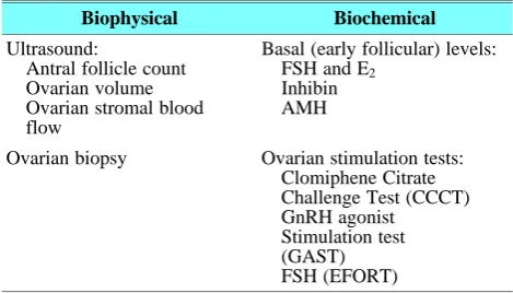
In general, ORT are either biophysical or biochemi-cal. (Table 1)4
This classification may be further classified as be-ing passive or dynamic ORT, or alternatively as bebe-ing direct or in direct ORT. Passive ORT includes: age, basal antral follicle count, basal ovarian volume, ova-Abstract
Objective: To evaluate the significance of conducting ovarian reserve testing and decide when to order this testing in the clinical setting. And also to give individualized counseling to patients re-garding the prognosis of infertility treatment based on their ovarian reserve tests, such as: basal antral follicle count, basal ovarian vol-ume, ovarian stromal blood flow, ovarian biopsy, basal serum Fol-licle Stimulating Hormone (FSH), basal serum estradiol, basal se-rum inhibin B, basal anti-Mullerian hormone sese-rum. Clomphine Cit-rate Challenge Test (CCCT), GnRH Agonist Stimulation Test (GA ST), and Exogenous FSH Ovarian Reserve Test (EFORT).
Method: Literature study on published studies about the meth-ods of ovarian reserve testing.
Conclusion: Ovarian reserve is an important key in initial as-sessment of the infertile patients and as a predictor of the success of infertility treatment. Currently, there is no single test which is high-ly reliable for assessing ovarian reserve. AMH serum may be a fu-ture hope and suggested as the best biomarker.
[Indones J Obstet Gynecol 2011; 35-4:235-40] Keywords: ovarian reserve test
Abstrak
Tujuan: Mengevaluasi pentingnya melakukan pemeriksaan ca-dangan ovarium dan memutuskan kapan pemeriksa ini harus di-lakukan di klinik. Pemeriksaan ini juga untuk memberikan konseling individu kepada pasien menyangkut prognosis penanganan infertili-tas berdasarkan hasil tes cadangan ovarium yang dilakukan, seper-ti: jumlah folikel antral basal, volume ovarium basal, aliran darah stroma ovarium, biopsi ovarium, serum Follicle Stimulating Hor-mone (FSH) basal, serum estradiol basal, serum inhibin B basal, se-rum hormon Anti-Müllerian basal, Clomphine Citrate Challenge Test (CCCT), GnRH Agonist Stimulation Test GnRH Agonist Stimu-lation Test (GAST), dan Exogenous FSH Ovarian Reserve Test (EF-FORT).
Metode: Telaah pustaka dari berbagai penelitian mengenai me-tode penilaian cadangan ovarium.
Kesimpulan: Cadangan ovarium merupakan suatu komponen penting dalam penilaian awal pasien infertil dan sebagai suatu prediktor keberhasilan penanganan infertilitas. Hingga saat ini, be-lum ada metode tunggal terbaik untuk menilai cadangan ovarium. Yang menjadi harapan di kemudian hari adalah pengukuran serum AMH diusulkan sebagai biomarker terbaik.
[Maj Obstet Ginekol Indones 2011; 35-4:235-40]
Kata kunci: tes cadangan ovarium
Correspondence: Nusratuddin Abdullah. Department of Obstetrics and Gynecology of Hasanuddin University, Dr. Wahidin Sudiro Husodo Hospital, Makassar. Telp.: 0411-585688.
rian stromal blood flow, ovarian biopsy, basal serum FSH, basal serum estradiol, basal serum inhibin B, basal anti-Mullerian hormone serum. Dynamic ORT includes: Clomiphene citrate challenge test (CCCT), GnRH agonist stimulation test (GAST), Exogenous FSH ovarian reserve test (EFORT). Direct ORT in-cludes: AFC, ovarian biopsy, serum inhibin-B, and basal serum anti-Mullerian hormone. Indirect ORT in-cludes: basal ovarian volume, ovarian stromal blood flow, basal serum FSH, basal serum estradiol, Clomi-phene citrate challenge test (CCCT), GnRH agonist stimulation test (GAST), Exogenous FSH ovarian re-serve test (EFORT).5
Table 1. Ovarian Reserve Tests.
Biophysical Biochemical
Ultrasound:
Antral follicle count Ovarian volume Ovarian stromal blood flow
Basal (early follicular) levels: FSH and E2
Inhibin AMH
Ovarian biopsy Ovarian stimulation tests: Clomiphene Citrate Challenge Test (CCCT) GnRH agonist
Stimulation test (GAST) FSH (EFORT)
PHYSIOLOGY OF OVARIAN RESERVE
Human follicles begin their development during the fourth month of fetal life and by the fifth month of intrauterine life, a maximum of approximately 7 mil-lion primordial eggs are present. At birth, 1-2 milmil-lions eggs are present and by puberty only around 25 per-cent eggs remain. About 400.000 of recruitable eggs in women’s reproductive life span to lose approxima-tely 1000 eggs each month either by atresia or entry into the growth phase. Only about 450 eggs will pro-gress to ovulation, which is finally followed by a de-cline to the menopause when only approximately 1000 eggs remain at an average age of 50-51 years. Only about 450 eggs will progress to ovulation.6,7
THE ASSESSMENT OF ORTs
OR Screening is a mechanism by which we can par-tially predict the reproductive potential in specific as well as oocytes potential in resulting in a healthy pregnancy. The number of tests being used to diag-nose or predict OR can simply be classified into two groups: Passive testing and dynamic testing. The goal of both approaches is to provide information regard-ing oocytes quality and quantity.5
FEMALE AGE
Ovarian reserve declines with age, but not uniformly. The effect of age on fertility is believed to arise from changes in both oocyte number and quality. Most stu-dies have found a greater frequency of cellular abnor-malities in oocytes from older women.8-13
Female age is the better predictor of egg quality, while basal FSH is the better predictor of egg quan-tity.7 In women over 40 years old, current success rates are overall low, even in women who have good OR with more number of eggs. In this case, quantity of eggs does not increase quality. On the contrary, young women with limited OR can have good success rates despite their limited cohort of eggs. Therefore, diminished OR in young women should not be used as an exclusion criterion for the likelihood of preg-nancy.14
AFC
AFC is defined as the total numbers of Antral Folli-cles Sized 2-5 or 2-10 mm being present in the ova-ries, which is detected by USG in early follicular pha-se.8 AFC numbers decline with age, from a range of 7-22 in early follicular phase in women aged 30 years to 2-7 in women aged 40 years.15 In a group of nor-mally cycling women with proven fertility, AFC also showed a strong correlation with age, declining slow-ly until age 37 and more rapidslow-ly there after.9,16,17
The normal AFC is 10-15 follicles over the two ovaries, and AFC is considered abnormal when less than 5-10. Other study suggested abnormal when AFC < 4 for one ovary and < 8 for both ovaries.2,18,19
AFC is single best predictor of response to ovarian stimulation, and superior to FSH.18 One study docu-mented that women with an AFC of less than 4 were 37 times more likely to have their cycle canceled and 8.7 times more likely not to be pregnant.7
The advantages of AFC provide a direct and also useful test to measure OR, as it has high specificity for predicting poor response to ovarian stimulation and failure to conceive (73-100% and 64-100% res-pectively). However, its low sensitivity (9-73% for poor response, and 8-33% for failure to conceive) making AFC limit its clinical utility. In addition, this kind of test is very much technically dependent and tends to be a wide range of inter and intra-observer variability.20
OVARIAN VOLUME
During a women’s life, ovarian volume changes from 0.7 cm3 at the age of 10 years to 5.8 cm3 at the age 18 years. However, at the age of 40 years, ovarian volume tends to decrease in size, and they decrease even further after menopause.21 These findings have led to the evaluation of ovarian volume as a useful marker of OR and menopausal status.22,23
clude ease of performance and incorporation into cli-nical practice, low interobserver variability, and the fact that the measurements remain relatively constant from cycle to cycle.19,25 Its main limitation at present is the lack of data regarding ovarian volume measu-rements in the general population, both fertile and in-fertile.4
OVARIAN STROMAL BLOOD FLOW (OSBF)
The rationale for this test is that the primordial folli-cles in the ovary have no independent capillary net-work, and therefore depend on their proximity to the stromal vessels for their supply of nutrients and hor-mones.4 By using Doppler color, ultrasound was able to detect and measure the ovarian stromal peak sys-tolic velocity (PSV). The ovarian stromal vascularity and AFC were decreasing with the advancing of age. The cut-off level for PSV was taken as 10 cm/s. When all the other predictive factors were held constant, the number of mature oocytes being retrieved increased by 7% for each additional 1 cm/s increase in ovarian PSV. A limitation of this technique is that it is very operator dependent.
OVARIAN BIOPSY
Histological Assessment of ovarian biopsy provides a direct assessment of follicular density. However ova-rian biopsy is not a reliable test to assess reproductive ageing on fertility, as there is a highly varied distri-bution of the follicles throughout the ovary. There-fore, the low density does not necessarily indicate a low ovarian reserve.3,26,27
Apart from being invasive with inherent risks and unknown future adverse effects on fertility, the main problem with this technique is there are no standard-ized measures for ovarian density among histopatho-logy laboratories, and may account for the large dis-crepancy in follicle numbers reported by various labo-ratories.4,28
FSH SERUM
Day 3 FSH is one of the most widely used measures of ovarian reserve and was proposed 20 years ago with the emergence of IVF.11,29,30 The test is an in-direct assessment of ovarian reserve which measures pituitary production of FSH in response to ovarian hormones.
Advancement of female age has been associated with a slow and steady compensatory elevation in FSH and a decrease in ovarian response.31 Persistent elevated basal FSH levels are consistent with dimin-ished ovarian reserve. However, some women expe-rience transient elevated basal FSH levels unrelated to their pool of primordial follicles.32
FSH measurements are typically combined with es-tradiol to enhance the sensitivity of testing. If the FSH or the estradiol level is high on day 3, patients are more likely to respond poorly to stimulate with gona-dotropins and have greatly reduced pregnancy rates. This is due to the aging follicle’s failure to produce adequate amounts of inhibin, a hormone that feeds
back to the pituitary to suppress FSH production.2,9 Basal FSH levels have been well studied, easily obtained, reliable, and inexpensive; and have been shown to be a superior predictor of IVF outcome compared with age alone. Some limitations of basal FSH levels in predicting OR include: relatively high incidence of false-positive values in younger women, high inter-cycle and inter-sample variability, different threshold levels used in different studies, and overlap between normal and abnormal levels especially in younger women.18
The cut-off values for basal FSH vary from 10.8 to 17 IU/l. The end points included pregnancy rates, ongoing pregnancy rates, number of oocytes or can-cellation rates.8 In general, a level between 5 and 10 mIU/ml is considered to be normal. Levels higher than 10 and 15 mIU/ml are considered borderline, and
≥ 16 mIU/ml are considered elevated, indicating di-minished success with infertility treatment.19 Further-more, another study confirmed a clear fall of preg-nancy rate when FSH level was > 20 ml/IU.4,33
Elevated basal FSH levels are indicative of dimin-ished ovarian reserve, and women with increased ba-sal FSH levels frequently have decreased oocytes be-ing retrieved in IVF program. On the contrary, of those who achieved pregnancy, about 98% had nor-mal findings on the OR tests.8
It appears that the highest value is of most prog-nostic significance.14 A meta-analysis of studies on basal FSH as a predictor of pregnancy confirmed a wide range of sensitivities (0.03-0.85) and specifici-ties (0.20-1.00).5,18,34
ESTRADIOL (E2)
The estradiol is elevated due to the elevation of FSH earlier in the cycle. Estradiol levels are commonly as-sessed during the early follicular phase of the men-strual cycle by infertility specialists as part of the hor-monal profiling component of the patient work-up, because it is a simple, inexpensive, and effective screening tool. High estrogen level on day 3 of cycle is indicative of Diminished Ovarian Reserve (DOR) even if the FSH level is completely normal. So it is very important to measure both day 3 FSH and estro-gen (E2) levels since an elevation in one or both tests may be equally predictive of decreased egg number and lower egg quality.35
Day 3 serum E2 levels may not be useful for ovar-ian reserve evaluation but they may help to indicate whether an ovulation induction should be cancelled. Therefore, they should be included in the female screening for infertility treatment. Normal Day 3 es-trogen levels vary from < 50 to < 80 pg/ml.2,9 Ideally, day 3 estrogen levels should be less than 50 pg/ml. Day 3 estrogen levels greater than 100 pg/ml are con-sidered abnormal.35
Those with PCOs can display a high estradiol since many antral follicles produce a bit of estradiol.7,36
INHIBIN
Inhibin is a glycoprotein hormone, which is produced by granulosa cells of preantral and early antral folli-cles. Inhibin levels vary during the menstrual cycle and is a direct measure of ovarian reserve when being assessed in the early follicular phase of the menstrual cycle.9 During a normal menstrual cycle, Inhibin B serum concentration increases gradually in the folli-cular phase to reach a peak at 7 days prior to the LH surge. It has been established that FSH secretion is controlled by Inhibin B and thus, the serum concen-trations of Inhibin B and FSH are inversely related, and at low serum levels of Inhibin B, FSH concen-tration goes up.2
Normal cut-off inhibin B levels range from ≥ 45-80 pg/ml. It has been suggested that women with low day 3 serum concentrations of inhibin-B have less ovarian reserve than women with high day 3 inhibin-B. Reduced secretion of inhibin B has been reported in women 35 years and older who have a normal men-strual cycle and a normal day 3 FSH level, compared with a similar group of women who were younger than 35 years. The data suggest that the serum inhibin B level might be an earlier predictor of loss of ovarian reserve when compared to the serum FSH level.1,5
One group of investigators demonstrated that wo-men with clinical evidence of diminished ovarian re-serve but a normal FSH level also had low inhibin-B production. The highest sensitivity (81%) and speci-ficity (81%) were obtained at a serum level of 56 pg/ml where the end-point was the number of oocytes collected.37
Inhibin-B testing involves a simple blood draw. However, routine testing for serum inhibin B levels is not recommended, due to limited availability of re-liable assays and controversy concerning its prognos-tic value.9
AMH
AMH is a glycoprotein growth factor and a member of the transforming growth factor-ß superfamily. An-tral follicles are considered to be the primary source of circulating AMH as they contain a large number of granulosa cells. Clinical data in IVF suggests that AMH is preferentially and constantly secreted by small rather than large antral follicles. Granulosa cells secrete AMH into both the bloodstream and follicular fluid. The number of small antral follicles is directly related to the total size of the primordial follicle pool. With the decrease in the number of antral follicles with age, AMH serum levels also become diminished and will invariably become undetectable near meno-pause.15
Range of AMH-1 3 ng/ml is concidered to be nor-mal, 0.3-0.9 ng/ml to be low, and less than 0.3 to be very low. AMH levels less than 0.3 - 0.5 ng/ml are associated with increased IVF cycle cancellation rates and fewer eggs retrieved from the ovaries. While AMH levels greater than 2.5 ng/ml are associated with greater number of eggs retrieved and a better fertility potential.38
Serum AMH levels in female are almost undetect-able at birth, and increase slightly around the age of
3 years. AMH levels continue to increase during pu-berty, then reach their peak after puberty and decrease thereafter.39 A progressive decline occurs throughout reproductive life as the follicular reserve becomes de-pleted39,40 and finally becomes undetectable after me-nopause.41,42
The potential advantages of AMH compared with other conventional markers of ovarian reserve inclu-de: it is the earliest marker to change with age; it has the least variability from cycle to cycle; it has the least variability when measured on different days of the same cycle.9,19
However, utility of AMH is limited in PCOs and obesity. In a recent series investigating AMH levels in women with PCOs, circulating levels of AMH are higher in women with PCOs than those without PCOs. This finding is thought to be indicative of oligo-ovulation and poor follicular development in polycys-tic ovaries.9,14,41,43,44 AMH levels have been found to be two to three times higher in PCOs women.5,18 The consistently positive correlation between AMH and PCOs may suggest a future role for this marker as a diagnostic tool. On the contrary, women who are overweight have 70% lower AMH levels than thin women, indicating that obesity may be associated with decreased ovarian reserve and/or with ovarian dysfunction.9
Serum AMH levels correlate strongly with the number of antral follicles and follicle depletion. AMH is thought to be the first ovarian reserve marker that decline at an earlier stage compared with FSH, estra-diol, and inhibin-B.19
The currently available literature indicates that AMH may be a superior marker for predicting ovarian response over either age of the patient, day 3 FSH, estradiol or inhibin B, whereas the vast majority of studies have found AMH and AFC to have similar predictive value for the poor response. Furthermore, several studies have shown that AFC and AMH mar-kers are equivalent in terms of their high accuracy in predicting ovarian reserve.19
CLOMPHINE CITRATE CHALLENGE TEST (CCCT)
Provocative tests of ovarian reserve, such as CCCT, GAST and EFORT are more sensitive indicators of ovarian reserve than basal tests. Among those, CCCT seems to be the best to predict ovarian reserve, and is an indirect testing of OR.9 This test involves the oral administration of 100 milligrams of clomiphene citrate on menstrual cycle days 5-9. Blood levels of FSH and estradiol are measured on cycle day 3 and the level of FSH is also measured on cycle day 10. The rationale behind the test is that once clomiphene citrate is administered, FSH and LH levels rise, fol-lowed by an increase in estradiol and inhibin. An ele-vated FSH (≥ 10 mIU/ml) on either day 3 and 10 indicates diminished ovarian reserve. The CCCT can help identify patients with diminished OR that was not detected by basal FSH measurements.
are relatively noninvasive; and rapid measures of ova-rian reserve. However, this test become more expen-sive since it used two blood draws instead of one, with a significant inter-cycle variability, and high in-tra-cycle variability.9
The basal FSH test and CCCT have high specificity (98% to 99% for each) as an assessment of reproduc-tive performance in infertile women and generate few false-positive results.12 However, the sensitivity of the tests is low (between 7% and 8% for basal FSH and between 25% and 40% for the CCCT). Such low sensitivity means that many women will not conceive after infertility treatment despite a normal test re-sult.12,45
Theoretically the CCCT was designed to detect low ovarian reserve that would not be discovered by a single FSH and/or E2 measurements.2
GONADOTROPHIN AGONIST STIMU-LATION TEST (GAST)
This test evaluates the estradiol serum concentration change from cycle day 2 to day 3 after administration of a GnRH agonist such as leuprolide acetate (Lup-ron) on day 2 or 3, causing a temporary increase in pituitary secretion of FSH and LH.4,46 A prompt es-tradiol response to GnRH agonist may be associated with a better ovarian reserve.8 In a significant study, the actual increase in estradiol concentration from day 2 to day 3 following GnRH agonist administration on day 2 was a better predictor of ovarian reserve than age and basal FSH.4
The sensitivity of GnRH agonist testing for preg-nancy is moderate (32% to 89%); specificity ranges from 79% to 97%).9,20
EXOGENOUS FSH OVARIAN RESERVE TEST (EFORT)
The exogenous FSH ovarian reserve test was designed to screen for good or poor response in IVF. The change of estradiol levels in response to 300 IU of FSH administered on cycle day 3 provides the dyna-mic component to this test and has recently been shown to demonstrate less intercycle variability than basal FSH levels. However, the value of this parame-ter as an independent variable predictive of IVF out-come has not been fully adminished, and the ability of this test to differentiate between normal and dimi-nished ovarian reserve appears to be limited.14
A recent randomized, prospective study in which basal and dynamic tests of ovarian reserve were com-pared revealed the EFORT as being the best predictor of ovarian reserve.47
CONCLUSION
Based on the advantages and limitations including sensitivity and specificity, inter and intra cycle vari-ability, ease and practice and affordable of every OR test, the most possible tests that could be performed in clinical setting are basal FSH, basal estradiol and basal AFC, basal AMH and CCCT.
AMH is suggested as the best OR test since it is the earliest marker to change with age and has the least variability from cycle to cycle, even when meas-ured on different days of the same cycle. However, the use of AMH is still limited in PCOs and obesity patients.
REFERENCES
1. Stovall DW, McGee EA. How chemotherapy harms ovarian function. SRM. 2010; 8(3): 21-8
2. Lifchez A. Infertility, Age and Ovarian Reserve.
3. Semary AMS. Determination Of Ovarian Reserve By Tran-svaginal Echographic Measurement Of Ovarian Volume and Antral Follicular Count. Cairo: AL Azhar University; 2007
4. Singh KL, Davies M, Chatterjee R. Fertility in female can-cer survivors: pathophysiology, preservation and the role of ovarian reserve testing. Hum Reprod Update. 2005; 11(1): 69-89
5. Abdel Sattar N, Tharwat A. Ovarian Reserve. ASJOG. 2005; 2: 338-43
6. Wallace WHB, Kelsey TW. Human Ovarian Reserve from Conception to the Menopause. Plos One. 2010; 5(1): 1-9 7. Toner JP. Ovarian Reserve, Female Age, and the Chance
for Successful Pregnancy. USA: Atlanta Center for Repro-ductive Medicine
8. Bukulmez O. Aricib A. Assessment of ovarian reserve. Current Opinion in Obstetrics and Gynecology: Lippincott Williams and Wilkins, 2004: 231-7
9. Butts SF, Seifer DB. 6 office tests to assess ovarian reserve, and what they tell you. American Society for Reproductive medicine 2008: 20(11)
10. Age and infertility in women. Fertil Steril. 2006; 86: 248-52
11. Muasher SJ, Oehninger S, Simonetti S, Matta J, Ellis LM, Liu H-C. The value of basal and/or stimulated serum go-nadotropin levels in prediction of stimulation response and in vitro fertilization outcome. Fertil Steril. 1988; 50: 298-307
12. Barnhart K, Osheroff J. Follicle stimulating hormone as a predictor of fertility. Curr Opin Obstet Gynecol; 1998: 227-32
13. Lim AS, Tsakok MFH. Age-related decline in fertility: a link to degenerative oocytes? Fertil Steril. 1997; 68: 265-71 14. Macklon NS, Fauser JM. Ovarian Reserve. Semin Reprod
Med. 2005; 23(3): 248-56
15. Broekmans FJ, Fauser BC. Ovarian Aging: Mechanisms and Clinical Consequences. Endocrine Reviews. 2009; 30 (5): 465-93
16. Ng EH, Yeung WS, Fong DY, Ho PC. Effects of age on hormonal and ultrasound markers of ovarian reserve in Chi-nese women with proven fertility. Hum Reprod. 2003; 18: 2169-74
17. Scheffer GJ, Broekmans FJ, Dorland M, Habbema JD, Looman CW, te Velde ER. Antral follicle counts by trans-vaginal ultrasonography are related to age in women with proven natural fertility. Fertil Steril. 1999; 72: 845-51 18. Maheshwari A, Bhattacharya S. Ovarian Aging and
Fertil-ity Bentham Science Publishers Ltd.; 2007
19. Maseelall PB. Ovarian Reserve Screening: An Update. Post Graduate Obstetric and Gynecology. 2011; 31(16) 20. Broekmans FJ, Fwee J, Hendricks DJ, Mol BW, Lambalk
CB. A systematic review of tests predicting ovarian reserve and IVF outcome. Hum Reprod Update. 2006; 12: 685-718 21. Pavlik EJ, DePriest PD, Gallion HH, Ueland FR, Reedy MB, Kryscio RJ, van NJ J. Ovarian volume related to age. Gynecol Oncol. 2000; 77: 410-2
22. Flaws JA, Langenberg P, Babus JK, Hirshfield AN, Sharara FI. Ovarian volume and antral follicle counts as indicators of menopausal status. Menopause. 2001; 8: 175-80 23. Flaws JA, Rhodes JC, Langenberg P, Hirshfield AN,
Kje-rulff K, Sharara FI. Ovarian volume and menopausal status. Menopause 7. 2000: 53-61
24. Gurgan T, Demirol A, Guven S. Ultrasonography evalua-tion of ovarian reserve. In: Rizk BRMB, editor. Ultrasono-graphy in Reproductive Medicine and Infertility: Cambrid-ge University Press; 2010: 213-9
25. Fanchin R, Schonauer LM, Righini C. Serum anti-Mulle-rian hormone is more strongly related to ovaanti-Mulle-rian follicular status than serum inhibin B, estradiol, FSH, and LH on day 3. Hum Reprod. 2003; 18: 323-7
26. Kupesic S, Kurjak A. Predictors of IVF outcome by three-dimensional ultrasound. Hum Reprod. 2002; 17: 950-5 27. Zaidi S, Usmani A, Ishrat S, Shokh. Ovarian reserve and
reproductive age. Pak J Med Sci. 2007; 23(3): 449-53 28. Tilly JL. Ovarian follicle counts-not as simple as 1, 2, 3.
Reprod Biol Endocrinol 2003; 1: 11
29. Scott RT, Toner JP, Muasher SJ, Oehninger S, Robinson S, Rosenwaks Z. Follicle stimulating hormone levels on cycle day 3 are predictive of in vitro fertilization outcome. Fertil Steril. 1989; 51: 651-4
30. Toner JP, Philiput CB, Jones GS, Muasher SJ. Basal follicle stimulating hormone level is a better predictor of in vitro fertilization outcome than age. Fertil Steril. 1991; 55: 784-91
31. Sills ES, Alper MM, Walsh AP. Ovarian reserve screening in infertility: practical applications and theoretical direc-tions for research. Euro J Obstet Gynecol Reprod Biol. 2009; 146: 30-6
32. de Koning CH, McDonnell J, Themmen AP. The endocrine and follicular growth dynamics throughout the menstrual cycle in women with consistently or variably elevated early follicular phase FSH compared with controls. Hum Reprod. 23: 1416-23
33. Van Rooij IA, de Jong E, Broekmans FJ, Looman CW, Habbema JD, te Velde ER. High follicle-stimulating hor-mone levels should not necessarily lead to the exclusion of subfertile patients from treatment. Fertil Steril. 2004; 81: 1478-85
34. Bancsi LF, Broekmans FJ, Mol BW, Habbema JD, te Velde ER. Performance of basal follicle-stimulating hormone in the prediction of poor ovarian response and failure to be-come pregnant after in vitro fertilization: a meta-analysis. Fertil Steril. 2003; 79: 1091-100
35. Ovarian Reserve Test. 2011
36. Lobo RA. Potential Options for Preservation of Fertility in Women. New Eng J Med. 2005; 353: 64-73
37. Ficicioglu C, Kutlu T, Demirbasoglu S, Mulayim B. The role of inhibin B as a basal determinant of ovarian reserve. Gynecol Endocrinol. 2003; 17: 287-93
38. Dahiya P, Dahiya K, Dhankhar R, Hooda N, Nayar KD. The Role of the Anti-Mullerian hormone in Female Fertil-ity; A review. J Clin Diagnostic Res. 2011; 5(2): 384-7 39. Lee MM, Donahoe PK, Hasegawa T, Silverman B, Crist
GB, Best S, Hasegawa Y, Noto RA, Schoenfeld D, Mac Laughlin DT. Mullerian inhibiting substance in humans: normal levels from infancy to adult hood. J Clin Endocrinol Metab. 1996; 81: 571-6
40. Guibourdenche J, Lucidarme N, Chevenne D, Rigal O, Ni-colas M, Luton D. Anti-Mullerian hormone levels in serum from human foetuses and children: pattern and clinical in-terest. Mol Cell Endocrinol. 2003; 211: 55-63
41. Marca AL, Orvieto R, Giulini S, Jasonni VM, Volpe A, De Leo V. Müllerian-inhibiting substance in women with polycystic ovary syndrome: relationship with hormonal and metabolic characteristics. Fertil Steril. 2004; 82: 970-1 42. Van Rooij IA, Tonkelaar I, Broekmans FJ, Looman CW,
Scheffer GJ, de Jong FH. Anti-mullerian hormone is a promising predictor for the occurrence of the menopausal transition. Menopause. 2004; 11: 601-6
43. Al-Qahtani A, Groome NP. Anti-Müllerian hormone: Cin-derella finds new admirers. J Clin Endocrinol Metab 2006; 91: 3760-2
44. Pellatt L, Hanna L, Brincat M. Granulosa cell production of anti-Müllerian hormone is increased in polycystic ova-ries. J Clin Endocrinol Metab. 2007; 92: 240-5
45. Jain T, Soules MR, Collins JA. Comparison of basal folli-cle-stimulating hormone versus the clomiphene citrate cha-llenge test for ovarian reserve screening. Fertil Steril. 2004; 82: 180-5
46. Sharara FI, Scott R, Seifer DB. The detection of diminished ovarian reserve in infertile women. Am J Obstet Gynecol. 1998; 179: 804-12
47. Galtier-Dereure F, De Bouard V, Picto MC. Ovarian re-serve test with the gonadotrophin-releasing hormone ago-nist buserelin: correlation with in-vitro fertilization out-come. Hum Reprod. 1996; 11: 1393-8