Aleksandra K. Majewska
1, Tomasz Janus
2, Elżbieta Ronin-Walknowska
1,
Tomasz Płonka
1, Krzysztof S. Borowiak
21
H Magnetic Resonance Spectroscopy of Urine
for the Assessment of Renal Dysfunction in Healthy
Pregnant Women*
1
H spektroskopia rezonansu magnetycznego moczu
w ocenie zaburzeń czynności nerek u zdrowych kobiet będących w ciąży*
1 Department of Maternal and Fetal Medicine and Gynecology, Pomeranian Medical University, Szczecin, Poland 2 Department of Forensic Medicine, Pomeranian Medical University, Szczecin, Poland
Abstract
Background. Pregnancy is associated with adaptive alterations affecting almost all organs and systems of the female body and is also a time when pathologies appear which would otherwise remain asymptomatic for many years. 1H Magnetic Resonance Spectroscopy (1H-MRS) of urine, a modern diagnostic method, can disclose discrete changes in organ function. It seemed interesting to search for pregnancy-dependent renal pathology in healthy women and thus prognosticate progression to overt disease after pregnancy.
Objectives. To determine whether physiological pregnancy is accompanied by alterations in renal structures detectable by 1H-MRS of urine and to assess the type and reversibility of alterations.
Material and Methods. Eighty women were enrolled in two groups: a study group (B) consisting of 40 healthy pregnant women and a control group (K) of 40 healthy nulligravida women. Enrollment criteria included normal health status, unrevealing physical and gynecological examination, and normal results of laboratory tests (complete blood count, urea, creatinine, uric acid, urinalysis). MRS of urine and laboratory tests were done in group B during each trimester (B1, B2, and B3, i.e. the 1st, 2nd, and 3rd trimesters, respectively) and six weeks after delivery (BP). In group K, the tests were done only once. The spectra were processed with MestReC software and then multidimen-sional statistical analysis using Unscrambler software was performed. The results were presented as the distribution of the variables in multidimensional space.
Results. The patterns in multidimensional space revealed a clustering of points when comparing the first and third trimesters of pregnancy and comparing the third trimester and the healthy non-pregnant women. Also, a partial clustering of points comparing healthy non-pregnant and pregnant women after the puerperium was noticeable.
Conclusions. Differences in 1H-MRS profiles of urine between healthy pregnant and non-pregnant women reflect alterations of specific structures of the kidney accompanying pregnancy. In the majority of patients the effect of pregnancy on renal tubules is transient and (presumably) does not lead to permanent lesions (Adv Clin Exp Med 2010, 19, 2, 177–183).
Key words: MRS, urine, reproductive and urinary physiology, kidney tubules.
Streszczenie
Wprowadzenie. Niejednokrotnie podczas ciąży pierwotnie fizjologicznej dochodzi do ujawnienia się chorób, któ-rych symptomy mogłyby pojawić się wiele lat później. 1H-MRS moczu można zaliczyć do najnowszych technik analitycznych, wystarczająco czułych, by wykryć dyskretne zmiany w funkcji narządów. Interesujące wydaje się poszukiwanie zmian czynności nerek związanych z ciążą fizjologiczną, których obecność mogłaby poprzedzać wystąpienie jawnej choroby nerek po okresie ciąży.
Adv Clin Exp Med 2010, 19, 2, 177–183 ISSN 1230-025X
oRIGINAl PAPERS
© Copyright by Wroclaw Medical University
Pregnancy is associated with adaptive altera-tions affecting almost all organs and systems of women. The maternal organism is thus optimally prepared for the growth and maturation of the fetus. Pregnancy is also a time when pathologies appear which otherwise would remain asymptom-atic for many years. Follow-up studies have shown that complications of pregnancy, such as abortion, premature delivery, pre-eclampsia, or low birth weight, often herald ischemic heart disease or cerebrovascular events as well as thrombo-embo-lic episodes in the case of severe pre-eclampsia. A prominent hypothesis explaining these associa-tions views pregnancy as a kind of stress [1].
Alterations in kidneys during physiological pregnancy are largely the consequence of hemody-namic changes. Increased cardiac ejection volume and total blood volume during gestation result in an almost 50% increase in renal perfusion and glomerular filtration rates [2], with faster creati-nine and urea clearances. Retention of sodium and water throughout pregnancy is caused by a syn-ergy between ADH and aldosterone. Glucosuria and minor proteinuria of 200–300 mg/24-h is frequently observed in healthy pregnant women because of increased glomerular filtration and reduced tubular reabsorption [2, 3]. All these changes are reversible and the situation returns to normal during the puerperium.
Considering the fact that modern diagnostic methods are sensitive enough to disclose discrete changes in organ function, it seemed interesting to search for pregnancy-dependent renal pathology in otherwise healthy females and thus to prognos-ticate progression to overt disease after pregnancy. Pregnant women would thus be screened and
fol-low-up, prevention, and early treatment could be started with measurable benefits.
1H magnetic resonance spectroscopy (1H-MRS)
of urine is among these modern analytical tech-niques. Changes in signal intensity in specific regions of the 1H-MRS urine profile provide
infor-mation about lesions involving specific renal struc-tures [4, 5, 8, 9]. From the clinical point of view,
1H-MRS of urine has several advantages, including
high sensitivity enabling the detection of metabo-lites at concentrations far below the thresholds of routine laboratory tests, noninvasiveness, repeat-ability, small sample volume (approx. 0.5 ml), simple sample processing, and short measurement times (5 min). Many substances can be finger-printed concurrently without the need for metab-olite pre-selection [4].
This study was undertaken to determine whether physiological pregnancy is accompa-nied by alterations in renal function detectable by
1H-MRS and to assess the type and reversibility of
the alterations.
Material and Methods
Eighty women seen at the Department of Maternal and Fetal Medicine and Gynecology, Pomeranian Medical University in Szczecin, Poland, were enrolled and allocated to two groups: a study group (B) consisting of 40 healthy pregnant women with single-fetus pregnancies (34 primi-gravida, 6 multiprimi-gravida, mean age: 27.6 years) and a control group (K) of 40 healthy nulligravida women (mean age: 27.5 years).
Enrollment criteria included normal health
Cel pracy. określenie, czy ciąża fizjologiczna powoduje możliwe do wykrycia za pomocą badania 1H-MRS moczu zmiany w strukturach nerek oraz jaki jest charakter i trwałość ewentualnych zmian.
Materiał i metody. Badaniami objęto 80 kobiet, podzielonych na dwie grupy: badaną (B) – 40 zdrowych ciężarnych i kontrolną (K) – 40 zdrowych nieciężarnych nieródek. Kryteria włączenia do badań dla obu grup obejmowały: dobry stan ogólny, brak odchyleń w badaniu fizykalnym oraz ginekologicznym, prawidłowe wyniki badań labo-ratoryjnych. W obu grupach pacjentek wykonano badania laboratoryjne krwi: oznaczenie morfologii, mocznika, kreatyniny i kwasu moczowego, badanie ogólne moczu i badanie moczu techniką 1H-MRS. Pomiary wykonywano w grupie B czterokrotnie: w każdym trymestrze ciąży oraz sześć tygodni po porodzie. W grupie kontrolnej badania wykonano raz. Widma rezonansowe moczu poddano obróbce w programie MestReC, a następnie statystycznej analizie wielowymiarowej (PCA) w programie Unscrambler. Wyniki przedstawiono w formie rozkładu zmiennych w przestrzeni wielowymiarowej w układzie przestrzennym.
Wyniki. Rozkład zmiennych w przestrzeni wielowymiarowej wykazuje tendencję do grupowania się punktów odpowiadających danym dla profili moczu z pierwszego i trzeciego trymestru ciąży w różnych obszarach przestrze-ni wielowymiarowej. Porównaprzestrze-nie rozkładu punktów odpowiadających danym z profili moczu zdrowych przestrze- nieciężar-nych oraz pacjentek po okresie połogu wskazuje na częściowe, aczkolwiek niezupełne nakładanie się zajmowanieciężar-nych przez nie obszarów.
Wnioski. Różnice w profilach 1H-MRS moczu między zdrowymi ciężarnymi a nieciężarnymi odzwierciedlają zmia-ny związane z ciążą obecne w specyficzzmia-nych strukturach nerki. Ciąża fizjologiczna wpływa obciążająco na cewki nerkowe, u większości pacjentek jest to jednak działanie przejściowe i prawdopodobnie nie prowadzi do trwałych uszkodzeń (Adv Clin Exp Med 2010, 19, 2, 177–183).
status, unrevealing physical and gynecological examination, and normal results of laboratory tests (complete blood count, urinalysis, creatinine, uric acid, urea). Patients with physiological pregnan-cies terminating with delivery of a healthy neo-nate were enrolled retrospectively to group B. No medication was administered during pregnancy. Fish and seafood (rich sources of trimethylamine-N-oxide (TMAo) producing false-positive results) were withdrawn from the diet seven days prior to urine collection. Women at risk of tubular lesions accompanying infection of the urinary tract or chronic renal disease, protracted antibiotic therapy, or administration of NSAIDs or other potentially nephrotoxic drugs were excluded. The following laboratory tests were performed in both groups: complete blood count, urea, creatinine, uric acid, urinalysis, and MRS of urine. The tests were done on four occasions in group B: during each trimester (B1, B2, B3, i.e. the first, second, and third trimesters,
respectively) and six weeks after delivery (BP). In
group K, the tests were done only on one occasion. The study was approved by the Ethics Committee of Pomeranian Medical University in Szczecin.
MRS of Urine
A 15-ml sample of urine (morning, midstream, sterile container) was collected for MRS and fro-zen immediately after collection to –70°C. Prior to spectroscopy, the urine was thawed and the pH was adjusted to 5.8. Spectra were obtained on a Bruker DPX-400 Avance instrument operating at 400 MHz and 295 K. The resolution was 0.15 Hz/ /point. Spectra were acquired with 64 scans and the chemical shift range was 12 ppm. The water signal was suppressed by presaturation (65 dB pulse power) and measurements were referenced to an external standard (DSS).
The resonance spectra were processed with MestReC software featuring Fourier transfor-mation, automatic phase correction, automatic baseline adjustment, and creatinine signal nor-malization and binned across 0.04 ppm intervals. Numeric data in the form of ASCII peaks rang-ing from 0.5–4.3 and 6.5–9.0 ppm were exported for multidimensional statistical analysis (princi-pal component analysis, PCA) with Unscrambler software. The results were presented as the dis-tribution of variables in multidimensional space. Computations were done in spectral regions typi-cal for amino acids + lactates, citrates + dimeth-ylamine, aliphatic amines + creatinine, and aro-matic compounds + hippurates.
The analyzed regions are known to change in signal intensity when lesions of specific renal structures occur (Table 1).
Results
Table 2 presents the mean values of the labora-tory indicators of renal function (urea, creatinine, uric acid) measured in the serum of patients in group B during each trimester of pregnancy and after the puerperium and in group K. Interpretation of the K and Bp values was based on the normal range for
the general population (urea: 17–43 mg/dl, creati-nine: 0.7–1.2 mg/dl, uric acid: 2.4–5.7 mg/dl). The
Table 1. Structures of the kidney which in case of their damage are responsible for changes in excretion of low molecular weight metabolites appearing in 1H-MRS urine
spectra [5].
Tabela 1. Struktury nerki, których uszkodzenie może powodować zwiększenie bądź zmniejszenie wydalania odpowiednich substancji drobnocząsteczkowych, co znaj-duje odzwierciedlenie w widmach 1H-MRS moczu [5]
low molecular me-tabolite
(Substancja drob- nocząsteczkowa
Effect
(Rezultat) Renal structure in-volved (Struktury nerki)
lactate
(Mleczan) increase S1/S2 regions pro-ximal tubule Citrate
(Cytrynian) decrease tubular mitochon-dria Dimethylamine
(DMA) increase renal medulla
Trimethylamine
N-oxide (TMAo) increase renal medulla Glycine
(Glicyna) increase pars recta S3 proxi-mal tubule Hippurate
(Hipuran) decrease convoluted proxi-mal tubule
Table 2. Mean concentrations of urea, creatinine, and uric acid in the serum of study groups (B1, B2, B3, and Bp – see
text for legend) and the control (K)
Tabela 2. Wartości średnie mocznika, kreatyniny, kwasu moczowego oznaczone w surowicy krwi pacjentek w gru-pie kontrolnej (K) oraz w grugru-pie badanej w poszczegól-nych trymestrach ciąży (B1, B2, B3) i po okresie połogu (Bp)
Group
(Grupa) Urea (Mocznik) [mg/dl] mean ± SD
Creatinine (Kreatynina) [mg/dl] mean ± SD
Uric acid (Kwas moczo-wy) [mg/dl] mean ± SD B1 19.28 ± 4.59 0.56 ± 0.08 2.82 ± 0.63
B2 16.4 ± 4.23 0.52 ± 0.08 3.15 ± 0.66
B3 17.59 ± 5.93 0.59 ± 0.13 3.69 ± 0.87
Bp 28.7± 6.85 0.79 ± 0.15 4.59 ± 0.94
concentrations reported in the literature as normal for pregnancy were adopted in the case of the B1, B2,
and B3 values (urea: 17.48 ± 3.21 mg/dl, creatinine:
0.46 ± 0.13 mg/dl, uric acid: 1.15–4 mg/dl) [2, 6, 7]. It can be seen that all concentrations were within the normal range (an enrollment criterion).
Figure 1 presents a matrix plot of urine spec-tra displaying intensity differences for chemical shifts. low-intensity bands are present at 0.5, 6.8, and 8.5 ppm.
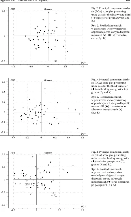
Multidimensional analysis (PCA) of the spec-tra produced a projection of points in multidimen-sional space. The analysis was restricted to the five main components responsible for 90% of the vari-ance. As the projection of points using XYZ coor-dinates was not legible enough, the points were paired (K with B1, K with B2, K with B3, K with BP)
to compare the results in the control group with those in the study group during each trimester and after the puerperium. The results are shown graph-ically using the XY coordinate system (PCA). only results disclosing differences between groups as to
1H-MRS profiles are displayed.
Figure 2 reveals point clustering in the first and third trimesters of pregnancy.
Point clustering in the third trimester and in the healthy non-pregnant women is evident in Figure 3.
Partial clustering of points in the healthy non-pregnant women and in the non-pregnant women after the puerperium is noticeable in Figure 4.
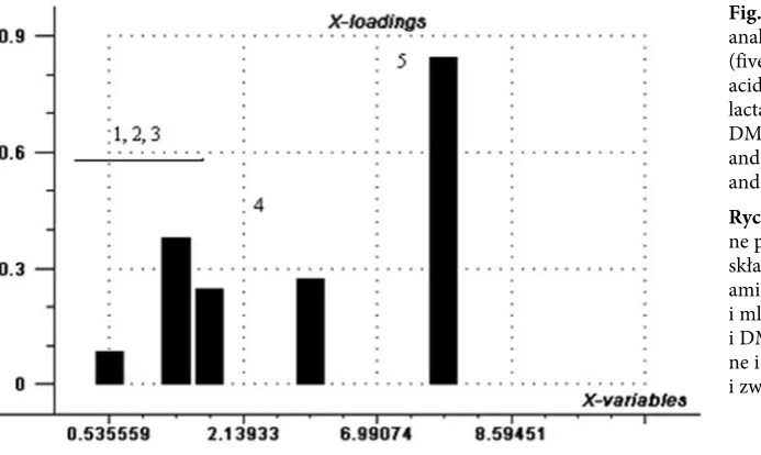
Statistical analysis was applied to determine the magnitude of differences between values (K, B1,
B2, B3, Bp) across spectral ranges representing
low-molecular-weight compounds. It appears from Fig. 5 that the differences were greatest for hip-purates + aromatic compounds and for lactates + amino acids.
Discussion
The main aim of this study was to determine whether physiological pregnancy, which is a kind of stress per se for the organism of a healthy woman, is also associated with structural changes in the kidneys. So far, no studies have been published on 1H-MRS of urine in healthy pregnant women.
In this study, five regions of 1H-MRS spectra of
urine were analyzed assuming, in line with current knowledge, that changes in signal intensity reflect lesions of specific renal structures. Thus increased or decreased excretion with urine of endogenous low-molecular-weight compounds producing pro-ton signals in a spectral region would identify the affected renal structure [8].
samples
chemical shifts (ppm)
matrix plot
Fig. 1.1H-MRS spectra of urine samples
Fig. 2. Principal component analy-sis (PCA) score plot presenting urine data for the first (
~
) and third (+) trimester of pregnancy (B1 and B3)Ryc. 2. Rozkład zmiennych w przestrzeni wielowymiarowej odpowiadających danym dla profili moczu z I (
~
) i III (+) trymestru ciąży (B1 i B3)Fig. 3. Principal component analy-sis (PCA) score plot presenting urine data for the third trimester (¿) and healthy non-gravida (+), groups (B3 and K)
Ryc. 3. Rozkład zmiennych w przestrzeni wielowymiarowej odpowiadających danym dla profili moczu z III (¿) trymestru oraz zdrowych nieciężarnych (+) (B3 i K)
Fig. 4. Principal component analy-sis (PCA) score plot presenting urine data for healthy non-gravida (¿) and after puerperium (£), groups (K and Bp)
Starting from the smallest chemical shift, the first spectral region analyzed here was that of amino acids (e.g. glycine, alanine) and lactates. These sub-stances are excreted with urine in larger quantities when dysfunction affects section S1/S2 and the straight S3 part of the proximal tubule [8, 9].
The next spectral region encompassed citrates and dimethylamine (DMA). Correlation has been demonstrated between decreased urinary excre-tion of citrates and mitochondrial lesions in the renal tubules [5, 8]. Increased urinary levels of DMA as well as of other aliphatic amines (trimeth-ylamine (TMAo), dimethylglycine (DMG)) have been observed in dysfunction of the renal medulla [5, 10, 11].
The largest chemical shift studied here came from protons of hippurates and aromatic com-pounds. Reduced excretion of hippurates has been observed in lesions of the proximal convoluted tubule [5, 12].
Principal component analysis (PCA) was applied instead of classical tests because of the expected discrete changes in renal structures. With this approach, one is able to observe relationships between spectral regions and to visualize profiles of low-molecular-weight compounds excreted with urine by healthy pregnant and non-pregnant.
No significant differences in urinary profiles were found during pregnancy and after the puer-perium which could convincingly reflect renal lesions. In other words, all variables obtained for the urinary profiles clustered in one area of the XY coordinate system. Interpretative difficulties were encountered in analyzing the distribution of points in multidimensional space for the groups. However, when the results were paired, differenc-es between the urinary profildifferenc-es for healthy preg-nant and non-pregpreg-nant women were revealed.
Moreover, the urinary profiles differed depend-ing on the trimester of pregnancy. These finddepend-ings showed that some profiles after the puerperium deviated from those of the control group (healthy non-pregnant women). The differences between the groups were most pronounced for two regions: hippurates + aromatic compounds and amino acids + lactates.
It is known from the literature that urinary levels of hippurates show significant inter-subject as well as diurnal intra-subject variability. The effect of diet cannot be neglected, considering that benzoic acid, widely used as a food preservative, is an important source of hippurates [5]. Moreover, increased urinary excretion of hippurates accom-panies the consumption of green and black tea as well as fat [13–15].
Due to the fact that the metabolic phenotype is the end-result of interactions between diet, life-style, environment, intestinal flora, and genetic factors [16], the role of each of these factors in the present study needs a separate discussion. The pregnant and non-pregnant patients of this study were healthy according to anamnesis, physical examination, and laboratory tests. Information on the diet, except for the consumption of fish (one or no meal per week), was not gathered. With regard to the environment, 97.2% of these subjects were residents of the province of West Pomerania. No medication was administered to the pregnant women except for vitamin prepa-rations. It is worth noting that non-pregnant women, as opposed to pregnant women, tend to pay less attention to lifestyle (beverages, stress, diet, physical activity) and their urinary profiles could be a consequence of this.
The present findings show the role of gestation as a stress factor for the kidneys. Presumably, minor
Fig. 5. Principal component analysis of chemical shifts (five intervals). 1 – amino acids, 2 – amino acids and lactates, 3 – citrates and DMA, 4 – aliphatic amines and TMAo, 5 – hippurates and aromatic compounds
structural changes appear in the course of pregnan-cy and disappear after delivery (i.e. in the samples collected 6 weeks after delivery of this study) or persist with partial regression. Further studies are needed to shed more light on this problem.
The authors concluded that differences in
magnetic resonance spectroscopy profiles of urine between healthy pregnant and non-pregnant women reflect alterations in specific structures of the kidney accompanying pregnancy. The effect of pregnancy on renal tubules is transient and (pre-sumably) does not lead to permanent lesions.
References
Samuels-Kalow ME, Funai EF:
[1] Is pregnancy a stress test? Contemporary oB/GYN 2007, 52, 10, 59.
Kempiak J:
[2] Zmiany ustrojowe w przebiegu ciąży. In: Położnictwo i ginekologia. Eds.: Bręborowicz GH. PZWl, Warszawa 2005, 41–53.
Cunningham FG, Leveno KJ, Bloom SL, Hauth JC, Gilstrap III LC, Wenstrom KD:
[3] Williams obstetrics 21st
edition. New York: McGraw-Hill 2005, 122–150.
Neild GH, Foxall PJD, Lindon JC, Holmes EC, Nicholson JK:
[4] Uroscopy in the 21st century: high-field NMR
spectroscopy. Nephrol Dial Transplant 1997, 12, 404–417.
Zuppi C, Messana I, Forni F et al.:
[5] 1H NMR spectra of normal urines: Reference ranges of the major metabolites. Clin Chim Acta 1997, 265, 85–97.
Sims EA:
[6] Renal function in normal pregnancy. Clin obstet Gynecol 1968, 11, 2, 461–472.
Lichodziejewska-Niemierko M, Rutkowski B:
[7] Nerki a ciąża. In: Nefrologia. Eds.: Książek A, Rutkowski B. Czelej, lublin 2004, 607–617.
Nicholson JK, Timbrell JA, Sadler PJ:
[8] Proton NMR spectra of urine as indicators of renal damage. Mercury-induced nephrotoxicity in rats. Mol Pharmacol 1985, 27, 644–651.
Hauet T, Baumert H, Gibelin H et al.:
[9] Noninvasive monitoring of citrate, acetate, lactate, and renal medul-lary osmolyte excretion in urine as biomarkers of exposure to ischemic reperfusion injury. Cryobiology 2000, 41, 280–291.
Janus T, Borowiak K, Rozwadowski Z, Machoy-Mokrzyńska A, Suchocka J:
[10] Changes in urine metabolic profiles
in patients with chronic intake of amphetamine and opiates revealed using 1H NMR spectroscopy with pattern recognition technique. Acta Toxicol 2006, 14, 1–2, 111–116.
Foxall PJD, Singer JM, Hartley JM et al.:
[11] Urinary proton magnetic resonance studies of early ifosfamide-induced
nephrotoxicity and encephalopathy. Clin Cancer Res 1997, 3, 1507–1518.
Halligan S, Byard SJ, Spencer AJ, Gray TJB, Harpur ES, Bonner FW:
[12] A study of the nephrotoxicity of three
cephalosporins in rabbits using 1H NMR spectroscopy. Toxicol lett 1995, 81, 15–21.
Zuppi C, Messana I, Forni F, Ferrari F, Rossi C, Giardina B:
[13] Influence of feeding on metabolite excretion
evi-denced by urine 1H NMR spectral profiles: a comparison between subjects living in Rome and subjects living at arctic latitudes (Svaldbard). Clin Chim Acta 1998, 278, 75–79.
Mulder TP, Rietveld AG, van Amelsvoort JM:
[14] Consumption of both black tea and green tea results in an increase
in the excretion of hippuric acid into urine. Am J Nutr 2005, 81, Suppl 1, 256–260.
Van Dorsen FA, Daykin CA, Mulder TP, Van Duynhoven JP:
[15] Metabonomics approach to determine metabolic
differences between green tea and black tea consumption. J Agric Food Chem 2006, 54, 18, 6929–6938.
Holmes E, Loo RL, Stamler J, Bictash M et al.:
[16] Human metabolic phenotype diversity and its association with diet
and blood pressure. Nature 2008, 453, 7193, 396–400.
Address for correspondence:
Aleksandra K. Majewska
Department of Maternal and Fetal Medicine and Gynecology Pomeranian Medical University
Unii lubelskiej 1 71-242 Szczecin Poland
Tel: +48 607 576 015 E-mail: [email protected]
Conflict of interest: None declared