Dr. Abbas Taher
Faculty of Dentistry, University of Kufa, Kufa, Iraq Email: [email protected]
Address for correspondence
Access this article online www.japer.in
CORM2 protects from myocardial ischemia reperfusion injury via
modulation of the inflammatory response and apoptosis
Introduction
A major cause of death in the world is the coronary
artery diseases. More than 6 millions people die each
year because of it. Re-introduction blood into an
ischemic organ needed to inhibit cellular loss, but this
can induce injury. This phenomenon is termed
myocardial reperfusion injury (1). Ischemia reperfusion
causes local cellular hypoxia that is accompanied by
inflammatory responses that lead to the recruitment of
leukocyte and subsequent damage (2). Ischemia has
been shown to lead to endothelial dysfunction with an
increase in permeability, an increase expression of
adhesion molecule and recruitment of leukocyte (3)
During ischemia there will be a process of catabolism for
adenine nucleotide, result in accumulation of
hypoxanthine,reintroduction of oxygen and (ROS)
formation. Which will upregulates the gene expression
for many inflammatory agents (e.g.,leukocyte adhesion
molecule and cytokines) and bioactive agent
(e,g.,endothiline,thromboxaneA2)while repressing other
"protective"gene products (e.g.,constitutive nitric oxide
synthase ,thrombomoduline)and bioactive agent
(e,g.,prostacycline ,nitric oxide )(4). Prolonged periods of
myocardial ischemia are related to an increase in the rate
of necrosis, whereas, paradoxically, reperfusion leads to
an augmentation in apoptosis(5,6). has raised particular
therapeutic interest because of its potent The
antiinflammatory and anti-oxidant activity for Carbon
monoxide (CO) gives it a good attention regarding its use
for therapeutic purposes, although it’s a toxic gas that
have the ability to impaire the respiratory system.
However, CO is also produced by the protein heme
oxygenase (HO) and as such functions as a potent
endogenous antioxidant that counteracts toxic effects of
ROS (7). CO inhibited the production of proinflammatory
cytokines, such as TNF-α, MIF and interleukin-1, from
macrophages (8). Carbone monoxide stimulated the
synthesis of the anti inflammatory cytokine
interleukin-10 (8). Exposure of macrophages to a low concentration
Research ResearchResearch
Research ArticleArticleArticleArticle
Background: Myocardial ischemia is one of the major clinical problems in the world. There are two forms of the cell injury occur during myocardial ischemia, which are necrosis and apoptosis. Reperfusion is very important to maintain the viability of the myocardial cells, but in the other hand reperfusion not free from hazardous effects and it is usually associated with what is called ischemia reperfusion injury.
Objective: This study was undertaken to assess the possible protective potential of Carbone monoxide releasing molecule-2 (CORM2) in regional myocardial ischemia reperfusion injury and apoptosis in male rats.
Methods: Twenty four adult male Swiss albino rats were divided into four groups (six rats per group). Sham , in a uniform manner, surgical procedure was done for the rats in all groups, and in this group there was no ligation for (LAD). Control group, rats were subjected for ligation of LAD for 30minutes then 3hr reperfusion. vehicle group, rats were subjected for ligation of LAD for 30minutes then 3hr reperfusion and received 0.5%DMSO before reperfusion time. Treatment group: reperfusion lasted for 3 hours after LAD ligation for half an hour, treatment with CORM2 at reperfusion time (8mg/kg I.V via the tail vein) have done to all rats.
Results: At the end of reperfusion animals sacrificed and cardiac TNF-α, IL-1β ,IL-6, ssDNA and plasma troponin I (cTnI) were measured. It has been found that CORM2 treated group showed a significant reduction (P< 0.05) in cardiac TNF-α, IL-1β, IL-6, ssDNA and plasma cardiac troponin I (cTnI) compared with the control group. Histopathological study found that treatment with CORM2 significantly (P<0.05) reduce the myocardial injury compared with the control group
Conclusion: CORM2 lead to reduction in regional myocardial ischemia reperfusion injury and apoptosis during ischemia via interfering with inflammatory pathway.
Keywords: CORM2, Myocardial ischemia, Reperfusion ABSTRACT
ABSTRACT ABSTRACT ABSTRACT Najah R Hadi*
Fadhil G Al-Amran1
Karrar A Muhsin2
Abbas Taher3
*Dept of Pharmacology and
Therapeutics, Faculty of Medicine, University of Kufa, Kufa, Iraq
1Department of Surgery,
Cariothorasic Section, Faculty of Medicine, University of Kufa, Kufa, Iraq.
2Dept of Pharmacology and
Therapeutics, Faculty of Medicine, University of Kufa, Kufa, Iraq
3Department of OMF Surgery,
Dean of Faculty of Dentistry, University of Kufa, Kufa, Iraq
of CO (250 ppm) as well as over expression of HO-1 in
these cells inhibited lipopolysaccharide- induced
production of granulocyte macrophage
colony-stimulating factor (GM-CSF). This effect of CO was
mediated by inhibition of the transcription factor NF-κB
(9-11). Carbone monoxide-donor pretreatment activated
progenitor cell and promoted vasculogenesis and the
formation of new cardiomyocytes after myocardial
infarction in rats(12). The HO-1 gene transfer has earlier
been shown to protect against angiotensin II-induced
cardiomyocyte apoptosis in vitro and against ischemia /
reperfusion (I /R) injury and cardiomyocyte apoptosis
after repeated I/R episodes in rat hearts in vivo (13).
Methods:
Material
CORM2 powder (pure) from (Sigma, USA), Xylazine
2% vials (RompunTM, Bayer AG, Leverkusen,
Germany), ketamine (Hikma, Jordan), ethanol (Fluka,
Switzerland) and normal saline (KSA). Rat 1β),
(IL-6), (TNF-α) ELISA kits ( Sigma, USA). Rat cTnI ELISA
(Life diagnostics Inc., USA). High Intensity Ultrasonic
Liquid Processor (Sonics & materials Inc., USA),
Vascular Clamp (Biotechno, Germany) and ventilator
(Harvard. USA).
Animal
Male swiss albino rats and their weight was between
(180-220 g) were purchased from Animal Resource
Center , National Center for Drug Control and
Research. The rats kept at 25˚C and 12-hour light-dark
cycle for two weeks with free access for water and
food. Animals had no manifestation of any illness upon
examination.
Study design
Rats were randomized into 4 groups 6 animals in each
group as follow:
1. group 1 (Sham) : the same anesthetic and surgical
procedures applied to rats of this group without
ischemia.
2. group 2 (Control) : (induced untreated group): Rats
underwent 30 min of LAD ligation followed by a 3hr of
reperfusion.
3. group 3 (vehicle) : Rats underwent 30 min of LAD
ligation and 3hr reperfusion and received 0.5%DMSO
before reperfusion time.
4. group 4 (CORM2 pre-treated) : in this group, all rats
undergo 30 min of ischemia and 3hr reperfusion with
CORM2 at reperfusion time was given (8mg/kg i.v via
the tail vein) .
Stock powder dissolved in100%
DMSO(dimethylsulfoxide) which then diluted to 10%
with saline to invivo use. It was prepared immediately
before use.(14,15)
Surgical LAD ligation
The rats were anesthetized by intraperitoneal
injection with a mixture of ketamine and xylazine in a
dose of 100mg/kg and 10 mg/kg respectively (16).
The trachea was intubated with a cannula and
ventilation was achieved by connecting the tube in the
trachea to the ventilator supplied with 100% oxygen
at a respiratory rate of 50/min with a tidal volume of
20 ml/kg body weight (17). Animal ventilated using a
small animal ventilator (Harvard Apparatus, Holliston,
MA, USA). Then a lateral thoracotomy approach is
done , heart was exposed and left anterior descending
branch of the left coronary artery was ligated, then
chest was closed. The survival rate following the
surgery was around 60%–70% and was equivalent
between groups. After 30 min reopen the chest and
the ligature removed at this point reperfusion started
which is last for 3hrs.
Collection of Samples
At the end of reperfusion The blood was collected
from the ventricular apical side. Hearts were cut from
their main arteries and rinsed with normal saline to
remove any debris, then stored in deep freeze at
(-20˚C to -80˚C). The ventricles were cut from
atrioventricular junction, each ventricle was divided
into 2 parts, apical part and basal part. The apical part
further more divide into two parts, one for
measurement of apoptosis level and the other was
fixed in 10% formalin and prepared for routine
histological processing by embedding in paraffin
were taken and stained with haematoxylin-eosin
(H&E).
Samples preparation
1-Preparation of Sample for TNF-α and IL-1β and
IL-6 measurements
Upper part of ventricles were rinsed with ice cold
saline, then homogenized in phosphate buffered saline
with a ratio of 1:10 (w/v) that contained protease
inhibitor cocktailand 1% Triton X-100 with ultrasonic
liquid processor . centrifugation for homogenate was done for 20 min at 4°C at 2,500 g. The supernatant
was collected for determination of TNF-α, 1β,
IL-6ELIZA kits(Sigma Aldrich, USA) (19).
2- cTn-I measurement
Blood was collected from the apex of each heart by a
syringe needle inserted directly to the heart. EDTA (22
mg/ml) tubes was used for blood samples collection,
mixed thoroughly and centrifuged at 3000 RPM for 15
min.
3-Detection of Apoptotic Cells by Foramamide
Formamide denaturation to DNA in apoptotic cells
and not in necrotic cells or in the cells with DNA
breaks in the absence of apoptosis was used in this
assay that denatured DNA with monoclonal antibody
to single-stranded DNA (ssDNA) was detected. The
used tissue for measurement of apoptosis level were
lysed using trypsin.
Statistical Analysis
By using SPSS 20.0 for windows, the analysis was
done statistically. An expert statistical advice was
consulted for tests used. Analysis of Variance
(ANOVA) was used for the multiple comparisons
among all groups followed by post-hoc tests using the
LSD method. Changes in the histopathology
differences in total score between more than 2 groups
was assessed by Kruskal-Wallis test, while
Mann-Whitney U test was used for the difference between 2
groups. In all tests, P< 0.05 was considered to be
statistically significant.
RESULTS
1- Effect of Carbone monoxide releasing
molecule-2 on the proinflammatory marker (TNF-α, IL-1β
and IL-6)
At the end of the experiment and in the control group ;
the levels of cardiac (TNF-α, IL-1β and IL-6) was
increased significantly (P<0.05) compared to sham
group.
The levels of cardiac (TNF-α, IL-1β and IL-6) of
CORM2 treated group was significantly lower(p<0.05)
than both control and vehicle groups. (Figure 1)
Figure 1: The means of cardiac (TNF-α,IL-1βand IL-6) levels (pg/mg) were found to be significantly elevated
(P˂0.05) in the control group and control vehicle compared with sham group . At the same time, cardiac
cytokines were significantly decreased (P˂0.05) in CORM2 treated group with respect to both control and
2-The effect of Carbone monoxide releasing molecule-2 on the cardiac troponin I
At the end of the experiment, the level of plasma
(cTnI) was significantly increased (P<0.05) in the
control group as compared with the sham group.
The plasma level of (cTnI) of CORM2 treated group
was significantly (p<0.05) lower than that of control
and control vehicle groups. (Figure 2)
Figure 2: The level of plasma cTnI (ng/ml) was significantly increased (P˂0.05) in the control group and control vehicle compared to the sham group . On
the other hand, cTn-I was significantly reduced (P˂0.05) in CORM2 treated group with respect to
both control or control vehicle groups.
3-Effect of Carbone monoxide releasing molecule-2 on
the level of apoptotic cell
At the end of the experiment, the result shows that
there is a significant increase (p<0.05) in apoptotic
cell in control, control v compared with the sham
group. CORM2 group show significant decrease
(p<0.05) in apoptotic cell compared with control and
control vehicle groups. CORM2 treated group showed
a significant increase (p<0.05) compared to the sham
group. (Figure 3)
Figure 3: The mean of cardiac apoptosis level
4-Effect of Carbone monoxide releasing molecule-2 on
the histopathological score
CORM2 treatment significantlyi mproved cardiac injury (P
< 0.05) compared to control group and the total severity
scores mean of this group showed 33.3% of the group had
no damage, 66.7% mild injury. Sham heart tissue showed
a normal cardiac structure. All rats in this group showed
normal hearts 100%. There was statistically insignificant
difference between control vehicle group and control
group (P=0.685) and the total severity scores showed
33.3% of the groups had severe cardiac injury and
66.7%had highly severe cardiac injury (Figure 4, 5)
Figure 4: Total myocardial damage scores
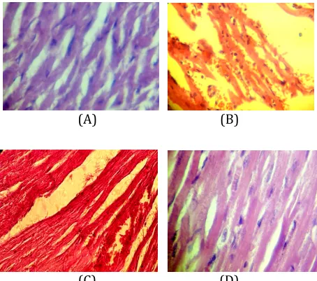
(A) (B)
(C) (D)
Figure (5): (A) cardiac section of normal rats with normal architecture. The section stained with Haematoxylin and Eosin(X40)(B) cardiac section showed interstitial edema, focal necrosis , contraction band,PMN infiltration, highly sever injury . The section stained with Haematoxylin and Eosin(C) cardiac section showed capillary compression sever injury.(D) cardiac section treated with CORM2. Haematoxylin and Eosin was used as staining agents (X 40).
Discussion
Myocardial damage resulting from ischemia-reperfusion
(I/R) is a major cause of morbidity and mortality in the
world, those myocardial I/R injuries result in cardiac
damages (20). Inflammatory responses have role in I/R
injury mediated by activation of cytokines and adhesion
molecules (21). Inflammatory cytokines, such as TNF-α,
IL-1β and IL-6 were shown to play key roles in the
pathophysiology of ischemia and reperfusion injury (22).
Many experimental studies have shown the specific and
independent role of exogenous CO in the modulation of
inflammation (23, 24). The existence of compounds based
on metal carbonyl (CORMs) that control the CO releases
in biological systems gives more opportunity to
investigate CO-mediated biological effects than before
(25). The vaso-active, antihypertensive, cardio protective
and anti-rejection effects of CORM-released CO in vivo
have been demonstrated (11, 26-30).In the present study,
CORM2 significantly reduced the elevation of
inflammatory mediators (TNF-α, IL-1β And IL-6) levels in
cardiac tissues ( p<0.05) compared to control group, gives
orientation about protective effect of CORM2 in
myocardial I/R injury. In our knowledge, there is no study
on CORM2 and (TNF-α, IL-1β And IL-6) However, Sawle et
al. (2005) show that CORM-2, a DMSO-soluble CO-RM,
gives an anti-inflammatory effect in an in vitro model of
LPS-stimulated macrophages (31). Bingwei et al. (2006)
show that In vivo application of CORM-2 in burn mice
markedly decreased the production of IL-1β and TNF-α in
BAL fluid.(32) CORM2 treatment rats shows a significant
reduction in cardiac injury (P < 0.05) compared to control
group and the total severity scores mean of this group
showed mild cardiac injury,also there is no study about
the effect of CORM2 on histopathological score. Bingwei et
al.( 2006) shows that PMN chemotaxis and infiltration in
the lung after thermal injury effectively prevents by the
use of CORM-2, with decrease in the production of
oxidants and reduction in tissue oxidative injury. CORM-2
usage in a model of burn mice reduces the accumulation of
PMNs , inhibits NF-κB activity and decreases the
production of inflammatory mediators.(32) Yunwei et al.
(2010) show that CORM2 can decrease the total severity
score of the histopathology by the decrease of PMN cell as
neutrophil in treated hepatic I/R injured rat.(14)
In the present study, we found that CORM2 treatment
significantly (P < 0.05) reduced the increasing plasma
levels of cTnI as compared with the control group.
According to our knowledge; there are no data available
about effect of CORM2 treatment on cTnI. However
Atsunori et al.,( 2010) find that CO treatment with
hydrogen decrease significantly cardiac troponin I in the
plasma (33).
Treatment of rats with CORM2 reduces apoptotic cell
significantly (P < 0.05) as compared with the control
group. Gunther et al., (2002) show that the cytoprotective
effects of CO have also been associated with inhibition of
apoptosis and up regulation of antiapoptotic proteins (34).
Guangwu et al., (2010) proved that CO delivery
exogenously in an in vivo by CORM-3 improved post
infarction LV remodeling and dysfunction and reduced
myocardial apoptosis (35). Recently Zrelli et al.,(2013)
that CORM2 synergistically strengthen the
antiapoptotic effects of hydroxylthirosol via
suppression of caspase-3 activation (36).
Limitation of the study
TUNEL technique was not used in this study to
differentiate between apoptotic and necrotic neuclei .
Conclusions:
We conclude that CORM2 has a cardioprotective
potential as it ameliorates myocardial ischemia
reperfusion injury via interfering with inflammatory
responses and apoptosis.
References
1. Keeley EC, Boura JA, Grines CL. Primary angioplasty versus intravenous thrombolytic therapy for acute myocardial infarction: a quantitative review of 23 randomised trials. Lancet 2003;361:13-20.
2. Boros P, Bromberg JS. New cellular and molecular immune pathways in ischemia/reperfusion injury. Am J Transplant 2006;6:652-8.
3. Frangogiannis NG, Smith CW, Entman ML. The inflammatory response in myocardial infarction. Cardiovasc Res 2002;53:31-47.
5. Anversa P, Cheng W, Liu Y, Leri A, Redaelli G, Kajstura J. Apoptosis and myocardial infarction. Basic Res Cardiol 1998;93:8-12.
6. Dumont EA, Reutelingsperger CP, Smits JF, Daemen MJ, Doevendans PA, Wellens HJ, et al. Real-time imaging of apoptotic cell-membrane changes at the single-cell level in the beating murine heart. Nat Med 2001;7:1352-5. 7. Nicoud IB, Knox CD, Jones CM, Anderson CD, Pierce JM,
Belous AE, et al. 2-APB protects against liver ischemia-reperfusion injury by reducing cellular and mitochondrial calcium uptake. Am J Physiol Gastrointest Liver Physiol 2007;293:623-30.
8. Otterbein LE, Choi AM. Heme oxygenase: colors of defense against cellular stress. Am J Physiol Lung Cell Mol Physiol 2000;279:1029-37.
9. Sarady JK, Otterbein SL, Liu F, Otterbein LE, Choi AM. Carbon monoxide modulates endotoxin-induced production of granulocyte macrophage colony-stimulating factor in macrophages. Am J Respir Cell Mol Biol 2002;27:739-45.
10. Pae HO, Oh GS, Choi BM, Chae SC, Kim YM, Chung KR, et al. Carbon monoxide produced by heme oxygenase-1 suppresses T cell proliferation via inhibition of IL-2 production. J Immunol 2004;172:4744-51.
11. Motterlini R, Clark JE, Foresti R, Sarathchandra P, Mann BE, Green CJ. Carbon monoxide-releasing molecules: characterization of biochemical and vascular activities. Circ Res 2002;90:17-24.
12. Lakkisto P, Kyto V, Forsten H, Siren JM, Segersvard H, Voipio-Pulkki LM, et al. Heme oxygenase-1 and carbon monoxide promote neovascularization after myocardial infarction by modulating the expression of HIF-1alpha, SDF-1alpha and VEGF-B. Eur J Pharmacol 2010;635:156-64.
13. Foo RS, Siow RC, Brown MJ, Bennett MR. Heme oxygenase-1 gene transfer inhibits angiotensin II-mediated rat cardiac myocyte apoptosis but not hypertrophy. J Cell Physiol 2006;209:1-7.
14. Wei Y, Chen P, de Bruyn M, Zhang W, Bremer E, Helfrich W. Carbon monoxide-releasing molecule-2 (CORM-2) attenuates acute hepatic ischemia reperfusion injury in rats. BMC Gastroenterol 2010;10:42.
15. Chen P, Sun B, Chen H, Wang G, Pan S, Kong R, et al. Effects of carbon monoxide releasing molecule-liberated CO on severe acute pancreatitis in rats. Cytokine 2010;49:15-23.
16. Wiedemann D, Schneeberger S, Friedl P, Zacharowski K, Wick N, Boesch F, et al. The fibrin-derived peptide Bbeta(15-42) significantly attenuates ischemia-reperfusion injury in a cardiac transplant model. Transplantation 2010;89:824-9.
17. Ding JW, Tong XH, Yang J, Liu ZQ, Zhang Y, Yang J, et al. Activated protein C protects myocardium via activation of anti-apoptotic pathways of survival in ischemia-reperfused rat heart. J Korean Med Sci 2010;25:1609-15. 18. Bancroft J.D., and Stevens A. Theory and practice of
histological techniques. Churchill Livingstone. Melbourne 1982; 2:125.
19. Zhang M, Xu YJ, Saini HK, Turan B, Liu PP, Dhalla NS. Pentoxifylline attenuates cardiac dysfunction and reduces TNF-alpha level in ischemic-reperfused heart. Am J Physiol Heart Circ Physiol 2005;289:832-9. 20. Ruiz-Meana M, Garcia-Dorado D. Translational
cardiovascular medicine (II). Pathophysiology of ischemia-reperfusion injury: new therapeutic options for acute myocardial infarction. Rev Esp Cardiol 2009;62:199-209.
21. Crawford MH, Grover FL, Kolb WP, McMahan CA, O'Rourke RA, McManus LM, et al. Complement and neutrophil activation in the pathogenesis of ischemic myocardial injury. Circulation 1988;78:1449-58. 22. Valen G, Yan ZQ, Hansson GK. Nuclear factor kappa-B and
the heart. J Am Coll Cardiol 2001;38:307-14.
23. Otterbein LE, Bach FH, Alam J, Soares M, Tao Lu H, Wysk M, et al. Carbon monoxide has anti-inflammatory effects involving the mitogen-activated protein kinase pathway. Nat Med 2000;6:422-8.
24. Goebel U, Siepe M, Mecklenburg A, Stein P, Roesslein M, Schwer CI, et al. Carbon monoxide inhalation reduces pulmonary inflammatory response during cardiopulmonary bypass in pigs. Anesthesiology 2008;108:1025-36..
25. Motterlini R, Mann BE, Johnson TR, Clark JE, Foresti R, Green CJ. Bioactivity and pharmacological actions of carbon monoxide-releasing molecules. Curr Pharm Des 2003;9:2525-39.
26. Motterlini R, Sawle P, Hammad J, Bains S, Alberto R, Foresti R, et al. CORM-A1: a new pharmacologically active carbon monoxide-releasing molecule. FASEB J 2005;19:284-6.
pharmaceuticals? Angew Chem Int Ed Engl 2003;42:3722-9.
28. Foresti R, Hammad J, Clark JE, Johnson TR, Mann BE, Friebe A, et al. Vasoactive properties of CORM-3, a novel water-soluble carbon monoxide-releasing molecule. Br J Pharmacol 2004;142:453-60.
29. Clark JE, Naughton P, Shurey S, Green CJ, Johnson TR, Mann BE, et al. Cardioprotective actions by a water-soluble carbon monoxide-releasing molecule. Circ Res 2003;93:2-8.
30. Guo Y, Stein AB, Wu WJ, Tan W, Zhu X, Li QH, et al. Administration of a CO-releasing molecule at the time of reperfusion reduces infarct size in vivo. Am J Physiol Heart Circ Physiol 2004; 286:1649-53.
31. Sawle P, Foresti R, Mann BE, Johnson TR, Green CJ, Motterlini R. Carbon monoxide-releasing molecules (CO-RMs) attenuate the inflammatory response elicited by lipopolysaccharide in RAW264.7 murine macrophages. Br J Pharmacol 2005; 145:800-10.
32. Sun B, Sun H, Liu C, Shen J, Chen Z, Chen X. Role of CO-releasing molecules liberated CO in attenuating leukocytes sequestration and inflammatory responses in the lung of thermally injured mice. J Surg Res 2007;139:128-35.
33. Nakao A, Kaczorowski DJ, Wang Y, Cardinal JS, Buchholz BM, Sugimoto R, et al. Amelioration of rat cardiac cold ischemia/reperfusion injury with inhaled hydrogen or carbon monoxide, or both. J Heart Lung Transplant 2010;29:544-53.
34. Gunther L, Berberat PO, Haga M, Brouard S, Smith RN, Soares MP, et al. Carbon monoxide protects pancreatic beta-cells from apoptosis and improves islet function/survival after transplantation. Diabetes 2002;51:994-9.
35. Wang G, Hamid T, Keith RJ, Zhou G, Partridge CR, Xiang X, et al. Cardioprotective and antiapoptotic effects of heme oxygenase-1 in the failing heart. Circulation 2010;121:1912-25.
36. Zrelli H, Wu C, Zghonda N, Shimizu H, Miyazaki H. Combined Treatment of Hydroxytyrosol With Carbon Monoxide-Releasing Molecule-2 Prevents TNF-α-Induced Vascular Endothelial Cell Dysfunction through NO production with Subsequent NFkB Inactivation.Biomed rest Int 2013;2013:912431.
How to cite this article: Najah R Hadi*, Fadhil G Al-Amran1, Karrar A Muhsin2, Abbas Taher3; CORM2 protects from myocardial ischemia reperfusion injury via modulation of the inflammatory response and apoptosis; J. Adv. Pharm. Edu. & Res. 2015: 5(1): 33-39.