M
ARYLAK
RASNOWSKA, M
ARYTAN
ITTNER−M
ARSZALSKA, R
YSZARDK
RASNOWSKI,
W
ACŁAWK
OPEĆ, A
NDRZEJF
AL, A
NNAB
IAŁEKEndothelin, Endothelial Cells,
and von Willebrand Factor in Peripheral Blood
of COPD in the Context of Hypoxemia
Endotelina, komórki śródbłonka oraz czynnik von Willebranda
a hipoksemia u pacjentów z przewlekłą obturacyjną chorobą płuc
Department of Internal Medicine and Allergology, Silesian Piasts University of Medicine in Wrocław, Poland
Adv Clin Exp Med 2007, 16, 3, 395–401 ISSN 1230−025X
ORIGINAL PAPERS
© Copyright by Silesian Piasts University of Medicine in Wrocław
Abstract
Background. The pulmonary endothelium plays a significant role in the pathobiology of chronic obstructive pul− monary disease (COPD), a chronic inflammatory disease of the respiratory tract in which increased activity or even damage to the endothelium due to chronic inflammation, hypoxemic stress, and/or angiogenesis may occur. The authors hypothesized that patients with COPD have increased concentrations of endothelin (ET−1) and von Willebrand factor (vWF) and/or circulating endothelial cell (CEC) counts, markers of endothelial activity and/or damage.
Objectives.The aim of the study was to assess the concentrations of ET−1 and vWF and the number of CECs in the peripheral blood of COPD patients compared with healthy individuals and to investigate if hypoxemia or ven− tilation disturbances affect these parameters.
Material and Methods.The study included 20 patients with COPD in stage III (according to GOLD) as well as 15 healthy individuals as controls. ET−1 and vWF were assessed by an immunoenzymatic method. Immuno− fluorescence using the specific monoclonal endothelium antibody CLB−HEC 19 MoAb was applied to evaluate CECs.
Results. The concentration of ET−1 was higher in COPD patients than in controls, medians being 1.2 (range: 1.0–3.8) and 0.44 (0.2–0.7) pg/ml (p< 0.001), respectively. The number of CECs was higher in COPD patients than in controls (mean: 1.5, range: 0.6–3.1 vs. mean: 0.44, range: 0.32–1.11 cells/ml, respectively, p< 0.001). The concentration of vWF in COPD patients did not differ from that in the controls. There was positive correlation between CEC count and the concentrations of both the studied factors (p < 0.05). The number of CECs in the patients peripheral blood correlated significantly with the pO2value (Rs: 0.36, p< 0.05).
Conclusions. The increased number of CECs and concentration of ET−1 in peripheral blood may result from endothelial damage or activation occurring in COPD (Adv Clin Exp Med 2007, 16, 3, 395–401).
Key words: COPD, endothelial cells, endothelin, vWF.
Streszczenie
Wprowadzenie. W przebiegu p.o.ch.p. dochodzi do aktywacji śródbłonka naczyniowego i uwolnienia jego media− torów, m.in. endoteliny 1, a równocześnie destrukcji z powodu towarzyszącego chorobie przewlekłego niedotle− nienia. Markerami uszkodzenia śródbłonka są: czynnik von Willebranda i krążące komórki śródbłonka (CEC).
Cel pracy.Ocena tych czynników w p.o.ch.p. w kontekście zaburzeń wentylacyjnych oraz hipoksemii.
Materiał i metody.Zbadano 20 pacjentów w III stadium p.o.ch.p. (według GOLD) oraz 15 osób zdrowych. ET−1 i vWF oznaczano we krwi metodą immunoenzymatyczną, CEC oceniano za pomocą metody immunofluorescen− cyjnej z wykorzystaniem swoistego przeciwciała monoklonalnego CLB−HEC 19. Wszyscy chorzy mieli oznacza− ne wskaźniki wentylacyjne płuc oraz mierzone gazy krwi arterializowanej włośniczkowej.
Inflammation is an important issue in the pathogenesis of chronic obstructive pulmonary disease (COPD). Cellular inflammatory infiltrates in COPD are dominated by neutrophils, T lym− phocytes (CD8+), and macrophages [1, 2]. Recruit−
ment of circulating cells to peripheral tissue (including the bronchi) occurs as an effect of adhe− sion between these cells and vascular endotheli− um. Thus the endothelium is not only a mechani− cal barrier between the blood stream and the ves− sel wall, but it also plays a role in immunological response, leukocyte mobilization to peripheral tis− sue during inflammation, and smooth muscle tonus in the respiratory tract. The markers of endothelial activity are endothelin (ET−1) and von Willebrand factor (vWF), secreted from the endothelium. ET−1 is a peptide controlling vascu− lar tonus able to initiate bronchoconstriction as well as proliferation of smooth muscles [3, 4]. Higher concentrations of ET−1 have been demon− strated in bronchial biopsies and BAL (bron− choalveolar lavage) fluids from asthmatic patients and COPD patients than in healthy individuals [5, 6]. vWF is also produced by endothelium and is also a marker of endothelial dysfunction in several vas− cular and non−vascular diseases. In acute or chron− ic hypoxemia the concentration of vWF in the peripheral blood increases. In some diseases, such as pulmonary hypertension, the vWF concentra− tion is a recognized indicator of vascular damage and appears together with an increased number of circulating endothelial cells (CECs) [8].
CECs have recently been evaluated and estab− lished as a novel marker of endothelial damage in a variety of disorders. Changes in the circulating population of endothelial cells can be found in hypertension, renal diseases, cytomegalovirus infection, septic shock, sickle cell anemia, and unstable angina and in renal transplant recipients [9–11]. Woywodt et al. recently established that elevated CEC number correlates with disease activity in antineutrophil cytoplasmatic antibody (ANCA)−associated vasculitis [12]. Though these conditions are diverse in their etiology and patho− biology, all share a common history of vascular injury.
The aim of this study was to investigate the markers of endothelial activity and damage, i.e. CEC counts and the concentrations of ET−1 and vWF in the peripheral blood of patients with
COPD. It is believed that such a complex experi− ment has not yet been published.
Material and Methods
The study involved 20 COPD patients in stage III (according to GOLD 2003) with a mean age of 69.5 ± 9.9 years. Disease duration and history of smoking are given in Table 1. Fifteen healthy indi− viduals, 9 men and 6 women with a mean age of 31.5 ± 9 years, were also included in the study as controls. Concomitant hypertension, diabetes, or glomerulonephritis were considered exclusion cri− teria in both groups. The members of the two groups were comparable with regard to gender, but not age, the mean age in the control group being much lower. Every patient gave his/her informed consent before inclusion to this study and the pro− tocol was approved by the Bioethics Committee of the Silesian Piasts University of Medicine in Wrocław.
Measurement of CECs
in Whole Blood
The procedure of counting circulating endo− thelial cells was performed according to van Mourik et al. [13] and Sbarbati et al. [14] with the present authors’ modification. The method for identifying CECs in blood samples involved Percoll density gradient centrifugation combined with immunofluorescent staining of subfractions.
Venous blood samples of 4.5 ml were drawn from the anticubital vein. The first 2 ml of the blood sample collected in the syringe was discard− ed as potentially polluted with endothelial cells from locally punctured tissues. The 4.5 ml of blood obtained was carefully mixed with 0.5 ml of 3.8% sodium citrate and gently stirred.
The blood samples were dripped over Percoll layers of 1.060 g/ml and centrifuged at 1000 ×gat room temperature for 10 minutes. The upper cell layer was collected and centrifuged at 400 ×gfor 10 minutes. The supernatant was discarded and the cellular pellet was resuspended in phosphate buffered saline (PBS) and 2% bovine serum albu− min (BSA), sedimented on glass coverslips by means of a cytocentrifuge (SHANDON, England), and processed for immunofluorescence. The cov−
CECs i stężeniem obu pozostałych czynników (p < 0,05). Liczba CEC we krwi obwodowej korelowała z warto− ściami stężenia parcjalnego pO2w arterializowanej krwi włośniczkowej (Rs = 0,36, p < 0,05).
Wnioski. Ocena krążących komórek śródbłonka i ET−1 w p.o.ch.p. może być jednym ze wskaźników uszkodzenia naczyń w tej chorobie (Adv Clin Exp Med 2007, 16, 3, 395–401).
erslips had been previously coated with 1% gelatin fixed with 0.5% glutardialdehyde.
Immunofluorescence
Every cell sample from the Percoll gradient was divided and sedimented on three coverslips. Two were randomly chosen and stained with MoAb CLB−HEC19. The third coverslip was stained with rabbit serum as a control. Each stain− ing was performed for 1 hour at room temperature. The slides were washed, fixed with methanol for 10 minutes, incubated with MoAbs, and washed once more. Then the slides were stained with flu− orescein isothiocynate (FITC)−conjugated polyva− lent goat antimouse Ig (1 : 80) (DACO, Denmark) and embedded in 87% glycerol in PBS (9 : 1) and 1 mg/ml para−phenylene diamide (pH 8.6).
The slides with fluorescent CECs were screened using a fluorescence microscope. Each slide previously stained with CLB−HEC19 MoAbs was screened for the presence of immunofluores− cent ECs. The sum of fluorescent cells on two slides (2/3 of the actual CEC number in one blood sample) was calculated and the result expressed as the num− ber of circulating ECs in one ml of whole blood.
Monoclonal Antibody
The CLB−HEC19 MoAb is one of a series of monoclonal antibodies raised against cultured HUVECs (human umbilical vein ECs). CLB−
HEC19 recognizes an EC−specific plasma mem− brane protein with an apparent molecular weight of 100,000. This antibody does not react with blood cells, including erythrocytes, lymphocytes, monocytes, granulocytes, and blood platelets, or with cultured fibroblasts and smooth muscle cells. The immunohistochemical analysis of multiple tis− sue samples shows also that MoAb CLB−HEC19 exclusively stains ECs.
The Measurement
of vWF Plasma Concentration
vWF was assayed in plasma using a commer− cially available immunoenzymatic kit (Assera− chrom, Diagnostica Stago, France). In this assay, rabbit antibody fixed on titration microplates binds vWF. Later, the remaining antigen determinants of vWF bind to another rabbit antibody (peroxydase labeled). This complex produces a color reaction in the presence of orthophenylenediamine and H202. The result is interpreted as the percentage of
the normal vWF concentration.
The Measurement
of Endothelin Plasma
Concentration
Endothelin was assayed in plasma using a commercially available immunoenzymatic me− thod (Biomedica Gruppe, Austria) according to the manufacturer’s protocol. In short, endothelin is bound in plasma by a polyclonal antibody previ− ously attached in the polystyrene wells of the titra− tion microplates. Bound endothelin is further detected by a mouse monoclonal antibody that in turn is bound by a peroxidase−labeled anti−mouse IgG. The complex polyclonal antibody – endothe− lin−labeled IgG is finally detected in an enzymatic reaction of peroxidase with the provided substrate. The color intensity after the reaction is directly proportional to the endothelin concentration.
Evaluation of gasometric parameters was car− ried out in capillary blood.
Statistical Analysis
The statistical significance of differences between groups regarding demographic data was determined using the Mann−Whitney nonparametric U−test. Apvalue < 0.05 was considered statistically significant. The concentrations of ET−1 and vWF and endothelial cell counts are expressed as medians and ranges. To compare values, the Spearman’s rank (Rs) correlation test was used for assessing correla− tion. Apvalue < 0.05 were accepted as statistically significant.
Table 1. Demographic data, airway parameters, and blood gases of the COPD patients
Tabela 2.Dane demograficzne, wyniki badań czynnoś− ciowych płuc i gazometrii u chorych na p.o.ch.p.
COPD patients (Chorzy na p.o.ch.p.) Demographic features
(Dane demograficzne)
sex M/F 12/8
age ± SD(years) 69.5 ± 9.9 duration of symptoms (years) 13.7 ± 5.7 history of smoking (pack−years) 38.1 ± 11.4 Airway parameters
(Wyniki badań czynnościo− wych płuc)
FEV1 l 0.94 ± 0.18
FVC l 1.38 ± 0.36
FEV1/FVC% 60.32 ± 12.70
Blood gases (Wyniki gazometrii)
pO2mm Hg 59.66 ± 11.68
Values are expressed as the mean ± SD.
Results
The demographic features, blood gases, and airway parameters of COPD patients are presented in Table 1. It was found that the concentration of ET−1 and the number of CECs in peripheral blood were higher in COPD patients than in healthy indi− viduals, whereas the activity of vWF did not differ in the two groups (Table 2). The calculated corre− lation was significantly positive between CEC count and vWF plasma level and between CEC count and ET−1 level. Correlation between CEC count and partial oxygen pressure in capillary blood existed as well; CEC count in the patients’ peripheral blood correlated significantly with the pO2value
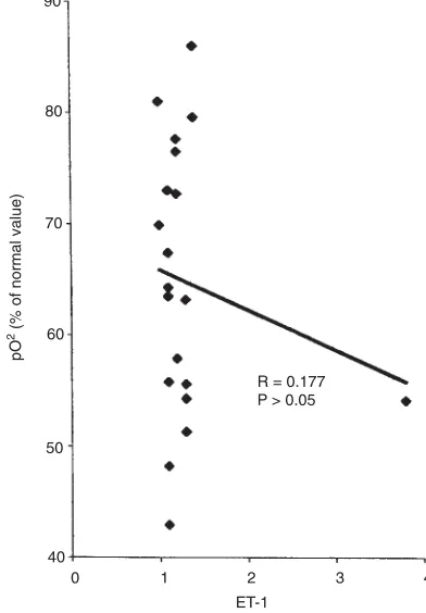
(Rs: 0.36, p < 0.05) (Fig. 1a). However, there was no correlation between ET−1 and the pO2 value
(Fig. 1b).
Discussion
These experiments demonstrated an increased number of CECs in the peripheral blood of patients with COPD compared with healthy controls. The interpretation of this finding is difficult at the moment, especially since to the authors’ knowl− edge there are no other published experiments addressing this issue. The demonstrated phenome− non does not have to be specific to patients with COPD and can be observed in patients with other respiratory diseases or in elderly persons. In arteri− al hypertension, chronic glomerulonephritis, and renal transplant rejection, increased CEC counts have been attributed to damage to the endothelium resulting from hypertensive stress and vascular stretching [8, 11, 15, 16]. It appears that the mech− anism leading to an increased number of CECs in COPD is distinct from that in hypertension or renal transplant rejection and mainly depends on chronic hypoxemia and, probably, angiogenesis The reason for the increase in angiogenesis needs to be further elucidated; however, it seems possible that chronic hypoxia in COPD can represent a causative or at least a conducive agent. The inverse correlation between oxygen partial pressure and CEC count in COPD patients supports this opinion.
Fig. 1a.Correlation between partial oxygen pressure and CECs in COPD patients
Ryc. 1a.Korelacja między parcjalnym ciśnieniem tlenu a krążącymi komórkami śródbłonka u pacjentów chorych na p.o.ch.p.
R2= 0.365
30 40 50 60 70 80 90 100
0 1 2 3
CEC
pO
(% of normal value)
2
Fig. 1b.Correlation between partial oxygen pressure and ET−1 in patients with COPD
Ryc. 1b.Korelacja między parcjalnym ciśnieniem tlenu a endoteliną−1 u pacjentów chorych na p.o.ch.p.
80
70
60
50
40
0 1 2 3 4
ET−1
pO
(% of normal value)
2
Hoshino and co−workers demonstrated that vascularization of the bronchi in asthmatics is much greater than in healthy controls [17]. Also in this case the exact mechanism remains obscure. Investigating the molecular basis for the phenom− enon they found that the concentrations of VEGF (vascular endothelial growth factor) and the VEGF receptor were much higher in bronchial biopsies from patients with asthma than in normal controls. Additionally, the number of cells expressing mRNA for VEGF inversely correlated with bronchial caliber and hyperresponsiveness. In con− clusion, VEGF seems to play an important role in angiogenesis and, consequently, in airway remod− eling in asthma. This evidence indicates that the most probable answer to the question about the origin of the circulating endothelial cells is that they come partly from the process of angiogenesis. The first stage of this process is characterized by loosening of intercellular tight junctions, which allows the cells to migrate and create new blood vessels [18]. The cascade of intercellular reactions and CEC response occurring mainly during hypoxia has been described in detail by Michiels and co−workers [15].
In the present study it was not possible to iden− tify the precise site of origin of the CECs in the COPD patients; the authors propose that one like− ly site is the pulmonary vasculature. This hypoth− esis is based on the observation that the CEC count correlates significantly with the severity of dis− eases (gasometric value) and in the patients of the present study the respiratory system, including the
pulmonary vascular bed, was the predominant site of the pathology. Bousssat and co−workers investi− gated the expression of VEGF, a potent growth and permeability factor of CECs, on cultured human bronchial epithelial cells and showed a sig− nificant increase in VEGF expression after hypox− ia. Increased levels of VEGF have been found to be associated with increased numbers of endothelial precursor cells in the peripheral circulation [19].
The increased number of CECs and its corre− lation with ET−1 concentration cast some light on the pathophysiology of bronchial narrowing in COPD. An elegant experiment by Celik and co− workers showed that in patients with pulmonary hypertension, concentrations of ET−1 in pul− monary arterial blood correlated with partial oxy− gen pressure [20]. At the same time, the ET−1 level in the peripheral blood was lower and correlation was not observed. This confirms the thesis about local ET−1 production and release.
vWF is a marker not only of pulmonary, but also of peripheral endothelial dysfunction. Increased concentrations of vWF were observed in acute bronchitis and in COPD. Chambers and col− leagues assumed that the vWF level can be a marker of a sub−clinical lung injury and may be used to predict faster impairment in pulmonary ventilation [21]. The concentration of vWF in plasma rises in patients with pulmonary, especial− ly primary, hypertension. Lopes and co−workers observed in a group of such patients that high lev− els of vWF correlated with shorter survival time [16]. This indicates that vWF may be not only
Table 2.Endothelin (ET−1), endothelial cells (CECs), and von Willebrand factor (vWF) in the COPD patients and controls
Tabela 2.Stężenie endoteliny (ET−1), czynnika von Willebranda i liczba krążących komórek śródbłonka u chorych na p.o.ch.p.
ET−1 CECs vWF
pg/ml N/ml %
COPD patients N 20 14 14
(Chorzy na p.o.ch.p.) Me 1.2 1.5 107.5
Min 1.0 0.6 92
Max 3.8 3.1 132
X± SD 1.38 ± 0.61 1.59 ± 0.89 110.48 ± 13.92
Control patients N 15 15 15
(Grupa kontrolna) Me 0.44 0.44 97.85
Min 0.2 0.,32 60
Max 0.7 1.10 110
X ± SD 0.45 ± 0.14 0.48 ± 0.3 102.8 ± 32.2
pvalue < 0.001 < 0.05 > 0.05
(Istotność statystyczna) N – number of patients. Me – median.
X ± SD– mean ± standard deviation. N/ml – number of CEC per ml.
N – liczba pacjentów. Me – mediana.
a disease marker, but also a prognostic factor. They confirmed this hypothesis in a follow−up study showing a relationship between vWF con− centration and FEV1decrease over a longer period
of time. Ferroni and coworkers also found an increase in vWF in COPD patients compared with healthy controls, but this difference did not reach statistical significance [22]. In the present experi− ments, no significant differences between healthy controls and patients were found. What was found was positive correlation between vWF concentra− tion and CEC count and between CEC count and endothelin concentrations.
Also found in the present study were a signif− icant increase in the number of CECs and the ET−1 concentration in COPD patients as well as a high− ly positive correlation between the number of endothelial cells and endothelin and vWF levels, which are recognized markers of endothelial activ− ity and/or damage. This indicates that in chronic obturative diseases the vascular endothelium can be affected. It will take further investigation to determine the exact origins of the CECs and the relationship of the increased number of these cells in angiogenesis.
Acknowledgements
We are grateful to Dr. J. A. van Mourik for presenting us with CLB−HEC19 MoAb.
References
[1] National Institutes of Health, National Heart, Lung and Blood Institute/WHO: Global Initiative for Chronic Obstructive Lung Disease. Global Strategy for the Diagnosis, Management and Prevention of Chronic Obstructive Lung Disease. NHLBI/ WHO Workshop Report, Publication No 97−4051A, 1997.
[2] Jeffery P:Structural and inflammatory changes in COPD: a comparison with asthma. Thorax 1998, 9, 129–136.
[3] Sun G, Stacey MA, Bellini A, Marini M, Mattoli S:Endothelin 1 induces bronchial myofibroblast differentia− tion. Peptides 1997, 18, 1449–1451.
[4] Kizawa Y, Ohuchi N, Saito K, Kusama T, Murakami H:Effects of endothelin and nitric acid on proliferation of cultured guinea pig. Comp Biochem Physiol C Toxicol Pharmacol 2001, 128, 4, 495–501.
[5] Redington AE, Springall DR, Ghatei MA:Endothelin in bronchoalveolar lavage fluid and its relation to airflow obstruction in asthma. Am J Resp Crit Care Med 1995, 151, 1034–1039.
[6] Trakada G, Marangos M, Spiropoulos K:Mechanism of endothelin 1 elevation in chronic obstructive pul− monary disease patients with nocturnal oxyhemoglobin desaturation. Respiration 2001, 68, 134–139.
[7] Siemiątkowski A: von Willebrand factor antigen as a prognostic marker in posttraumatic acute lung injury. Haemostasis 2000, 30, 189–195.
[8] Dignat−George F, Sampol J:Circulating endothelial cells in vascular disorders: New insights into an old concept. Eur J Haematol 2000, 65, 2150–2220.
[9] Murunga M, Fulton B, Bullock R:Circulating endothelial cells in patients with septic shock. Am J Respir Crit Care Med 2001, 163, 195–200.
[10] Solovey A, Lin Y, Browne P, Choong S, Wayner E, Hebbel RP:Circulating activated endothelial cells in sick− le cell anemia. N Engl J Med 1997, 337(22), 1584–1590.
[11] Woywodt A, Schroeder M, Gwinner W, Mengel M, Jaegger M, Schwarz A, Haller H, Haubitz M:Elevated numbers of circulating endothelial cells in renal transplant recipients. Transplantation 2003, 76(1), 1–4.
[12] Woywodt A, Streiber F, Regelsberger H:Circulating endothelial cells as markers for ANCA−associated small vessel vasculitis. Lancet 2003, 306, 206–210.
[13] Sbarbati R, De Boer M, Marzilli M, Scarlattini M, Rossi G, van Mourik JA: Immunologic detection of endothelial cells in whole blood. Blood 1991, 77, 764–769.
[14] van Mourik JA, Leeksma OC, Reinders J H de Groot PG, Zandbergen−Spaargaren J:Vascular endothelial cells synthesize a plasma membrane protein indistinguishable from the platelet membrane glycoprotein IIa. J Biol Chem 1985, 260, 11300–11306.
[15] Michiels C, Arnould T, Remacle J:Endothelial cell responses to hypoxia: initiation of a cascade of cellular inter− actions. J Biochim Biophys Acta 2000, 1497, 1–10.
[16] Lopes AAB, Maeda NY, Goncalves RC et al.:Endothelial cells dysfunction correlates differentially with sur− vival in primary and secondary hypertension. Am Heart J 2000, 139, 618–623.
[17] Hoshino M, Nakamura Y, Hamid QA:Gene expression of vascular endothelial growth factor and its receptors and angiogenesis in bronchial asthma. J Allergy Clin Immunol 2001, 107, 1034–1038.
[18] Mul F P, Zuurbier A E, Janssen H, Calafat J, van Wetering S, Hiemstra PS, Roos D, Hordijk PL:Sequential migration of neutrophils across monolayers of endothelial and epithelial cells. J Leukoc Biol 2000, 68, 529–537.
[19] Boussat S, Eddahibi S, Coste A, Fataccioli V, Gouge M, Housset B, Adnot S, Maitre B:Expression and reg− ulation of vascular endothelial growth factor in human pulmonary epithelial cells. Am J Physiol Lung Cell Mol Physiol 2000, 279, 371–378.
[21] Chambers DC, Boldy DA, Ayres JG:Chronic respiratory symptoms, von Willebrand factor and longitudinal decline in FEV1. Resp Med 1999, 93, 726–733.
[22] Ferroni P, Basil S, Martini F et al.:Soluble P−selectin as a marker of platelet hyperreactivity in patients with chronic obstructive pulmonary disease. J Investig Med 2000, 48L, 21–27.
Address for correspondence:
Maryla Krasnowska
Department of Internal Medicine and Allergology Silesian Piasts University of Medicine
Traugutta 57a/59 50−454 Wrocław Poland
Conflict of interest: None declared