J
OLANTAS
ACZKO1, M
AŁGORZATAD
ACZEWSKA2, J
ULITAK
ULBACKA1,
A
GNIESZKAC
HWIŁKOWSKA1, T
ERESAB
ANAŚ1Proliferating Cell Nuclear Antigen in Neoplastic PC12
and Normal 3T3 Balb Cells After Photodynamic Therapy
Aktywność w komórkach linii nowotworowej PC12 i prawidłowej
3T3 Balb pod wpływem terapii fotodynamicznej
1 Department of Medical Biochemistry, Silesian Piasts University of Medicine in Wrocław, Poland 2 Department of General Zoology, Zoological Institute University of Wroclaw, Poland
Adv Clin Exp Med 2006, 15, 2, 241–245 ISSN 1230−025X
ORIGINAL PAPERS
Abstract
Background.Photodynamic treatment (PDT) is an emerging therapeutic procedure for the management of cancer, based on the used of photosensitizers, compounds that generate highly reactive oxygen species (ROS) on irradia− tion with visible light.
Objectives.Study of the effect of photodynamic therapy with photosentilizer application (Photofrin II) and laser light on proliferative activity of neoplastic and normal cell lines.
Material and Methods.Effect of photodynamic therapy with photosensitizer application (Photofrin II) and laser light of 632.8 nm wavelength on proliferative activity of neoplastic PC12 and normal 3T3 Balb cell lines was studied. The experiment was conducted at different irradiation times and different times of incubation with photosensitizer. The cells proliferation was analyzed by immunocytochemical method ABC with monoclonal antibodies anti−PCNA.
Results.Decrease in the number of proliferating cells was shown in both neoplastic and normal cell lines. Differences in the proliferating cells number between the two lines were insignificant. In neoplastic line cells PC12 the greatest drop in proliferative activity was found after 1 h and 3 h incubation with photosensitizer at 10−minute irradiation time. In normal line cells 3T3 Balb lowered proliferative activity was noticed after 1 h and 24 h incubation with photosensitiz− er at 10−minute irradiation, and after 24 h incubation with photosensibilizator at 5−minute irradiation. No significant decrease in proliferating cells number was observed in neoplastic and normal lines without irradiation, mainly after 0 h and 24 h incubation periods. Greater decrease in proliferative activity in PC12 cells was observed after 3 h incubation without irradiation, as compared to 3T3 Balb cells.
Conclusions.The obtained results suggest that photosensitizer (Ph II) has significant influence on proliferative activi− ty of neoplastic and normal cells (Adv Clin Exp Med 2006, 15, 2, 241–245).
Key words: photodynamic therapy, PCNA, Photofrin II.
Streszczenie
Wprowadzenie.Terapia fotodynamiczna (PDT) jest nowoczesną i obiecującą metodą wykrywania i zwalczania nowotworów, prowadzącą do rozpadu komórek na drodze fotodynamicznego utleniania. Terapia polega na selek− tywnym zatrzymywaniu w tkance nowotworowej fotouczulacza, który po aktywacji światłem powoduje powsta− wanie reaktywnych form tlenu (RFT). Pod wpływem RFT w komórce pojawia się stres oksydacyjny, a w rezulta− cie następuje śmierć komórki.
Cel pracy. Ocena aktywności proliferacyjnej komórek linii nowotworowej PC12 i prawidłowej 3T3 Balb pod wpływem terapii fotodynamicznej
Materiał i metody.Zbadano wpływ terapii fotodynamicznej z wykorzystaniem fotofrinu II (Ph II), wzbudzanego świa− tłem czerwonym o długości fali λ= 632,8 nm, na aktywność proliferacyjną komórek nowotworowych PC12 i prawi− dłowych 3T3 Balb. Zastosowano różne czasy naświetlania i różne czasy inkubacji z fotouczulaczem. Aktywność pro− liferacyjną komórek oceniono metodą immunocytochemiczną ABC z zastosowaniem przeciwciał anty−PCNA.
Photodynamic therapy (PDT) is a therapeutic method directed at neoplastic cells destruction. The factors causing cells disintegration observed during PDT are reactive oxygen species (ROS), emerging during interaction of halogen lamp light or low−power laser light with photosensitizing dye inserted into the cells [1, 2]. Reactions induced in photodynamic therapy are also called photosensitive processes of the first and second types. Type one, the so called free radical mecha− nism, leads, among others, to superoxide and hydroxyl radicals’ formation, during the direct reaction of light excited photosensitizer with neo− plastic tissue. This process takes place in condi− tions of low oxygen concentration in cellular environment [3]. The second type involves singlet oxygen created when photosensitizer in triplet state transmits energy on oxygen molecule. Reactions of singlet oxygen with other molecules result in cellular structures impairment [4, 5]. Access of an appropriate quantity of dye and oxy− gen into tumour cells (greater than into normal cells) is possible, among others, due to intensive tumour vascularization [6]. The effect of PDT action on blood vessels is their contraction induced by inhibition of nitric oxide formation and release, different cytokine types, as well as the increase of vessel walls permeability and blood flow decrease in neoplastic tissue [7–10].
PDT affects all the intracellular structures. One of the important morphological effects is al− teration in cell shape, which unequivocally reflects the therapy factors impact on cytoskeleton pro− teins [11]. In studies conducted on animal cells DNA fragmentation (both nuclear and mitochon− drial) has been shown, which could bring about impairment in genetic information. It is strictly conditioned by physicochemical properties of an applied photosensitizer [10, 12, 13].
Cellular structures disintegration and genetic information modulation induced by PDT diverts neoplastic cells into death pathway [14]. Nume− rous studies prove that tumour cells exposition on PDT may lead to their death through two separate processes: apoptosis and necrosis [15–18], which is visualized by decrease in proliferative activity of cells.
The aim of the study was the assessment of proliferation activity of neoplastic and normal
cells subjected to photodynamic therapy at differ− ent incubation time (0, 1, 3 and 24 hours) with photosensitizer Photofrin II (30 µg/ml of Ph II) and different times of irradiation (10 and 15 min− utes) with laser light of 632.8 nm wavelength using He−Ne laser (helium−neon laser).
Material and Methods
Cell Lines Culture
The experiments were carried out on two cell lines: normal 3T3 Balb (mouse embrional fibrob− last−like cells) and neoplastic cell line PC12 (pheochromocytome cells from rat adrenal gland). Cells were obtained as a gift from the Department of Histology and Embryology.
The cells were harvested in cell culture medi− um MEM (Sigma−Aldrich) with 3% glutamine, 10% fetal calf serum and antibiotics: gentamycin (100 µg/ml) and penicillin (100 UI/ml). The cell lines were incubated at 37°C with 5% CO2. Cells
were collected from the culture flasks and distrib− uted on the eight−well plate. Subsequently the cells were incubated with 30 µg/ml of photosensitizer – Photofrin II (Ph II) (QLT Phototherapeutics, Inc. Vancouver, Canada).
The cells underwent the following incubation times with Ph II: 0 h, 1 h, 3 h, and 24 h. Irradiation time with laser light of 632.8 nm wavelength was 5 and 10 minutes. The same incubation times with Ph II were applied for non−irradiated cells.
The cells proliferation was analyzed immuno− cytochemically (ABC method) with the usage of Monoclonal Mouse Anti−Proliferating Cell Nuclear Antigen (DAKO).
Immunocytochemical Studies
Slides with microculture were fixed in buffered formalin for studies with immunoperoxidase method ABC. Endogenous peroxidase was blocked with 1% solution of H2O2. After repeated rinsing in PBS,
non−specific protein binding was blocked in Protein Block Serum Free (DAKO). The slides were incu− bated with an antibody against proliferating cell nuclear antigen (PCNA – clone PC10) in commer−
świetlania. Dla obu linii nie zaobserwowano żadnego znaczącego zmniejszenia aktywności bez naświetlania po in− kubacji z fotouczulaczem dla 0 i 24 godzin. Największe zmniejszenie wartości indeksu proliferacyjnego obserwo− wano dla inkubacji po 1 godzinie dla PC12 oraz dla inkubacji po 1 i 3 godzinach dla 3T3 Balb.
Wnioski.Uzyskane wyniki sugerują, że zastosowany fotouczulacz (Ph II) ma znaczący wpływ na aktywność pro− liferacyjną komórek prawidłowych i nowotworowych (Adv Clin Exp Med 2006, 15, 2, 241–245).
cially available concentration (1 : 50), during 30 min at a room temperature, then rinsed in PBS, and a bio− tinilated antibody was superimposed (LSAB 2 KIT, DAKO). After PBS rinsing the slides were incubat− ed with streptavidin−peroxidase complex (LSAB 2 KIT, DAKO). Immunocytochemical reaction was triggered with diaminobenzadine (DAB) solution. Then the slides were rinsed with running water and dehydrated in a graded alcohol series. Positive and negative controls were included in each experiment but cut off for PCNA for the sake of low importance for presented analysis. Proportional assessment of proliferative activity of the cells (for a 100 cells in the field of vision) was analyzed in the light micro− scope Olympus BHS with Nomarski appendage with the lens of 20 xs zoom.
Results
The number of proliferating cells, both in nor− mal 3T3 Balb and neoplastic PC12 lines decreased under the influence of the applied factors of pho− todynamic therapy. Only minor differences in pro− liferative index between normal and neoplastic cells after PDT application were observed.
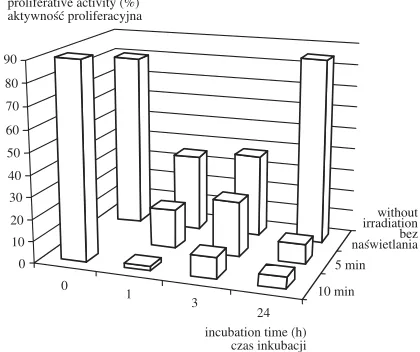
In neoplastic cell line (PC12) the greatest drop in proliferative activity was found after 1 h and 3 h incubation with photosensitizer at 10−minute irra− diation time (Fig. 1).
In normal cell line 3T3 Balb lowered prolifer− ative activity was shown after 1 h and 24 h incu− bation with photosensitizer at 10−minute irradia− tion time, and after 24 h incubation with photosen− sibilizator at 5−minute irradiation (Fig. 2).
In non−irradiated cells decrease in proliferat− ing cells both neoplastic and normal was not sig− nificant, which was evident mainly after 0 h and 24 h incubation. However, the greatest drop in pro− liferative activity was observed in PC12 cells after 3 h incubation without irradiation in comparison to 3T3 Balb cells (Fig. 1, 2).
Discussion
The mechanism of photodynamic reaction in tissues is not fully elucidated [5, 19]. The purpose of PDT is selective destruction of neoplastic tissue resulting in decreased proliferative activity of the cells. Many studies have shown that applied in PDT photosensitizers are absorbed better by can− cer than normal cells [13, 20]. The results of this study did not show significant differences in pro− liferative activity drop between normal and neo− plastic cell lines, which suggests similar affinity for the applied photosensitizer in both PC12 and 3T3 Balb lines.
The obtained results showed the greatest drop in proliferative index in PC12 cells after 1 h and 3 h incubation with photosensitizer at 10−minute irra− diation, and in normal cells after 1 h incubation and the same irradiation time.
An astonishing observation was the increase in proliferative activity in control neoplastic and nor− mal cells after 24 h incubation with photosensitiz− er without irradiation. It could be assumed that this period allows for removal of the photosensitizer from the cells or its degradation. However, more studies are needed to explain this phenomenon. Photodynamic therapy in vivo starts after 24–72 hours, depending on the administration way and a photosensitizer type. It seems that in vitrocondi− tions (monolayer) allow for faster photosensitizer
Fig. 1. Proliferative activity of neoplastic cell line (PC12) after photodynamic therapy
Ryc. 1. Aktywność proliferacyjna komórek linii nowo− tworowej (PC12) pod wpływem terapii fotodynamicznej
0 1 3 24 10 min 5 min without irradiation bez naświetlania 0 10 20 30 40 50 60 70 80 90
proliferative activity (%) aktywność proliferacyjna
incubation time (h) czas inkubacji
Fig. 2. Proliferative activity of normal cell line (3T3 Balb) after photodynamic therapy
Ryc. 2. Aktywność proliferacyjna komórek linii prawidłowej 3T3 Balb pod wpływem terapii fotodynamicznej 0 1 3 24 10 min 5 min 0 10 20 30 40 50 60 70 80 90
proliferative activity (%) aktywność proliferacyjna
without irradiation bez naświetlania
penetration into cells, so the optimal exposition time of cells subjected to PDTin vitrois not com− parable with in vivoconditions.
In normal 3T3 Balb cells decreased prolifera− tive index was shown after 1 h and 24 h incubation with photosensitizer at 10−minute irradiation, and after 24 h incubation with photosensibilizator at 5−minute irradiation (Fig. 2). In vivostudies have shown photosensitizers distribution also in healthy tissues, e.g. in liver or skin cells, which indirectly confirms possibility of photosensibilizator (Ph II) accumulation in normal line cells. Uehara and Inokuchi also obtained significantly lower PCNA indicator than in control group in the vascular cell at 24 h after PDT.
Photosensitizer’s distribution within an organ− ism is carried out mainly viablood system, proba− bly with the help of different protein carriers, but mostly lipid carriers [11, 21]. The carriers’ charac− ter is conditioned by the used photosensitizer’s properties (hydrophilic, hydrophobic, amphiphi− lic). Hydrophobic dyes show greater efficiency and selectivity of absorption by neoplastic tissue. The involvement of cancer tissue lipoprotein (LDL) receptors in the accumulation of medium and high hydrophobic photodyes has been shown [22]. Complex receptor−LDL−photosensitizer in PDT is absorbed via endocytosis. High amounts of photosensibilizator can be inserted into cells with the usage of LDL [13]. Kessel’s studies have shown that the pattern of HpD (porphyryn mix−
ture) distribution in lung tumour is correlated with the number of LDL receptors in various tissues [15]. LDL catabolism increases mainly in cells with high proliferative activity, which could also concern healthy cells with high proliferative activ− ity, e.g. fibroblasts. Therefore, it could be assumed that lipoprotein receptors involvement may affect better photosensitizer absorption and transport into cells [13]. Used in this study normal cell line 3T3 Balb is also characterized by high proliferative activity.
So far investigations on PDT have been main− ly conducted on neoplastic cell lines; fewer studies have concerned normal cell lines or tumours in vivo. In none of the studies by now, proliferative activity under PDT influence has been compared in normal and neoplastic cell lines, with simulta− neous application of the same photosensitizer, laser light of the same wavelength and irradiation time. Comparative studies carried out so far have been focused rather on death type analysis (apop− tosis, necrosis) of normal and cancerous lines [23]. The results of this study with the application of photosensitizer (Ph II) and laser light (of the same wavelength and irradiation time for both types of cells) did not show significant differences in PDT impact on the cells between normal and neoplastic lines. The obtained results confirm the importance of the comparison between neoplastic cell lines and normal cell lines characterized by low proliferative index.
References
[1] Luksiene Z: Photodynamic therapy: mechanism of action and ways to improve the efficiency of treatment. Medicina 2003, 39(12), 1137–1150.
[2] Mang TS:Laser and light sources for PDT: past, present and future. Photodiagn Photodyn Ther 2004, 1, 43–48.
[3] Kim SY, Kwon OJ, Park JW: Inactivation of catalase and superoxide dismutase by singlet oxygen derived from photoactivated dye. Biochimie 2001, 83, 437–444.
[4] Ohse T, Nagaoca S, Arakawa Y, Kawakami H, Nakamura K: Cell death by reactive oxygen species generat− ed from water−soluble cationic metalloporphyrins as superoxide dismutase mimics. J Inorg Biochem 2001, 85, 201–208.
[5] Cases A, Perotti C, Fukuda H, del C. Batlle AM: Photodynamic therapy of activated and resting lymphocytes and its antioxidant adaptive response. Lasers Med Sci 2002, 17, 42–50.
[6] Rockson SG, Lorenz DP, Cheong W−F, Woodburn KW: Photoangioplasty: An emerging clinical cardiovascu− lar role for photodynamic therapy. Circulation 2000, 102(5), 591–596.
[7] Korbelik M, Parkins CS, Shibuya H, Cecic I, Stradford MRL, Chaplin DJ:Nitric oxide production by tumour tissue: impact on the response to photodynamic therapy. Br J Cancer 2000, 82(11), 1835–1843.
[8] Ali SM, Olivo M:Mechanisms of action of phenanthroperylenquinones in photodynamic therapy. Int J Oncol 2003, 22, 1181–1191.
[9] Yang Y, Sharma R, Sharma A, Avasthi S, Avasthi YC: Lipid peroxidation and cell cycle signalling: 4−hydrox− ynonenal, a key molecule in stress mediated signalling. Acta Biochim Polon 2003, 50(2), 319–336.
[10] Almeida RD, Manadas BJ, Carvalho AP, Duarte CB:Intracellular mechanisms in photodynamic therapy. Biochim Biophys Acta 2004, 1704, 59–86.
[11] Saczko J, Kulbacka J, Chwilkowska A, Lugowski M, Banas T: Levels of lipid peroxidation in A549 cells after PDTin vitro.Ann Acad Med Bialocent 2004, 49, 82–84.
[13] Girotti AW:Photosensitized oxidation of membrane lipids: reaction pathways, cytotoxic effects, and cytoprotec− tive mechanisms. J Photochem Photobiol 2001, 63: 103–113.
[14] Patito IA, Rothmann C, Malik Z: Nuclear transport of photosensitizers during photosensitization and oxidative stress. Biol Cell 2001, 93, 285–291.
[15] Kessel D, Luo Y: Mitochondrial photodamage and PDT−induced apoptosis. J Photochem Photobiol 1998, 42, 89–95.
[16] Plaetzer K, Kiesslich T, Verwanger T, Krammer B: The modes of cell death induced by PDT: an overview. Med Lasser Appl 2003, 18, 7–19.
[17] Wyld L, Reed MW, Brown NJ:Differential cell death response to photodynamic therapy is dependent on dose and cell type. Br J Cancer 2001, 84, 1384–1386.
[18] Kaneko T, Chiba H, Yasuda T, Kusama K: Detection of photodynamic therapy−induced early apoptosis in human salivary gland tumour cells in vitroand in mouse tumour model. Oral Oncol 2004, 40(9), 787–792.
[19] Behrend L, Henderson G, Zwacka RM:Reactive oxygen species in oncogenic transformation. Biochem Soc Trans 2003, 31, 1441–1444.
[20] Konan YN, Gurny R, Alleman E: State of the art delivery of photosensitizers for photodynamic therapy. J Photochem Photobiol 2001, 66, 89–106.
[21] Allison RR, Downie GH, Cuenca R, Hu XH, Childe CJH, Sibata CH: Photosensitizers in clinical PDT. Photodiagn Photodyn Ther 2004, 1, 27–42.
[22] Abuja PM, Albertini R: Methods for monitoring oxidative stress, lipid peroxidation and oxidation resistance of lipoproteins. Clin Chim Acta 2001, 306, 1–17.
[23] Macrobert AJ, Speight PM, Bennet JH:Induction of apoptotic cell death by photodynamic therapy in human keratinocytes. Arch Oral Biol 1998, 43(2), 143–149.
Address for correspondence:
Jolanta Saczko
Department of Medical Biochemistry Silesian Piasts University of Medicine Chałubińskiego 10
50−368 Wrocław Poland
e−mail: [email protected] Conflict of interest: None declared Received: 24.06.2005
Revised: 16.09.2005 Accepted: 24.10.2005
Praca wpłynęła do Redakcji: 24.06.2005 r. Po recenzji: 16.09.2005 r.