Ugur Keklikci
1, A–F, Izzet Yavuz
2, A–C, E, F, Selcuk Tunik
3, B, C, E,
Zelal Baskan Ulku
4, A–C1,, Sedat Akdeniz
5, A–DOphthalmic Manifestations in Patients
with Ectodermal Dysplasia Syndromes
1 Department of Ophthalmology, Dicle University, Faculty of Medicine, Diyarbakir, Turkey 2 Department of Pediatric Dentistry, Dicle University, Faculty of Dentistry, Diyarbakir, Turkey 3 Department of Histology and Embryology, Dicle University, Faculty of Medicine, Diyarbakir, Turkey 4 Department of Prosthetic Dentistry, Dicle University, Faculty of Dentistry, Diyarbakir, Turkey 5 Department of Dermatology, Dicle University, Faculty of Medicine, Diyarbakir, Turkey
A – research concept and design; B – collection and/or assembly of data; C – data analysis and interpretation;
D – writing the article; E – critical revision of the article; F – final approval of article; G – other
Abstract
Background. Ectodermal dysplasia (ED) is a disorder that results from abnormal formation of at least two of the four major ectodermal derivatives in the developing embryo. The ectoderm of the embryo forms the skin, teeth, hair and nails, sweat glands and part of the eyes.
Objectives. The aim of this article is to reveal ophthalmologic symptoms and signs as multidisciplinary, reliable criteria for ectodermal dysplasia.
Material and Methods. In this retrospective study, 24 patients with ED were analyzed from the recorded data. Ophthalmological examination of the patients, who had previously received the diagnosis of ED in the dental department, was done. During the examination, ocular symptoms related to tear film, corneal changes, lacrimal duct, periorbital hyperpigmentation, alteration lashes and eyebrows were evaluated.
Results. The age ranged between 3–45, and the mean and standard deviation (Mean ± SD) was 15.8 ± 7.4 years. The number of males was 13 (54.2%) and females, 11 (45.8%). Eighteen patients (75.0%) suffered from ocular com-plaints related to the ocular surface.In 11 of the patients with ED, there were dry eye symptoms. While the mean age of cases with eye involvement was 17.5, it was 23.1 in cases with dry eye symptoms.
Conclusions. In the study, it was observed that, in patients with ED, ocular complaints, particularly dry eye symp-toms, may increase as age advances (Adv Clin Exp Med 2014, 23, 4, 605–610).
Key words: dry eye, ectodermal dysplasia, ocular involvement.
Adv Clin Exp Med 2014, 23, 4, 605–610 ISSN 1899–5276
ORIGINAL PAPERS
© Copyright by Wroclaw Medical University
Ectodermal dysplasia (ED) is a disorder that results from abnormal formation of no less than two of the four major ectodermal derivatives in the developing embryo. The ectoderm of the embryo forms the skin, teeth, hair and nails, sweat glands and part of the eyes [1]. As it has earlier been re-ported, the first recorded ED cases seem to have been described in 1792 [2]. However, over 200 dif-fering pathologic clinical conditions have been de-termined and defined ever since; these conditions are relatively rare, as being between 1 in 10,000 and 1 in 100,000 births [3–5]. Hypohidrotic ectodermal dysplasia (HED), ectrodactyly-ectodermal dyspla-sia-clefting (EEC) syndrome
ankyloblepharon-ectodermal dysplasia-clefting (AEC) syndrome and hidrotic ectodermal dysplasia are the most fre-quently encountered syndromes [6].
to occur in ED, but how common and the specific ways the eyes are involved is not fully understood. Eye problems do seem to differ between the dif-ferent types of ED and may vary in severity. As al-ready discussed, the structures of the eye formed from the ectoderm are affected by ED. The func-tion and comfort of the eye is therefore disturbed. Alterations of the eyebrows and lashes are men-tioned in combination with several ectodermal dysplasia syndromes [6, 9]. Most recently, Callea et al. [10] havereported infantile bilateral glauco-ma in a child with ectoderglauco-mal dysplasia.
EEC syndrome is a rare autosomal-dominant disease which has a penetrance of 95% and variable expression within one family. It has been mapped to the gene locus 7ql 1.2-q21.3 [11, 12]. The syndrome is characterized by a clefting deformity of the hands and/or feet (ectrodactyly-lobster claw deformity), ectodermal dysplasia, and cleft lip and palate [13]. The etiology of ocular surface disease in EEC syn-drome still remains unclear. The reported ophthal-mologic manifestations are strabismus, telecanthus, fused lids at birth, blepharophimosis, entropion, absence of eyelashes, bilateral eyelid cysts, agenesis of lacrimal puncta, dacryocystitis, blepharitis, con-junctivitis, deficient meibomian gland function, and corneal limbal deficiency [13]. Disorders of the tear film and deformities of the meibomian glands are described for the EEC syndrome. Anomalies of the lacrimal system are seen in patients with EEC syn-drome. Meibomian gland deformities have so far only been described in patients with EEC syndrome. There is evidence that, on the basis of the similar pa-thology, other ectodermal dysplasias should also be combined [6, 14, 15].
The aim of this study is to establish easily de-tectable ophthalmologic symptoms and signs as re-liable criteria for ectodermal dysplasia syndromes.
Material and Methods
In this retrospective study, 24 patients with ED, evaluated between January 1997 and January 2012 at the Department of Ophthalmology Clinic, were ana-lyzed from the recorded data. The diagnosis of the pa-tients had previously been made in the dental depart-ment. For ophthalmologic evaluation, however, the patients were referred to our clinic. During the exam-ination, ocular symptoms related to tear film, corne-al changes, lacrimcorne-al duct, periorbitcorne-al hyperpigmenta-tion, alteration lashes and eyebrows were evaluated.
The photographs of those patients who had agreed that images of the face, teeth, hands and eyelids could be included into the study were documented.
The study was performed according to the guidelines of the Declaration of Helsinki.
Statistical Analysis
Mean and standard deviation (x– ± SD) were calculated for continuous variables. All categorical variables were presented as number of patients and percentages. Statistical evaluations were analyzed through use of the statistical package SPSS 15.0 for Windows (SPSS Inc., Chicago, IL, USA).
Results
In the whole series of 24 patients with ED, the age ranged between 3–45, and the mean and stan-dard deviation (Mean ± SD) was 15.8 ± 7.4 years. The number of males was 13 (54.2%) and females, 11 (45.8%). The mean age of the cases with ocular complaints was 17.5, while it was 23.1 in those who had dry eye symptoms.
Twenty-two patients included in this study were suffering from HED. There were 2 patients with EEC syndrome. All of the cases had dental as-pects of different degrees, conically shaped teeth, and dental ageneses presenting maxillary retru-sion, maxillary hypotrophy and forward-upward-displaced-protused mandible (Fig. 1).
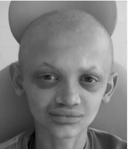
Eighteen patients (75.0%) were suffering from ocular complaints related to the ocular surface. They reported irritation, tearing, epiphora, pho-tophobia, redness and recurrent inflammations of the lids. The ocular findings of the patients are shown in Table 1. Periorbital hyperpigmentation and eyebrow anomalies were detected in 88.9% of cases, and lashes anomalies in 83.3%(Fig. 2).
There were ocular symptoms in 18 of those with ED. Dry eye complaints in 9 of these patients were moderate, a Schirmer I test was over 10 mm, and the tear film break-up time (BUT) was be-tween 8–12 s. In 2 cases, there were severe dry eye findings, the Schirmer I test was below 5 mm in both eyes, and BUT was short by 5 s. There was
positive fluorescein staining.In both eyes of these patients, there was corneal opacity, and the mean age of these patients was 37.5 years.
Two patients with EEC had lacrimal duct anomaly. In a female patient of 3.5 years old, there was lacrimal system agenesis, and upper and lower puncta were nonexistent (Figure 3). In a male pa-tient of 3 years old, however, the upper punctum was nonexistent, and there was lacrimal system hy-poplasia (Fig. 4). In both patients, there were cleft lip and palate, as well as ectrodactyly in hands and feet (Fig. 5a, b).
Fig. 2. Trichodysplasia and Protuberant lip seems
Fig. 3. Upper and lower puncta are inexistent in EEC case
Fig. 4. Early DCR was performed on the patient with lac-rimal duct hypoplasia, cleft lip and palate of 3 years case
Table 1. Ocular findings of ocular involvement with ED patients (18 cases)
Periorbital hyperpigmentation 16 cases (88.9%)
Eyebrows anomaly 16 cases (88.9%)
Lashes anomaly 15 cases (83.3%)
Dry eye symptoms 11 cases (61.1%)
Lacrimal duct anomaly 4 cases (22.2%)
Corneal Opacity 2 cases (11.1%)
Fig. 5. Ectrodactyly in hands (a); Ectrodactyly in feets (b) a)
Discussion
It is now explicitly known that HED is most-ly inherited as an X-linked recessive trait. In these cases, only in male patients can the disorder be ful-ly expressed. Female cases who carry onful-ly a copy of the disease gene (heterozygote carriers) are like-ly to present with some signs and findings associ-ated with the disorder. There are also reports that have been documented in literature suggesting that HED seems to have been inherited as an au-tosomal recessive genetic trait, in which the disor-der is fully expressed in both male and female cas-es [9, 16, 17]. In the current study, the number of female cases (11) is seen to be lower than that of males (13 cases).
In his study, Kaercher [6] reported that 94.4% of the patients suffered from dry eye symptoms, reduction of eyebrows was seen in 94.4%, and the lashes were altered in 91.6%. In our study, how-ever, ocular symptoms were detected in 18 cases (75.0%). In cases with ocular symptoms, perior-bital hyperpigmentation (88.9%), eyebrow anoma-lies (88.9%), lashes anomaanoma-lies (83.3%) and dry eye symptoms (61.1%) were observed. Dry eye symp-toms were observed to exist in 11 patients in our study; in 9 of these, a Schirmer I test was found to be within normal limits and BUT was shorter than normal (8–12 s). The values obtained in our study were found to be partly lower with respect to those by Kaercher [6]. Patients in our study (mean age 15.8 years) were younger than those in Kaerch-er’s (mean age 21.9 years)[6]. We observed that ocular complaints increased together with age. The mean age of patients with ocular complaints was 17.5, whereas it was 23.1 in the group having dry eye symptoms, suggesting that ocular complaints and dry eye symptoms increase together with age. In the literature, severe ocular symptoms are de-scribed for adult patients with ectodermal dyspla-sia [6, 11].
Kaercher [6] reported corneal opacity as 19.4%. We determined keratopathy in two pa-tients (11.1%). The mean age of these papa-tients was higher (37.5 years) with respect to the other pa-tients (15.8 years). Therefore, we are of the opin-ion that the development of keratopathy would be seen in older ages. Some earlier studies have re-ported that keratopathy is seen in advanced ages [6, 11]. In both patients, the Schirmer I test was be-low 5 mm, and BUT was short by 5 s in both eyes. Although the Schirmer I test was 5/6 mm and BUT 6/6 s in one patient, he had no dry eye complaints. The authors attributed the corneal abnormalities to an unstable tear film due to a lack of lipid sec-ondary to absence of meibomian glands and goblet cell abnormalities [15, 18–20].
There is evidence that keratopathy in ectoder-mal dysplasia syndromes is the result of an altered lipid layer of the tear film. It is, in fact, an indirect consequence of the meibomian gland alteration [6, 21]. The age-dependent appearance of keratopathy would favor this hypothesis. Besides that, there are cases with an early onset of keratopathy. Baum re-ports that ectodermal dysplasia is a possible prima-ry developmental defect in the corneal epithelium during embryogenesis, and that corneal alterations are a primary sign of ectodermal dysplasia syn-dromes, starting with an epithelial insufficiency and ending with stromal scarring and vasculariza-tion [22]. Corneal changes in EEC may have a dif-ferent presentation. For instance, limbal stem cell deficiency (LSCD) seems to play a role in the eti-ology of this keratopathy. Among other risk fac-tors for the severity and progression of the disease are recurrent infections from lacrimal drainage obstruction and tear film instability [18]. Most re-cently, Di Lorio [23] has described the ocular phe-notype of 23 cases through EEC syndrome. Accord-ing to what they reported in their study, EEC results from heterozygous missense mutations in the DNA- -binding domain of the p63 gene, which is known to be a crucial element during embryogenesis and stem cell differentiation of stratified epithelia. Four-teen cases (61%) were diagnosed with LSCD as evi-denced by the absence of limbal palisades of Vogt on slit-lamp examination. They hypothesized that p63 mutation resulted in LSCD, leading to pro-gressive keratopathy, the major cause of visual morbidity in EEC patients. It is still unclear why, in many cases, the corneal epithelium remains in-tact, whereas the stroma, which is of non-ectoder-mal origin, shows opacifications [6].
In our study, lacrimal duct complaints were seen in 4 patients (22.2%); two of them were males and two females. There was epiphora and recur-rent infection. Silicone tube was applied to a male patient of 4 years. Dacryocystorhinostomy (DCR) was performed on a female patient of 24 years due to chronic dacryocystitis. The other two patients were those with EEC syndrome.
non-familiar, sporadic EEC syndrome are usually pre-sented as affected more severely [12]. Two cases in our group were sporadic cases, and there were not any siblings in their family.
The shortened BUT assessed on several occa-sions suggested marked instability of the tear film, as-suming that the absence of meibomian glands would lead to a deficiency in the lipid layer. The other is the absence of meibomian glands as first described in 1984 [11]. The absence of the meibomian glands leads to an instability of the tear film, with a normal Schirmer test but pathological BUT. Lemp [24] re-ported that patients with absent or deficient meibo-mian gland function should be categorized in the evaporative group of dry eye syndromes [15]. Mat-sumoto et al.[13] measured the tear evaporation rate to clarify the changes in tear quality. They reported that the tear evaporation rate was still higher com-pared to that in patients with obstructive meibomian gland dysfunction. In addition, Mathers [25] report-ed that the tear evaporation rate was three times higher in patients with meibomian gland disease.
Lemp [24] presumed that EEC syndrome be-longed to the evaporative type of dry eyes. Mat-sumoto et al. [13] reported that increased tear evaporation is one of the most important process-es in the pathogenprocess-esis of dry eye and keratopathy in this rare syndrome. In our patients with EEC,
there was no keratopathy. One of the patients was male (3 years) and the other was female (3.5 years). In both patients, there was a lacrimal duct anom-aly. In one patient, the absence of bilateral lower and upper punctus, and in the other patient, the absence of bilateral upper punctus were seen. Al-though the Schirmer I test was found to be within normal limits in both patients, BUT was 7/8 s. in one patient and 8/8 s. in the other. In both patients, there was recurrent infection, and the meibomian orifices were of normal appearance. There were no dry eye symptoms in the patients. We think that the absence of dry eye symptoms may be related to the nonexistence of tear drainage. Early DCR was performed on the patient of 3 years. The other was followed up. Cleft palate-lip and ectrodactyly were detected in both patients.
Patients who receive a diagnosis of ED should be assessed in terms of ocular findings. In pa-tients with ED, degradation arises in the tear lip-id layer due to alterations in the meibomian gland, on the basis of which dry eye and ocular surface symptoms are seen. When ED is diagnosed, ocular symptoms that disturb patients do not exist. How-ever, with increasing age, ocular symptoms, espe-cially dry eye symptoms, increase. As soon as the dry eye symptoms occur, conservative treatment should be initiated and followed up.
References
[1] Hill VA, Nischal KK, Collin JR, Harper JI: An unusual ectodermal dysplasia with unique eye defects. Br J Dermatol 2005, 152, 365–367.
[2] Abadi B, Herren C: Clinical treatment of ectodermal dysplasia: a case report. Quintessence Int 2001, 32, 743–745.
[3] Lamartine J: Towards a new classification of ectodermal dysplasia. Clin Exp Dermatol 2003, 28, 351–354.
[4] Buyse M: Birth Defects Encyclopedia. Volume I. Ames, IA: Blackwell Scientific Publications, 1990.
[5] Priolo M, Lagana C: Ectodermal dysplasias: a new clinical-genetic classification. J Med Genet 2001, 38, 579–585.
[6] Kaercher T: Ocular symptoms and signs in patients with ectodermal dysplasia syndromes. Graefes Arch Clin Exp Ophthalmol 2004, 242, 495–500.
[7] Callea M, Faletra F, Maestro A, Verzegnassi F, Rabusin M, Vinciguerra A, Radovich F, Clarich G, Yavuz I, Tumen EC: Dental Phenotype in a Patient With Hypohidrotic Ectodermal Dysplasia And Severe Immunodeficiency. J Int Dent Med Res 2011, 4, 17–20.
[8] Akgun MO, Sabuncuoglu F, Altun C, Guven G, Basak F: Multidisciplinary Treatment Approach of Patient With Ectodermal Dysplasia. J Int Dent Med Res 2010, 3, 141–145.
[9] Yavuz I, Baskan Z, Ulku R, Dulgergil TC, Dari O, Ece A, Yavuz Y, Dari KO: Ectodermal dysplasia: Retrospective study of fifteen cases. Arch Med Res 2006, 37, 403–409.
[10] Callea M, Vinciguerra A, Willoughby CE, Deroma L, Clarich G: Infantile bilateral glaucoma in a child with ectodermal dysplasia. Ophthalmic Genet 2013, 34, 58–60.
[11] Mondino BJ, Bath PE, Foos RY, Apt L, Rajacich GM: Absent meibomian glands in the ectrodactyly, ectodermal dysplasia, cleft lip palate syndrome. Am J Ophthalmol 1984, 97, 496–500.
[12] Kasmann B, Ruprecht KW: Ocular manifestations in a father and son with EEC syndrome. Graefes Arch Clin Exp Ophthalmol 1997, 235, 512–516.
[13] Matsumoto Y, Dogru M, Goto E, Endo K, Tsubota K: Increased tear evaporation in a patient with ectrodactyly-ectodermal dysplasia-clefting syndrome. Jpn J Ophthalmol 2004, 48, 372–375.
[14] Ireland A, Meyer DR: Ophthalmic manifestations of ectrodactyly-ectodermal dysplasia-clefting syndrome. Ophthalmic Plast Reconstr Surg 1998, 14, 295–297.
[15] Ota Y, Matsumoto Y, Dogru M, Goto E, Uchino Y, Endo K, Tsubota K: Management of evaporative dry eye in ectrodactyly-ectodermal dysplasia-clefting syndrome. Optom Vis Sci2008, 85, 795–801.
[17] Pinherio M, Freire-Maia N: Ectodermal displasias: a clinical classification and a causal review. Am J Med Genet 1994, 53, 153–162.
[18] Felipe AF, Abazari A, Hammersmith KM, Rapuano CJ, Nagra PK, Peiro BM: Corneal changes in ectrodactyly-ectodermal dysplasia-cleft lip and palate syndrome: case series and literature review. Int Ophthalmol 2012, 32, 475–480.
[19] Soker S, Keklikci U, Mese A, Akkus M, Nergiz Y: Conjunctival impression cytology in patients with ectodermal dysplasia. Turk Arch Ped 2012, 47, 72–77.
[20] Saw VP, Dart JK, Sitaru C, Zillikens D: Cicatrising conjunctivitis with anti-basement membrane autoantibodies in ectodermal dysplasia. Br J Ophthalmol 2008, 92, 1403–1410.
[21] Wilson FM, Grayson M, Pieroni D: Corneal changes in ectodermal dysplasia. Am J Ophthalmol 1973, 75, 17–27.
[22] Baum JL, Bull MJ: Ocular manifestations of the ectrodactyly, ectodermal dysplasia, cleft lip-palate syndrome. Am J Ophthalmol 1974, 78, 211–216.
[23] Di Iorio E, Kaye SB, Ponzin D, Barbaro V, Ferrari S, Böhm E, Nardiello P, Castaldo G, McGrath JA, Willoughby CE: Limbal stem cell deficiency and ocular phenotype in ectrodactyly-ectodermal dysplasia-clefting syndrome caused by p63 mutations. Ophthalmology 2012, 119, 74–83.
[24] Lemp MA: Report of the National Eye Institute/Industry Workshop on clinical trials in dry eyes. CLAO J 1995, 21, 221–232.
[25] Mathers WD: Ocular evaporation in meibomian gland dysfunction and dry eye. Ophthalmology 1993, 100, 347–351.
Address for correspondence:
Ugur KeklikciDepartment of Ophthalmology Dicle University Faculty of Medicine TR-21280, Diyarbakir
Turkey
Tel.: + 90 412 248 80 01 E-mail: [email protected]
Conflict of interest: None declared