R E S E A R C H A R T I C L E
Open Access
Modeling hormonal and inflammatory
contributions to preterm and term labor
using uterine temporal transcriptomics
Roberta Migale
1, David A. MacIntyre
1*, Stefano Cacciatore
1, Yun S. Lee
1, Henrik Hagberg
2,3, Bronwen R. Herbert
1,4,
Mark R. Johnson
1,4, Donald Peebles
5, Simon N. Waddington
6,7and Phillip R. Bennett
1*Abstract
Background:Preterm birth is now recognized as the primary cause of infant mortality worldwide. Interplay between hormonal and inflammatory signaling in the uterus modulates the onset of contractions; however, the relative contribution of each remains unclear. In this study we aimed to characterize temporal transcriptome changes in the uterus preceding term labor and preterm labor (PTL) induced by progesterone withdrawal or inflammation in the mouse and compare these findings with human data.
Methods:Myometrium was collected at multiple time points during gestation and labor from three murine models of parturition: (1) term gestation; (2) PTL induced by RU486; and (3) PTL induced by lipopolysaccharide (LPS). RNA was extracted and cDNA libraries were prepared and sequenced using the Illumina HiSeq 2000 system. Resulting RNA-Seq data were analyzed using multivariate modeling approaches as well as pathway and causal network analyses and compared against human myometrial transcriptome data.
Results:We identified a core set of temporal myometrial gene changes associated with term labor and PTL in
the mouse induced by either inflammation or progesterone withdrawal. Progesterone withdrawal initiated labor without inflammatory gene activation, yet LPS activation of uterine inflammation was sufficient to override the repressive effects of progesterone and induce a laboring phenotype. Comparison of human and mouse uterine transcriptomic datasets revealed that human labor more closely resembles inflammation-induced PTL in the mouse.
Conclusions:Labor in the mouse can be achieved through inflammatory gene activation yet these changes are not a requisite for labor itself. Human labor more closely resembles LPS-induced PTL in the mouse, supporting an essential
role for inflammatory mediators in human“functional progesterone withdrawal.”This improved understanding of
inflammatory and progesterone influence on the uterine transcriptome has important implications for the development of PTL prevention strategies.
Keywords:Preterm labor, Inflammation, Progesterone, Myometrium, Transcriptome, RNA-Seq
* Correspondence:[email protected];[email protected] 1Imperial College Parturition Research Group, Institute of Reproduction and Developmental Biology, Imperial College London, Hammersmith Campus, London, United Kingdom
Full list of author information is available at the end of the article
Background
Preterm labor (PTL), defined as labor onset before 37 weeks of gestation, is the leading cause of neonatal mortality in children under 5 years of age, with incidence rates continu-ing to rise in both developcontinu-ing and developed countries [1]. PTL is a complex syndrome resulting from the activation of multiple pathological processes; however, infection and inflammation are implicated in the majority of cases of very early PTL in humans, whereas they are not in most late PTL cases [2, 3]. Regardless of the causal factor(s) deter-mining the timing of labor onset, both preterm and term parturition involves common phenotypic changes, includ-ing cervical remodelinclud-ing and dilatation, fetal membrane rup-ture, and uterine contractions that collectively permit the expulsion of the fetus [4].
The transition of the uterus from a quiescent to con-tractile organ results from a complex interplay between inflammatory and hormonal signals yet the relative contribution of each to the contractile phenotype remains poorly understood. In the mouse, parturition at term ap-pears to be initiated by fetal lung maturation, which drives steroid receptor cofactor-dependent secretion of surfactant proteins into the amniotic fluid, migration of activated macrophages into the uterus, and induction of the inflam-matory transcription factor nuclear factor kappa light chain enhancer of activated B cells (NFkB). This leads to the up-regulation of inflammatory and contraction-associated genes within the uterus, including those encoding for the principal prostaglandin synthase enzyme, cyclooxygenase-2 (COX-2). Increased levels of prostaglandin F2 alpha (PGF2α) cause luteolysis and subsequent progesterone
withdrawal, leading to further activation of pro-labor path-ways [5–8]. However, stimulation of uterine contractions and preterm birth can be achieved in the mouse in the absence of progesterone withdrawal using bacteria or bacteria-derived products (e.g., lipopolysaccharide, LPS) [9, 10]. We have also recently shown that uterine acti-vation of the inflammatory transcription factor activa-tor protein 1 (AP-1), and not NFkB, is sufficient to stimulate inflammatory and contractile gene transcrip-tion and pathways causing PTL in the mouse at gesta-tion day 16, when circulating progesterone levels are high [11–13]. While these findings indicate that patho-physiological PTL and spontaneous term labor in the mouse involve modulation of different hormonal and inflammatory pathways, their convergence in a common contractile phenotype suggests overlap and redundancy in the underlying mechanisms of parturition.
As in the mouse, human labor is accompanied by infil-tration of immune cells into gestational tissues and release of inflammatory mediators that promote cervical ripening and myometrial activation [14]. Unlike most other mammals, this occurs in the presence of high levels of circulating progesterone, suggesting that inflammatory
pathway activation in the human uterus may play a dom-inant role in promoting the laboring phenotype [15–17]. However, although progesterone concentrations in women remain high beyond labor, a number of non-mutually ex-clusive hypotheses have been formulated to explain how a
“functional”withdrawal of progesterone may still precede inflammatory activation at term and signal parturition on-set in humans: (1) catabolism of progesterone in the uterus into metabolites that regulate relaxant pathways in-dependent of progesterone receptor (PR) action [18]; (2) altered expression of PR isoforms that drive upregulation of inflammatory mediator expression [19–21]; (3) changes in cofactor protein levels affecting PR transactivation [22]; and (4) inflammation-induced trans-repression of PR by NFkB [23]. In addition, recent mouse model investigations of immune cells in gestational tissues suggest that parturition-associated inflammation may not be causa-tive of labor, but may be involved in the amelioration of host response to pathogens and the activation of tissue repair mechanisms necessary for remodeling of the uterus following delivery [24–28].
Many of the hormonal and inflammatory pathways proposed to regulate term and preterm human partur-ition can be recapitulated in the mouse. Inhibpartur-ition of the PR by the antagonist pharmaceutical drug RU486 (mife-pristone) robustly induces PTL in the mouse and the same molecule acts as an effective abortifacient in humans [29, 30]. Infection and/or inflammation are frequently as-sociated with human PTL, accounting for 30 % of all cases [2, 31]. Activation of cytokine and inflammatory pathways associated with human PTL are similarly stimulated in infection/inflammation-induced PTL mouse models [11, 12, 32–34]. The use of these mouse models also permits temporal characterization of parturition-associated gene expression changes in the uterus, which are mani-fested prior to the acquisition of the contractile phenotype. Previous time-course studies of gene expression changes in the murine pregnant uterus using microarray have been highly informative yet limited by the comparatively low spe-cificity, sensitivity, and resolution of hybridization-based techniques [35–37]. Furthermore, practical and ethical rea-sons limit the feasibility of conducting similar studies in the human and thus studies on gestational tissues (myome-trium, cervix, and chorioamniotic membranes) have been limited to the comparison of samples collected before and after labor onset [38–44]. An absence of time-course data from human studies has further limited our knowledge of large-scale gene expression changes involved in initiating the onset of labor, most of which are likely to be manifested some time prior to labor.
(3) RU486-induced progesterone withdrawal leading to PTL, to identify transcriptomic changes specific to each model and the core set of genes essential to parturition in all circumstances. We assess the relative contribution of progesterone-regulated and inflammatory-regulated transcription pathways to the mechanism of labor in each of the murine models and compare these changes to human uterine transcriptome data.
Methods
Murine studies and ethics statement
All animal studies were performed under UK Home Of-fice License 70/6906 in accordance with the UK Animals Scientific Procedures Act of 1986 and with approval of the Imperial College and University College London Ethical Review Committees. Virgin female CD1 outbred mice were time mated and the presence of a copulatory plug was considered day 0 of gestation. Mice were housed in open cages at 21 ± 1 °C under a constant 12 h light-dark cycle regimen, with ad libitum access to standard rodent food and water. Unless otherwise stated, each experiment represents a minimum of four bio-logical replicates.
Murine models of preterm labor
For the infection/inflammation-induced PTL murine model, CD1 pregnant mice received, at gestational day 16 (E16), a subcutaneous injection of morphine (2.5 mg/kg) 20 min before the surgery. A laparotomy was performed under general anesthesia using isoflurane as previously de-scribed [11, 12]. Uterine horns were exteriorized and 20μg (25μL total volume in phosphate-buffered saline [PBS]) of
Escherichia coli LPS serotype O111:B4 (Sigma) or sterile PBS was injected into the upper right uterine horn between the first and the second sacs without entering the amniotic cavity. Recovery following surgery was remotely monitored via an infrared CCTV camera system until tissue collection or the onset of spontaneous delivery. Onset of labor was defined as delivery of the first pup. For the noninflam-matory model of PTL induced by chemical progester-one withdrawal, CD1 pregnant mice received, at E16, a 150μg subcutaneous injection of RU486 (Sigma) or vehicle (20μl dimethyl sulfoxide [DMSO]) as described previously [11, 45]. Animals were monitored by infrared CCTV prior to tissue collection or the onset of labor.
Tissue collection
Briefly, tissue samples from the upper right uterine horn were excised and immediately snap-frozen on dry ice be-fore being stored at−80 °C until processing for RNA ex-traction. For the gestational model, samples were harvested at E14, E16, E18, and during active term labor (LAB). For the RU486-induced PTL model, tissue was collected 6 h and 18 h post injection (hpi) or during
active labor (20 ± 1.6 hpi, mean ± SD), corresponding re-spectively to 30 %, 90 %, and 100 % of the time between the injection and the onset of PTL. Similarly for the LPS-induced PTL model, myometrium was collected 2 h and 6 h following PBS or LPS injection and during active labor (7 ± 2 hpi, mean ± SD), which again was designed to correspond to 30 %, 90 %, and 100 % of the time interval between injection and PTL onset.
RNA isolation and sequencing
Frozen myometrium tissue was pulverized in liquid nitro-gen and RNA was extracted in lysis buffer ML (guanidi-nium thiocyanate) using the NucleoSpin® miRNA kit (Macherey-Nagel) with DNase treatment following manu-facturer’s instructions. RNA quality was assessed for all samples using an RNA 6000 Nano Kit and Bioanalyzer (Agilent). Samples with an RNA integrity number (RIN) >8 were used for RNA-Seq library preparation. cDNA libraries with an insert length of 300 bp were prepared from 2μg of purified large RNA fraction (>200 nucleotides) using a Tru-Seq Stranded mRNA Sample preparation kit (Illumina). Samples were enriched for poly-A mRNA using oligo-dT-coated magnetic beads. Following purification, mRNA was randomly fragmented at 94 °C for 8 min to obtain frag-ments of around 200 bp while minimizing bias at the 3' end of transcripts. First-strand complementary DNA synthesis was performed using random primers (Illumina) and SuperScript II Reverse-Transcriptase (Invitrogen) followed by second strand synthesis with RNaseH and DNA poly-merase I (Illumina). Adapters provided were used to tag each sample. Compatibility between adapters was checked with Illumina Experiment Manager to allow subsequent pooling of three samples in each lane during sequencing. cDNA libraries were amplified by 10 cycles of PCR. The quality of each library was evaluated on a 2100 Bioanalyzer (Agilent) followed by paired-end sequencing (2 × 100 bp) on an Illumina HiSeq 2000. Sample preparation and run was randomized by model and treatment.
Processing of RNA-Seq data
RNA-Seq reads were aligned to the reference genome of
Mus musculus GRcm38 from ENSEMBL release 73
file 2. While no outliers were detected in the term gestation model, two samples were classified as outliers (P< 0.01, Bonferroni post hoc test) in the RU486- and LPS-induced PTL models (6 h DMSO and 6 h LPS) and excluded from further analysis.
Differential expression analysis of gene expression data
Gene read counts were normalized for the library size using TMM scaling implemented in the Bioconductor package followed by transformation into log2counts per million. Differentially expressed genes (DEGs) were identified in R using the negative binomial differential expression method edgeR [52]. P values were adjusted for multiple testing with the Benjamini–Hochberg cor-rection and a false discovery rate (FDR) cutoff of 0.001 was used. The full gene expression dataset is reported in Additional file 3.
Pathway analysis
Biological functions enriched with DEGs were identified using two manually curated databases of signaling and metabolic pathways represented on networks (Process Network Analysis by MetaCore®, Thomson Reuters [53]) or on canonical pathways maps (canonical pathways analysis by Ingenuity® Pathway Analysis [IPA®], QIAGEN Redwood City).
Process Network Analysis was performed to identify biological functions significantly enriched by DEGs. A threshold P value less than 0.05 (−log10= 1.3) was used as a cut-off. Overrepresented canonical pathways were also identified using IPA (QIAGEN Redwood City), which, in addition to identifying enriched biological functions, allows prediction of the activation status for each specific pathway. Canonical pathways were consid-ered significantly enriched when P< 0.05 and activated/ inhibited when Z-score≥|2.0|.
Causal Network Analysis
IPA was used to identify upstream regulators potentially responsible for the observed gene expression changes. Putative upstream regulators were predicted using IPA Causal Network Analysis by calculating a regulation Z-score and an overlapPvalue, which predict the activation or inhibition state of regulators based on downstream DEGs and known directionality of expression [54]. Up-stream regulators with overlapP≤0.05 and an IPA activa-tion Z-score≥|2.0| were considered significantly activated/ inhibited. The overlap P value (calculated used Fisher’s exact test) threshold was used to identify possible upstream regulators based on significant overlap between DEGs and known targets regulated by a given upstream regulator. The activation Z‐score was used to infer likelihood of activation status of upstream regulators based on evidence from DEGs in our dataset for which the direction of the observed
expression change was consistent with that expected from the literature.
Multivariate data analysis
For multivariate analysis, genes with zero read counts were excluded and the remaining genes were mean-centered and unit variance-scaled. Principal component analysis (PCA) was performed on transcriptome changes using the stand-ard algorithm as implemented in the prcomp function in-cluded in the R library stats. Transcriptome changes specifically associated with progression of gestation charac-terizing each murine model were identified and modeled using orthogonal signal correction-partial least squares (OSC-PLS) regression modeling [55]. OSC-PLS is a super-vised method that aims to maximize the variance between groups in the output data (i.e., score). It also provides as output an indication (i.e., loadings) of the importance of the variables in the regression. Analyses were carried out in R and OSC-PLS modeling was applied to expression levels of 4142 genes (FDR < 0.001), including those changing in at least one pairwise comparison within a model. Loadings values indicating the relative importance of each gene to each murine model of gestation are reported in Additional file 4. Model goodness of fit was assessed by calculating the correlation coefficients (r) using Pearson linear correlation andPvalues were also calculated for each prediction. Hier-archical clustering was performed using Ward’s method [56]. The number of clusters was calculated using the Silhouettes algorithm [57].
Analysis of human myometrium RNA-Seq data
A human myometrium RNA-Seq dataset deposited by Chan et al. [40] was re-analyzed in our study to permit comparison of our murine transcriptome data with relevant human data. This dataset was derived from samples of lower segment human myometrium collected dur-ing Caesarean section at term (38–40 weeks of gestation) from women in active, spontaneous labor (n= 5, IL) and from women not in labor undergoing elective Caesarean section (n= 5, NIL). Women in the IL group required Cae-sarean section for undiagnosed breech or cephalopelvic dis-proportion, without labor augmentation. Count data were accessed at the NCBI Gene Expression Omnibus database using the series accession number [GEO: GSE50599] and genes differentially expressed when comparing IL versus NIL were identified using edgeR with an FDR value <0.001.
PTL model. These gene sets were further analyzed using OSC-PLS modeling to test for congruence between human and murine datasets across a time course lead-ing to the acquisition of a laborlead-ing phenotype. In the mouse, term labor occurred at gestational day E18.5, corresponding to a human term gestation of approxi-mately 270 days or 38.5 weeks. E18 corresponded to 263 days or 37.5 weeks of human gestation and thus, in the absence of labor, was considered equivalent to the NIL human sample group (term, not in labor). LPS and RU486 treatments led to PTL at E16 within 7 and 20 h following injection, respectively, corresponding to 234 days or 33 weeks of human gestation (preterm). Temporal gene expression changes relative to each murine model of gestation were alternatively used as a training set against which the human datasets were inter-rogated using OSC-PLS modeling. Pearson correlation scores andPvalues were derived for each test. A positive Pearson score was indicative of a positive correlation be-tween genes similarly changing as a function of gestational age in the mouse and in the human samples. Loadings derived from the OSC-PLS model were indicative of the importance of variables (genes) for discrimination between different sample groups. Hierarchical clustering of loadings was then performed to identify profiles consist-ent between human and murine gestational models and to identify gene clusters similarly expressed across gestational time course in both organisms.
Results
Myometrial transcriptome modeling of term and preterm labor induced by progesterone withdrawal or
inflammation
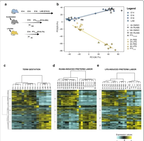
Using RNA-Seq we undertook temporal transcriptome profiling on mouse myometrial samples collected across a time series from animals experiencing spontaneous term labor, or PTL following pharmacologically induced (RU486) progesterone withdrawal, or inflammation-in-duced (LPS) PTL (Fig. 1a). In normal mouse gestation, cir-culating progesterone levels peak on E16 before falling to the lowest level on E18 [13]. We therefore characterized the myometrial transcriptome in this model on E14, E16, E18 prior to labor, and E18.5 after the onset of spontan-eous labor (postdelivery of the first pup). In the PTL models, LPS and RU486 induced labor onset 7 and 20 hours posttreatment respectively.
Because gene expression changes underpinning the acquisition of the laboring phenotype occur across dra-matically different time scales in each of the models, we next assessed transcriptome profiles at time points pro-portionally comparable on the time course to parturition in each model. Samples were thus collected at 2 and 6 hpi of LPS, and 6 and 18 hpi of RU486. PCA of myome-trial transcriptome data revealed that both spontaneous
term labor and PTL following progesterone withdrawal involve similar trajectories of transcriptome changes pre-ceding labor. However, a divergent trajectory of myome-trial transcriptome changes was observed in animals undergoing inflammation-induced PTL (Fig. 1b). Broad visualization of each individual transcriptome signature indicated consistency within each experimental group; however, in animals delivering at term, there was variation in the myometrial transcriptome just prior to labor on E18, with two samples clustering with E16 and one sample clustering with LAB (Additional file 5). The majority of uterine transcriptome changes were observed to occur between 6 and 18 hpi in the RU486-induced PTL model and between 2 and 6 hpi in the LPS-induced PTL model.
We next performed hierarchical clustering analysis on the 4142 genes differentially expressed (FDR < 0.001) in at least one condition within each of the murine models. Venn diagrams depicting the total number of DEGs for each comparison within each model were also generated (Additional file 6). Comparisons not exhibiting signifi-cant gene changes (6 h DMSO vs 6 h RU486 and 6 h LPS vs LABLPS) were excluded from further analyses. A characteristic change in the myometrial transcriptome was detected with increasing gestation and spontaneous term labor onset (Fig. 1c). Three main clusters of gene expression patterns were identified, representing genes either increasing (Cluster 1) or decreasing (Cluster 3) with proximity to labor, or genes exhibiting variable change (Cluster 2). Similar clusters were observed in animals undergoing RU486-induced PTL (Fig. 1d). In contrast, unique transcriptome changes were identified in animals experiencing LPS-induced PTL (Fig. 1e). Cluster 1 could be subdivided into two groups: (1) genes whose expression increased only with the onset of labor (Cluster 1a) and (2) genes whose expression increased immediately after ad-ministration of LPS or PBS but decreased following labor onset (Cluster 1b), likely to represent those genes affected by the laparotomy procedure rather than the treatment it-self. Consistent with normal term and RU486-induced labor, expression of Cluster 3 genes in the LPS model decreased with approaching labor. Cluster 2 genes were uniquely upregulated in the LPS model just prior to labor onset. Process Network Analysis [58] (MetaCore®, Thomson Reuters) of Cluster 2 genes showed enrichment of pathways associated with cellular adhesion – platelet endothelium–leucocyte interactions (P= 8.70 × 10⁻⁹), pro-teolysis and connective tissue degradation (P= 1.53 × 10⁻⁶), and enrichment of multiple gene pathways associated with inflammation (immune response – TH17-derived cytokines, P= 2.25 × 10⁻³; inflammation– IL-6 signaling,
P= 3.57 × 10⁻³; inflammation–kallikrein–kinin system,
P= 3.99 × 10⁻³; and immune response – phagocytosis,
Identification of a core set of differentially expressed genes associated with term and preterm labor
To identify sets of genes mutually regulated between models, or unique to each of them, we evaluated the overlap between DEGs detected in the following pairwise
comparisons: E16 vs E18, E16 vs LAB, and E18 vs LAB for the term gestation model; 6 h RU486 vs 18 h RU486, 6 h RU486 vs LABRU486, and 18 h RU486 vs LABRU486 for the RU486-induced PTL model; and 2 h LPS vs 6 h LPS, 2 h LPS vs LABLPS, and 6 h LPS vs LABLPSfor the E14 E16 E18
RU486/DMSO
LPS/PBS
LAB (E18.5)
PC1(30.7%)
PC2(23.3%)
−40 −20 0 20 40 60
−80 −60 −40 −20 0 20 40
E14 E16 E18 LAB Legend
6h DMSO 6h RU486 18h DMSO 18h RU486
PTL
2h PBS 2h LPS 6h PBS
PTL 6h LPS
a
b
18h R
U486
PTL
PTL
PTL
PTL
PTL
18h R
U486
18h
R
U486
18h
R
U486
6h R
U486
6h DMSO
18h DMSO
6h DMSO
6h R
U486
18h DMSO
18h DMSO
18h DMSO
6h DMSO
6h R
U486
6h
R
U486
6h LPS
6h LPS
PTL
PTL
PTL
6h LPS
PTL
6h PBS
6h PBS
6h PBS
6h PBS
2h PBS
2h PBS
2h PBS
2h PBS
2h LPS
2h LPS
2h LPS
2h LPS
− 0 +
e
c
d
E14
E14
E14
E14 E16 E16 E16 E16 E18 E18 E18 E18 LAB LAB LAB LAB
Expression values
TERM GESTATION RU486-INDUCED PRETERM LABOR LPS-INDUCED PRETERM LABOR
6h
PTL (E16+20h) E16
18h
PTL (E16+7h) E16
2h 6h
3
2
1b
1a
3
2
1
3
2
1
Fig. 1Transcriptome changes associated with term gestation, RU486-, and lipopolysaccharide (LPS)-induced preterm labor (PTL) in the mouse myometrium.aMyometrium samples were collected from three mouse models of gestation: term labor (blue), progesterone withdrawal/RU486-induced PTL) (gray), and inflammatory/LPS-induced PTL (yellow).bPrincipal component analysis of transcriptome changes represented along principal component (PC)1 (30.7 % of total variance) and PC2 (23.3 % of total variance). Both term labor and RU486-induced PTL exhibit similar trajectories of transcriptome changes whereas animals experiencing LPS-induced PTL undergo a divergent trajectory.c–eHeatmaps representing hierarchical clustering analysis performed on differentially expressed genes (false discovery rate < 0.001) for unsupervised identification of gene expression patterns characterizing mouse myometrium collected from term labor (c), RU486-induced PTL (d), and LPS-induced PTL (e). The number of clusters was calculated using the Silhouettes algorithm. (n≥4 mice for each experimental group).DMSOdimethyl sulfoxide,LAB
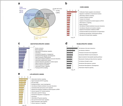
LPS-induced PTL model (Fig. 2a). Canonical pathway analysis of the DEGs was then performed using IPA (QIAGEN Redwood City, www.qiagen.com/ingenuity) to enable identification of groups of biological functions similarly activated or inactivated across these compari-sons. A total of 46 canonical pathways were detected as significantly overrepresented in all models of parturition (Additional files 8 and 9). Strong similarity in enrich-ment profiles characterizing genes changing with spon-taneous labor and RU486-induced PTL were observed. Several enrichment profiles specific to LPS-induced labor were identified and included pathways involved in
bacterial and viral pattern recognition, interferon sig-naling, death receptor sigsig-naling, and acute phase re-sponse signaling. A core set of gene changes (95 upregulated, 148 downregulated, Additional file 10) were identified across all models. Process networks analysis on this core set showed enrichment of signaling pathways involved in tissue remodeling (P= 1.12 × 10⁻³), muscle contraction (P= 1.499 × 10⁻²), cell proliferation (P= 1.94 × 10⁻⁷), apoptosis (P= 9.190 × 10⁻³), and NFkB and mitogen-activated protein kinase (MAPK) signaling (P= 9.186 × 10⁻³and 1.747 × 10⁻²respectively, Fig. 2b). This group included genes that have been shown to be
a
307 401
286 303
70 63
95 148
46 42
1104 744
TERM
GESTATION RU486-INDUCED
PTL
LPS-INDUCED PTL
c
d
e
0 2 4 6 8
0 2 4 6
b
CORE GENES
GESTATION-SPECIFIC GENES
LPS-SPECIFIC GENES
LPS-SPECIFIC GENES
TERM GESTATION-SPECIFIC GENES
RU486-SPECIFIC GENES
[734] [894]
[1315] [997] 341
260 [813] [872]
0 2 4 6 8
CORE GENES
RU486-SPECIFIC GENES
Fig. 2Identification of core gene sets differentially expressed in spontaneous term labor and preterm labor (PTL) models. (a) Venn diagram representing overlap between differentially expressed genes (DEGs) as detected in term labor (blue), and RU486-induced (gray) and lipopolysaccharide (LPS)-induced (yellow) PTL models. Process Network Analysis (Metacore®) was performed on the core set, including those genes similarly changing in all models of parturition (b), on genes changing only in the term labor model (c), genes exclusively changing with RU486 treatment (d), and on genes exclusively changing following LPS treatment (e). Bar length indicates significance and is equal to the negative logarithm of enrichmentPvalue. AP
associated with myometrial activation and contraction, including increased levels of prostaglandin-endoperoxide synthase 2 (Ptgs2), prostaglandin E synthase 2 (Ptges2) [59], and apolipoproteins [60] as well as decreased levels of potassium voltage-gated channels [61], G-protein coupled receptors, and leukemia inhibitory factor receptor, consistent with a compensatory role in the attenuation of myometrial sensitivity to contraction-promoting signals as labor approaches [62–65]. In addition, numerous novel candidates were identified that may play an important, previously unrecognized, role in promoting myometrial contractions, including components of the cytoskeleton machinery, transporters, and alternative potassium channel isoforms. Similar analyses of genes exclusively modulated throughout normal murine gestation (341 upregulated, 260 downregulated, Fig. 2c) showed enrichment of several bio-logical functions associated with pregnancy, such as muscle contraction pathways, including genes encoding for G-protein coupled receptors, guanylate cyclase-activating protein, and dystroglycan; genes involved in gonadotropin regulation : (i.e., family members of the AP-1 family such as JunB, Fos, and FosB), chemotaxis, and inflammation [i.e., chemokine (C-X-C motif ) ligand (Cxcl) 1, Cxcl2,
Cxcl10,Cxcl16, andTNF-α] (Additional file 11).
We next examined those genes changing specifically in the RU486-induced PTL model (286 upregulated, 303 downregulated, Fig. 2d). DEGs unique to this model were primarily associated with tissue remodeling (cell adhesion and proteolysis), consistent with the role of the progester-one/PR axis in regulating extracellular matrix integrity [66]. These genes included those encoding for collagen structural proteins (Col5a2 and Col6a4) and cadherins (Cdh4, Cdh13,Cdh17, Pcdha11, Pcdha12) as well as re-modeling enzymes such as metalloproteinases (Mmp2,
Mmp20,Mmp27) and several genes encoding for Adam (a disintegrin and metallopeptidase domain) family members (Adam5,Adam10, andAdam19; Additional file 12).
Analysis of DEGs in the LPS model showed that ap-proximately 80 % (1848/2312) of gene changes were unique to this model and included genes belonging mostly to inflammatory pathways such as interferon signal-ing (i.e.,Irf7,Stat1,Stat2, andStat5,iNOS, andCaspase-1), Notch signaling (i.e., platelet-derived growth factor B [Pdgfb] andPdgfr), platelet endothelium–leucocyte interac-tions (i.e.,P-selectin,E-selectin,L-selectin, andCD47), apop-tosis mediated by NFkB (including the genes C-iap1, Tnfrsf17, Il-15ra, and I-κb), and immune response TH17-derived cytokines pathways (Fig. 2e; Additional file 13).
Progesterone withdrawal induces two waves of myometrial gene expression changes preceding spontaneous term labor
Since mice laboring at term or preterm following RU486 treatment exhibited the largest number of shared DEGs,
we next sought to investigate the extent and stage at which progesterone signaling was exercising its greatest effect on gene expression. Genes differentially expressed between E16 and E18 and between E18 and LAB were compared with those changing in response to RU486 from 6 h to 18 h, which represented the time frame encompassing the majority of gene changes in this model. Two sets of genes were identified, one in-cluding 55 % of the genes (187/342) changing between E16 and E18 and 18 h post-RU486 treatment (early changing genes), and a second group including 63 % of the genes (295/466) similarly changing between E18 and labor and 18 h post-RU486 treatment (late changing genes) (Fig. 3a, FDR < 0.01). Process networks analysis of early progesterone withdrawal response genes showed that these genes are primarily involved in extracellular matrix remodeling (P= 6.16 × 10⁻³), cell adhesion (P= 6.61 × 10⁻³), and blood vessel morphogenesis (P= 1.37 × 10⁻²) (Fig. 3b). Late response genes are involved in muscle contraction (P= 3.10 × 10⁻⁴), regulation of cytoskeleton rearrangement (P= 1.96 × 10⁻³), and potassium transport (P= 9.87 × 10⁻³) (Fig. 3c). These data provide evidence that progesterone withdrawal mediates two waves of gene expression in the uterus, involved firstly in tissue remodeling, presumably for preparation of the uterus for labor, and subsequently for activation of the uterine contractile machinery. Causal Network Analysis by IPA was then used to identify poten-tial upstream regulators of DEGs detected in each pairwise comparison (Fig. 3d). As expected, the main upstream regulator predicted to modulate gene changes associated with RU486-induced progesterone withdrawal was mife-pristone (RU486, Z score6h RU486 vs 18h RU486= 6.6, IPA Causal Network Analysis, Fig. 3d and Additional file 14). RU486 was also predicted to be a major upstream regula-tor of DEGs between E16 and E18 (Z scoreE16 vs E18= 2.9) and DEGs between E18 and LAB (Z scoreE18 vs LAB= 2.2), albeit to a lesser extent. Other top predicted regulators of transcriptional networks in the spontaneous term labor model included calcium/calmodulin-dependent protein kinase I (CaMKI) and epidermal growth factor (EGF) and its receptor (EGFR).
Identification of time-associated gene expression varia-tions associated with contractile phenotype acquisition during preterm and term labor
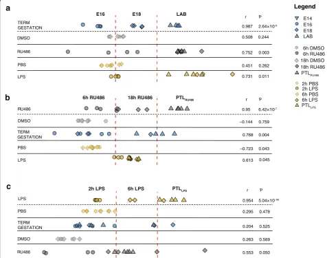
between the gestation time and the predicted scores of the test sets to indicate positive or no correlation between the training and the test set. First, the RU486 and LPS models were used as test sets against genes changing as a function of time in mice spontaneously laboring at term (Fig. 4a). A strong positive correlation was found with the training in both cases (r= 0.752,P= 0.003 andr= 0.731,P= 0.011, re-spectively) indicating consistency between major gene changes associated with progression of gestation and those changes observed with premature parturition induced by RU486 and LPS. Similar results were observed when the RU486 model was used as the training set (Fig. 4b) with a strong positive correlation found between the normal ges-tation model (r= 0.768,P= 0.004) and to a lesser extent the LPS model (r= 0.613,P= 0.045). In contrast, when the LPS model was used as the training set (Fig. 4c) no signifi-cant correlation was found with either RU486 (r= 0.553,
P= 0.05) or normal gestational (r= 0.204, P= 0.525) models, providing evidence that temporal gene changes observed in LPS-induced PTL are unique to this model.
Temporal transcriptome changes in murine term and preterm labor correlate with human labor-associated myometrial gene changes
While animal models of parturition and PTL are regularly used to infer functional mechanisms in human pathology, the reliability of such inference on a global transcriptome scale has yet to be examined. We therefore compared the transcriptome data generated from each of the murine models used in our study with available human uterine transcriptome data [40]. This study was chosen because it represents the first and only RNA-Seq study published to date using pregnant human myometrium collected at term (>37 weeks) before and during labor. This dataset provides transcriptome changes characterizing human myometrium collected during Caesarean section in women following spontaneous labor (n=5, IL) onset, and from women not in labor (n=5, NIL), between 38 and 40 weeks of gestation undergoing elective Caesarean section.
First, edgeR analysis was repeated to identify DEGs with a homogenous FDR threshold of less than 0.001,
a
b
E18 vs LAB E16 vs E18
113 23 [273] [69]
6h RU486 vs 18h RU486
c
d
787 834
72 80 6
0
9 5 145 41
[144] [322]
[998] [1117]
6h RU486 vs 18h RU486 E16 vs E18 E18 vs LAB
Upstream
Regulators Depth
Activation Z-score
0 +6.6
mifepristone CaMK I
mifepristone EGF
EGFR
a
b
E18 vs LAB E16 vs E18
113 23 [273] [69]
6h RU486 vs 18h RU486
c
d
787 834
72 80 6 0
9 5 145 41
57 237
[144] [322]
[998] [1117]
6h RU486 vs 18h RU486 E16 vs E18 E18 vs LAB
Upstream
Regulators Depth
Activation Z-score
0 +6.6
mifepristone CaMK I
mifepristone EGF
EGFR
Fig. 3RU486-induced progesterone withdrawal anticipates gene changes observed prior and during labor onset in term gestation.aVenn diagram representing overlap between gene changes characterizing term gestation (E16 vs E18 and E18 vs LAB) and genes changing between 6 h and 18 h following RU486 treatment. Two groups of genes were identified, one including 55 % of the genes (187/342) changing between E16 and E18 and 18 h post-RU486 treatment (highlighted inred) and a second group including 63 % of the genes (295/466) similarly changing between E18 and LAB and 18 h post-RU486 (highlighted inyellow).bProcess Network Analysis performed on differentially expressed genes similarly changing between E16 and E18 and between 6 h RU486 and 18 h RU486; and (c) between E18 and LAB and 6 h RU486 and 18 h RU486. Bar length indicates significance and is equal to the negative logarithm of enrichmentPvalue. APvalue cut-off less than 0.05 (−log10P> 1.3) was applied and is indicated by thedashed line.
which was applied to both human and murine datasets. We identified 357 DEGs shared between mouse and hu-man datasets that were selected for further analysis (Fig. 5a, Additional file 15). OSC-PLS modeling was then used to investigate the relationship between time-associated gene changes observed in mouse myometrium and changes associated with the acquisition of a laboring phenotype in human myometrium. Gene expression variations across term murine gestation, and following RU486 or LPS treat-ment in the mouse, served as training sets against which the corresponding gene changes in human myometrium were tested (Fig. 5b). The OSC-PLS model showed clear separation between human samples collected prior to and following the onset of labor when each of the murine models was alternatively used as the training set. These
results indicate the existence of temporal similarities in gene changes characterizing murine and human myome-trial evolution to a contractile phenotype regardless of the nature of the gestation (term or preterm). The strongest correlation was observed between human and LPS-induced PTL gene changes. We then examined the relative contri-bution of each gene to the gestational models by analyzing corresponding loading scores using hierarchical clustering analysis (Fig. 5c, Additional file 16). Human transcriptome changes associated with labor were mostly similar to those gene changes observed in LPS-induced PTL in the mouse, whereas orthologous DEGs in RU486-induced PTL clus-tered with spontaneous mouse term labor. Temporal dy-namics of these gene clusters were then examined in each of the murine models of parturition and human datasets
DMSO
RU486
PBS
LPS
0.987
0.508
0.752
0.451
0.731 2.64×10
0.244
0.003
0.262
0.011
r P
a
E16 E18 LAB
PBS
LPS
−0.723
DMSO −0.144 0.759
0.043 TERM
GESTATION 0.768 0.004
TERM GESTATION
b
0.613 0.045 r P
r
RU486 0.95 6.42×10
PTLRU486 6h RU486 18h RU486
DMSO
RU486
0.263
0.553 0.204 0.525
0.569
0.050
PBS 0.295 0.478
c
r P0.954 5.04×10 LPS
2h LPS 6h LPS PTLLPS
E14 E16 E18 LAB
Legend
6h DMSO 6h RU486 18h DMSO 18h RU486
PTLRU486
2h PBS 2h LPS 6h PBS
PTLLPS 6h LPS
TERM GESTATION
Fig. 4Orthogonal signal correction-partial least squares (OSC-PLS) analysis illustrates gene expression changes associated with progression of term gestation and preterm labor (PTL) models. This OSC-PLS analysis describes correlations between time-associated gene changes characterizing term gestation (blue), RU486 (gray), and lipopolysaccharide (LPS) models (yellow).aTerm gestation was used as training set and a positive correlation was found when using either RU486 or LPS models as test sets.bUsing the RU486 model as a training set, a positive correlation was found with both term and LPS models.cWhen the LPS model was used as the training set, no significant correlation was found with either RU486 or term labor models. Correlation coefficients (r) were calculated using Pearson linear correlation (P< 0.05).DMSOdimethyl sulfoxide,LAB
2874
357 612
c
a
b
r=0.887
Human in Labour (IL) Human not in Labour (NIL)
Key
r=0.744
r=0.921
P=0.001
P=0.014
P=0.0001 E16 E18 LAB
6h RU486 18h RU486 PTL
2h LPS 6h LPS PTL
d
ROCESS NETWORKS CLUSTER 1
CLUSTER 4 CLUSTER 3
CLUSTER 8 CLUSTER 6 CLUSTER 2
CLUSTER 7 CLUSTER 5
TERM
GEST RU486 LPS HUMAN
8 7 6 5 4 3 2 1 TERM
GEST RU486 LPS NIL vs IL
8 7 6 5 4 3 2 1
TERM
GEST 486 LPS HUMAN
e
[969][3231]
(Fig. 5d). Process Network Analysis (Fig. 5e) of genes down-regulated throughout gestation (Cluster 1) showed high representation in cell adhesion pathways. Genes upregu-lated throughout gestation, belonging to Cluster 6, were strongly enriched in immune response, chemotaxis, and cell proliferation pathways. Two other clusters, 4 and 7, are noteworthy since the direction of labor-associated changes in the human is opposite to all three murine models.
Discussion
We provide a detailed description of transcriptome changes characterizing the transition of the uterus from a quiescent to a contractile phenotype in three different models of murine parturition: term gestation, PTL induced by functional progesterone withdrawal (RU486), and PTL induced by inflammation (LPS). Further, we compared changes in the mouse myometrial transcriptome with those observed in the human myometrium with labor onset. Our results reveal the relative contribution of hor-monal and inflammatory signals to labor onset, and provide further insights into the biology of parturition.
The murine models examined in our study are widely used by the research community to mimic pathophysio-logical processes of human parturition [32, 67]. Progester-one withdrawal, be it either functional as in the human, or systemic, as in most other mammals, results in the release of the progesterone block and activation of uterine contrac-tions [45, 68]. Infection and/or inflammation are frequently associated with human PTL, accounting for 30 % of all cases [2, 31]. Therefore, bacteria-derived molecules such as LPS are often used to mimic infection/inflammation-in-duced PTL in animal models [12, 32], which occurs without changes in the levels of circulating progesterone [10, 69]. While the majority of very early PTL cases in humans are associated with infection/inflammation, most late PTL cases are not [3]. Here we show that normal murine labor at term and RU486-induced PTL exhibited similar global gene expression changes, while PTL induced by LPS ex-hibited a distinctive transcriptome profile. These results are consistent with previous studies indicating differences
between the underlying mechanisms of inflammation- and progesterone withdrawal-induced PTL and spontaneous term labor [9, 10, 36].
By identifying genes differentially expressed throughout gestation, our study revealed that the great majority of gene changes are observed before the onset of labor in the mouse. This highlights the importance of temporal tran-scriptome profiling approaches. Previous human or animal studies comparing samples collected prior to and follow-ing labor provide very limited insight into transcriptome changes required specifically for labor onset. Many gene expression changes observed in samples collected during labor likely occur as a consequence of labor or may be due to activation of uterine involution and be erroneously as-sumed as necessary for the onset of labor [70]. To control for this in our study, samples obtained from each animal model at labor onset were collected immediately following the birth of the first pup.
Cross-comparison of DEGs identified in each model of parturition allowed identification of a “core” gene set likely to be representative of transcriptome changes re-quired for both term and PTL. These genes are overrep-resented in pathways associated with cell proliferation, adhesion and tissue remodeling, ion transport, muscle contraction, and NFkB and MAPK signaling. Transcrip-tome changes unique to the onset of normal term labor were predominately related to muscle contraction, chemo-taxis, and inflammation, indicating that the process of nor-mal labor involves an inflammatory component. These data are consistent with the concept that spontaneous labor in the mouse is initiated by fetal signals that induce progester-one withdrawal via uterine NFkB activation and subsequent inflammatory pathway activation [5, 6]. However, although spontaneous labor is associated with a chemokine-driven influx of inflammatory cells into the myometrium, deple-tion of various immune cells subsets does not block labor [24–26, 28]. Moreover, activation of pro-inflammatory pathways is sufficient but not required for cervical ripening [28, 71, 72]. These data allude to the possibility that inflam-matory pathway activation in the mouse uterus at the time (See figure on previous page.)
of labor is a side effect of the mechanism leading to proges-terone withdrawal. Further evidence for this was generated when we examined those genes changing specifically in the RU486-induced PTL model. Our analyses show that term gestation and RU486-induced PTL models share around 60 % of similarly regulated genes, of which the majority are prematurely modulated by RU486 treatment prior to labor onset in this model. DEGs unique to this model were, however, primarily associated with tissue remodeling (cell adhesion and proteolysis), consistent with the role of progesterone/PR axis in regulating extracellular matrix integrity [66]. The absence of inflammatory gene network upregulation in the uterus following RU486-induced labor, taken together with previous studies showing that knockout of PR in myocytes does not affect inflammatory gene expression [66] and that RU486-induced labor does not involve infiltration of macrophages or neutrophils [70], provides further evidence that the acquisition of a contractile phenotype can be attained in the absence of inflammatory gene activation or synthesis of pro-inflammatory cytokines.
While activation of inflammation via progesterone withdrawal does not appear to be a requisite for acquisi-tion of a contractile phenotype in the mouse, much evidence suggests that inflammation can still promote a laboring phenotype in pathological situations, particularly infection/inflammation-induced PTL. For example, bac-teria and their products can robustly induce preterm birth in the absence of progesterone withdrawal [10, 69]. We have also previously shown that myometrial inflammatory and contractile gene activation via AP-1 is sufficient to cause PTL in the mouse at gestation day 16, when circu-lating progesterone levels are high [11]. In vitro studies have also shown that LPS may directly promote myocyte contraction via activation of the Rho/ROCK signaling pathways [73]. In this study we identified a set of genes uniquely upregulated with increasing proximity to labor following LPS treatment that were overrepresented in multiple inflammatory pathways. While these changes may represent unique mechanisms of contractile activa-tion via inflammatory gene modulaactiva-tion, they may alterna-tively represent unrelated acute inflammatory response to LPS treatment [36] and still harbor the potential to cause maternal and, importantly, fetal damage [9, 74–78].
We identified two sets of genes shared between normal and RU486 models, whose expression changed either early (E16 vs E18) or late (E18 vs LAB) in gestation. Process Net-work Analysis showed that the earlier responding genes were primarily involved in tissue remodeling, cell adhesion, and blood vessel morphogenesis, whereas the later responding genes were involved in muscle contraction, regulation of cytoskeleton rearrangement, and potassium transport. These data provide evidence that progesterone withdrawal mediates two waves of gene expression in the
uterus involved firstly in tissue remodeling, presumably for preparation of the uterus for labor, and subsequently for activation of the uterine contractile machinery.
Considering E16 as the start point of the time-course experiment, gene expression changes associated with ongoing gestation were acquired across a different time scale in each mouse model. OSC-PLS modeling of tem-poral gene changes observed throughout murine gestation allowed the investigation of similarities in the transcrip-tome profiles underlying labor onset. This analysis showed that gene changes acquired throughout a term gestation also occur in a similar fashion in the RU486- and LPS-induced PTL models, although within a shorter time frame. Second, by using each murine model alternatively as a training set, it was possible to test whether similar or different genes were important for the acquisition of a la-boring phenotype in each model. These results showed that similar transcriptome changes govern the onset of both term and RU486-induced PTL. In contrast, time-dependent transcriptome changes induced by LPS treat-ment did not correlate with term gestation or RU486 models, suggesting the existence of a specific set of genes controlling the onset of labor in the LPS model.
We identified two other clusters, 4 and 7, that comprised genes whose direction of change within the gestational time course in the human was opposite to all three murine models. We speculate this may be because the human sam-ples were collected well after the onset of labor, and thus may reflect gene expression changes induced by labor itself or by the initiation of involution mechanisms necessary for remodeling the uterus post-delivery [40]. Additionally, we showed that those genes similarly modulated in the LPS-induced murine PTL model and in human labor (Cluster 5) were enriched in proteolysis and cell proliferation pathways. These data indicate that a significant percentage of gene ex-pression changes observed in human at labor onset are likely to represent the activation of involution pathways ra-ther than pathways involved in activation of myometrial contractions.
Comparison between murine models of labor with human-derived data highlighted the existence of similar-ities in temporal gene changes acquired towards labor. Moreover, these data provide further evidence that the mechanism underlying human labor onset involves progesterone signaling that may occur via functional progesterone withdrawal mediated by changes in PR iso-form expression [19, 20, 80], but point toward a more prominent role for inflammatory pathway activation in human labor onset [81].
Conclusions
Our data are concordant with the paradigm that murine labor is initiated by progesterone withdrawal through fetal signaling that involves induction of inflammatory transcription factors and subsequent inflammatory gene activation in the uterus. However, parturition can be in-duced by progesterone withdrawal alone and inflammation is not required for the acquisition of the laboring phenotype in the mouse. Conversely, LPS activation of inflammation is sufficient to override the repressive effects of high circulat-ing concentrations of progesterone and induce labor in the mouse. Human myometrial transcriptome changes at the time of labor most closely resemble the mouse LPS model, indicating a dominant role for inflammatory pathways in human labor. These results have important implications for the understanding of the underlying mechanism of labor onset and the development of future prevention strategies for preterm birth.
Additional files
Additional file 1List of the total number of reads generated for each sample using RNA-Seq. (XLSX 10 kb)
Additional file 2:Supplementary methods used for outlier detection. (DOCX 15 kb)
Additional file 3:Complete RNA-Seq dataset as Excel file. (XLSX 34680 kb)
Additional file 4:Loadings derived from the OSC-PLS modeling of the 4142 DEGs detected in three models of parturition (term gestation, RU486-induced PTL, and LPS-induced PTL). (XLSX 256 kb)
Additional file 5:PCA of transcriptome profiles of murine myometrium collected throughout (a) term gestation, (b) following DMSO/RU486 intraperitoneal injection, and during RU486-induced preterm labor (LABRU486), (c) following PBS/LPS intrauterine injection and during
LPS-induced PTL.n≥4 biological replicates for each experimental group. (PDF 949 kb)
Additional file 6:Venn diagrams depicting number of genes differentially expressed in all pairwise comparisons across term gestation (a), RU486 (b), and LPS models (c). FDR < 0.001.n≥4 biological replicates for each experimental group. (PDF 1024 kb)
Additional file 7:Gene Ontology Process Network Analysis by Metacore® performed on genes exclusively changing 6 h following LPS treatment and during LPS-induced PTL but not changing throughout term gestation or the RU486-induced PTL model. Bar length indicates significance and is equal to the negative logarithm of enrichmentPvalue. APvalue cut-off less than 0.05 (−log10P> 1.3) was applied and is
indicated by the dashed line. (PDF 119 kb)
Additional file 8:Canonical pathways enrichment calculated by Ingenuity Pathway Analysis. A total of 46 pathways were detected as significantly enriched with genes differentially expressed in the pairwise comparisons indicated in the first row of the table (P< 0.05). First column on the left indicates Gene Ontology name associated with the canonical pathway. Log10Pvalues are reported for each pathway. (XLS 38 kb)
Additional file 9:Canonical Pathway enrichment by IPA®. DEGs as detected in the following pairwise comparisons were analyzed by IPA to identify canonical pathways highly represented within each comparison: 2 h LPS vs 6 h LPS, 2 h LPS vs LABLPS, 6 h RU486 vs 18 h RU486, 6 h
RU486 vs LABRU486, 18 h vs LABRU486, E16 vs E18, E16 vs term labor (LAB),
and E18 vs LAB. Hierarchical clustering of significantly enriched pathways based on their activation Z-score was used to assess for similarity in enrichment profiles between different models. Activation Z-score > |2|,
P< 0.05. (PDF 196 kb)
Additional file 10:List of core set of genes associated with all models of parturition. (XLS 258 kb)
Additional file 11:List of genes exclusively changing throughout normal murine gestation. (XLS 584 kb)
Additional file 12:List of genes exclusively changing in the RU486-induced PTL model. (XLS 573 kb)
Additional file 13:List of genes exclusively changing in the LPS-induced PTL model. (XLS 1767 kb)
Additional file 14:Z-scores derived from Causal Network Analysis by IPA®. (XLS 75 kb)
Additional file 15:Ensembl gene annotation for the 357 gene orthologs differentially expressed across murine gestation and during human labor. (XLSX 72 kb)
Additional file 16:Loadings values derived from the OSC-PLS modeling of the 357 human–mouse gene orthologs. (XLSX 39 kb)
Abbreviations
ADAM, a disintegrin and metallopeptidase domain; AP-1, activator protein 1; CaMKI, calcium/calmodulin-dependent protein kinase I; Cdh, cadherins; COX-2, cyclooxygenase-2; Cxcl, chemokine (C-X-C motif) ligand; DEGs, differentially expressed genes; DMSO, dimethyl sulfoxide; E, embryonic day; EGF, epider-mal growth factor; FDR, false discovery rate; hpi, hours post injection; IL, in labor; IPA, Ingenuity® Pathway Analysis; Irf, interferon; LAB, active term labor; LPS, lipopolysaccharide; MAPK, mitogen-activated protein kinase; Mmp, me-talloproteinase; NFkB, nuclear factor kappa light chain enhancer of activated B cells; NIL, not in labor; OSC-PLS, orthogonal signal correction-partial least squares; PBS, phosphate-buffered saline; PCA, principal component analysis; PDGF, platelet-derived growth factor; PGF2α, prostaglandin F2 alpha; PR,
Acknowledgements
This work was supported by the Wellcome Trust (grant DSRR_P24225), the Arthur Haffenden Fund, and Genesis Research Trust. DAM is supported by a Career Development Award from the Medical Research Council (MR/L009226/1).
Availability of data and materials
The murine datasets supporting the conclusions of this study are included within the article (Additional file 3). The human datasets re-analyzed in this study can be accessed at NCBI Gene Expression Omnibus database [GEO: GSE50599].
Authors’contributions
RM, DAM, and PRB conceived and designed the experiments. RM, DAM, YL, BRH, and SNW collected samples and performed experiments. RM, DAM, SC, and PRB performed the data analysis, and generated the figures and tables. RM, DAM, and PRB wrote first draft of the manuscript. RM, DAM, SC, HH, MRJ, DP, SNW, and PRB critically reviewed the data and the manuscript. All authors read and approved the final manuscript.
Competing interests
The views expressed in the submitted article are of the authors and not an official position of the institutions or funders. The authors declare that they have no competing interests.
Author details
1
Imperial College Parturition Research Group, Institute of Reproduction and Developmental Biology, Imperial College London, Hammersmith Campus, London, United Kingdom.2Perinatal Center, Department of Clinical Sciences, University of Gothenburg, Gothenburg, Sweden.3Centre for the Developing Brain, Division of Imaging Sciences and Biomedical Engineering, King’s College London, King’s Health Partners, St. Thomas’Hospital, London, United Kingdom.4Academic Department of Obstetrics and Gynaecology, Chelsea and Westminster Hospital, London, United Kingdom.5UCL Centre for Perinatal Brain Protection & Repair, Institute for Women’s Health, University College London, London, United Kingdom.6Gene Transfer Technology Group, Institute for Women’s Health, University College London, London, United Kingdom.7Antiviral Gene Therapy Research Unit, Faculty of Health Sciences, University of the Witswatersrand, Johannesburg, South Africa.
Received: 19 March 2016 Accepted: 1 June 2016
References
1. Liu L, Oza S, Hogan D, Perin J, Rudan I, Lawn JE, et al. Global, regional, and national causes of child mortality in 2000-13, with projections to inform post-2015 priorities: an updated systematic analysis. Lancet. 2015;385(9966):430–40.
2. Goldenberg RL, Culhane JF, Iams JD, Romero R. Epidemiology and causes of preterm birth. Lancet. 2008;371(9606):75–84.
3. Goldenberg RL, Andrews WW, Hauth JC. Choriodecidual infection and preterm birth. Nutr Rev. 2002;60(5 Pt 2):S19–25.
4. Romero R, Mazor M, Munoz H, Gomez R, Galasso M, Sherer DM. The preterm labor syndrome. Ann N Y Acad Sci. 1994;734:414–29. 5. Condon JC, Jeyasuria P, Faust JM, Mendelson CR. Surfactant protein
secreted by the maturing mouse fetal lung acts as a hormone that signals the initiation of parturition. Proc Natl Acad Sci U S A. 2004;101(14):4978–83. 6. Gao L, Rabbitt EH, Condon JC, Renthal NE, Johnston JM, Mitsche MA, et al.
Steroid receptor coactivators 1 and 2 mediate fetal-to-maternal signaling that initiates parturition. J Clin Invest. 2015;125(7):2808–24.
7. Montalbano AP, Hawgood S, Mendelson CR. Mice deficient in surfactant protein A (SP-A) and SP-D or in TLR2 manifest delayed parturition and decreased expression of inflammatory and contractile genes. Endocrinology. 2013;154(1):483–98.
8. Sugimoto Y, Yamasaki A, Segi E, Tsuboi K, Aze Y, Nishimura T, et al. Failure of parturition in mice lacking the prostaglandin F receptor. Science. 1997; 277(5326):681–3.
9. Filipovich Y, Klein J, Zhou Y, Hirsch E. Maternal and fetal roles in bacterially-induced preterm labor in the mouse. Am J Obstet Gynecol. 2015; 214(3): 386.e1–386.e9.
10. Hirsch E, Muhle R. Intrauterine bacterial inoculation induces labor in the mouse by mechanisms other than progesterone withdrawal. Biol Reprod. 2002;67(4):1337–41.
11. MacIntyre DA, Lee YS, Migale R, Herbert BR, Waddington SN, Peebles D, et al. Activator protein 1 is a key terminal mediator of inflammation-induced preterm labor in mice. FASEB J. 2014;28(5):2358–68.
12. Migale R, Herbert BR, Lee YS, Sykes L, Waddington SN, Peebles D, et al. Specific lipopolysaccharide serotypes induce differential maternal and neonatal inflammatory responses in a murine model of preterm labor. Am J Pathol. 2015;185(9):2390–401.
13. Virgo BB, Bellward GD. Serum progesterone levels in the pregnant and postpartum laboratory mouse. Endocrinology. 1974;95(5):1486–90. 14. MacIntyre DA, Sykes L, Teoh TG, Bennett PR. Prevention of preterm labour
via the modulation of inflammatory pathways. J Matern Fetal Neonatal Med. 2012;25 Suppl 1:17–20.
15. Murr SM, Stabenfeldt GH, Bradford GE, Geschwind II. Plasma progesterone during pregnancy in the mouse. Endocrinology. 1974;94(4):1209–11. 16. Sanyal MK. Secretion of progesterone during gestation in the rat. J Endocrinol.
1978;79(2):179–90.
17. Tan H, Yi L, Rote NS, Hurd WW, Mesiano S. Progesterone receptor-A and -B have opposite effects on proinflammatory gene expression in human myometrial cells: implications for progesterone actions in human pregnancy and parturition. J Clin Endocrinol Metab. 2012;97(5):E719–730.
18. Mitchell BF, Wong S. Changes in 17 beta,20 alpha-hydroxysteroid dehydrogenase activity supporting an increase in the estrogen/ progesterone ratio of human fetal membranes at parturition. Am J Obstet Gynecol. 1993;168(5):1377–85.
19. Merlino AA, Welsh TN, Tan H, Yi LJ, Cannon V, Mercer BM, et al. Nuclear progesterone receptors in the human pregnancy myometrium: evidence that parturition involves functional progesterone withdrawal mediated by increased expression of progesterone receptor-A. J Clin Endocrinol Metab. 2007;92(5):1927–33.
20. Mesiano S, Chan EC, Fitter JT, Kwek K, Yeo G, Smith R. Progesterone withdrawal and estrogen activation in human parturition are coordinated by progesterone receptor A expression in the myometrium. J Clin Endocrinol Metab. 2002;87(6):2924–30.
21. Pieber D, Allport VC, Hills F, Johnson M, Bennett PR. Interactions between progesterone receptor isoforms in myometrial cells in human labour. Mol Hum Reprod. 2001;7(9):875–9.
22. Condon JC, Jeyasuria P, Faust JM, Wilson JW, Mendelson CR. A decline in the levels of progesterone receptor coactivators in the pregnant uterus at term may antagonize progesterone receptor function and contribute to the initiation of parturition. Proc Natl Acad Sci U S A. 2003;100(16):9518–23. 23. Kalkhoven E, Wissink S, van der Saag PT, van der Burg B. Negative
interaction between the RelA(p65) subunit of NF-kappaB and the progesterone receptor. J Biol Chem. 1996;271(11):6217–24.
24. Rinaldi SF, Catalano RD, Wade J, Rossi AG, Norman JE. Decidual neutrophil infiltration is not required for preterm birth in a mouse model of infection-induced preterm labor. J Immunol. 2014;192(5):2315–25.
25. Filipovich Y, Agrawal V, Crawford SE, Fitchev P, Qu X, Klein J, Hirsch E. Depletion of polymorphonuclear leukocytes has no effect on preterm delivery in a mouse model of E. coli-induced labor. Am J Obstet Gynecol. 2015; 213(5):697.e1–697.e10.
26. Menzies FM, Higgins CA, Shepherd MC, Nibbs RJ, Nelson SM. Mast cells reside in myometrium and cervix, but are dispensable in mice for successful pregnancy and labor. Immunol Cell Biol. 2012;90(3):321–9.
27. Care AS, Ingman WV, Moldenhauer LM, Jasper MJ, Robertson SA. Ovarian steroid hormone-regulated uterine remodeling occurs independently of macrophages in mice. Biol Reprod. 2014;91(3):60.
28. Timmons BC, Mahendroo MS. Timing of neutrophil activation and expression of proinflammatory markers do not support a role for neutrophils in cervical ripening in the mouse. Biol Reprod. 2006;74(2):236–45.
29. Cameron IT, Michie AF, Baird DT. Therapeutic abortion in early pregnancy with antiprogestogen RU486 alone or in combination with prostaglandin analogue (gemeprost). Contraception. 1986;34(5):459–68.
30. Baulieu EE. Contragestion and other clinical applications of RU 486, an antiprogesterone at the receptor. Science. 1989;245(4924):1351–7. 31. Romero R, Espinoza J, Kusanovic JP, Gotsch F, Hassan S, Erez O, et al. The preterm
parturition syndrome. BJOG. 2006;113 Suppl 3:17–42.