www.ijper.org
Evaluation of in vitro Anticancer Activity of Vulpinic
Acid and its Apoptotic Potential Using Gene
Expression and Protein Analysis
Nil Kılıç1, Mehmet Kürşat Derici2, İlker Büyük3, Semra Soydam Aydın4, Sümer Aras3, Demet Cansaran-Duman1,*
1Ankara University, Biotechnology Institute, System Biotechnology Advance Research Unit, Tandogan, Ankara, TURKEY. 2Kırıkkale University, Faculty of Medicine, Medical Pharmacology, Kırıkkale, TURKEY.
3Ankara University, Faculty of Science, Department of Biology, Ankara, TURKEY.
4Turkish Medicines and Medical Device Agency, Ministry of Health of Turkey, Ankara, TURKEY.
ABSTRACT
Lichens and their secondary metabolite are still among the many unexamined natural sources in the drug industry. This study was designed to evaluate the cytotoxic effects of vulpinic acid lichen secondary metabolite and 6.25, 12.5, 25, 50, 100, 200 and 400
µM concentrations that treat cancer cell lines (CaCo2, HepG2, Hep2C, RD Wehi) and normal cells (Vero and L929) by MTT assay. The aim of this study was to determine the apoptotic effect of vulpinic acid on a molecular level. The determination of apoptotic molecular patterns of vulpinic acid was performed on western blot and quantitative real-time PCR (qRT-PCR) analysis. In our study, transcriptome changes on both gene and protein levels showed similar results. The determination of, mRNA levels of Bax, Bcl-2
and P53 genes were showed by qRT-PCR in cancer and normal cell lines. The results of the study showed that IC50 value of vulpinic acid altered the mRNA levels of Bax, Bcl-2 and P53 genes in all examined cancer cells when compared to the untreated cells. When the mRNA levels of the examined genes were compared, it was observed that
Bax gene showed more expression increase in all cell lines when compared to Bcl-2 and
P53 genes. This is the first evaluation of the apoptotic effect of vulpinic acid secondary
metabolite on mRNA levels. The current study highlights some points regarding vulpinic acid and its use for cancer treatment.
Key words: Vulpinic acid, Apoptotic effect, qRT-PCR, Western blot.
DOI: 10.5530/ijper.52.4.73 Correspondence: Dr. Demet Cansaran-Duman, Associate Prof. Ankara University, Biotechnology Institute, TURKEY. Phone: +90 312 2125820 E-mail: dcansaran@yahoo. com
INTRODUCTION
Cancer, caused by uncontrolled division of cells in the body, is a serious public health problem and a very common disease in the world. Cancer disease is significantly influenced by genetic, physical and socio-economic conditions.1 In recent years, cancer has steadily increasedas a health problem throughout the world sincemore effective treatments have not still been found.2,3 Treatments like surgery, chemotherapy and radiotheraphy are fre-quently used methods for cancer therapy. Many laboratories and studies focus on the search for an effective solution of this serious
Submission Date: 18-10-2017; Revision Date: 05-12-2017; Accepted Date: 18-04-2018
have identified approximately 1000 differentsecondary metabolites of the lichen species. Lichens and their sec-ondary metabolite are still among the many unstudied natural sources in the pharmaceutical sector.5,6,7 One of the secondary metabolites obtained from lichen is vul-pinic acid and it was first isolated in 1831 by Bebert.8 In 1925, vulpinic acid was isolated by Mazza from pulvinic acid derivative.9 With bright yellow appearance, vulpinic acid has toxic effects on reindeer if it consumes very high concentrations of Letharia vulpina.8 Vulpinic acid is referred to by IUPACas 3-hydroxy-5-oxo-4-phenyl-3-2-(2H)-furan, from which phenylacetate is formed and it has a molecular weight of 322.32 g.mol-1 and a chemical structure of C19H14O5.8
There are various studies related to the determination of anticancer activity of lichen secondary metabolites in the literature.10-16 As a result of these studies, lichen secondary metabolites may provide important results related withits use as candidate drug molecules. Usnic acid and its enantiomers are the most extensively stud -ied lichen secondary metabolites.17,18,19 Vulpinic acid has also been examined for malignant mesothelioma cells MM98, vulvar carcinoma cells A431 and Keratinocytes HaCaT cells by Burlando et al.20 According to their study results, vulpinic acid extracted from Letharia vulpine shows minimum toxic effect when compared with usnic acid, salazinic, gyrophoric and evernic acid.20 The potential use of vulpinic acid and gyrophoric acid for cosmetic purposes was evaluated against the harmful effects of UVB radiation, and 2,5 J/cm2 UVB radiation was applied to human keratinocyte (HaCaT) cell for the determination of the activity.21 After that, 25, 50, 100, 200, 400 and 800 μM vulpinic acid and gyrophoric acid concentrations were applied on the exposed and unexposed cells to UVB radiation. The effect of UVB radiation was determined by MTT, apoptosis and cyto-skeleton alteration assay. When the effect of the two lichen secondary metabolites was compared, vulpinic acid showed more protective effects on HaCaT cells exposed with UVB radiation.21 Koparal (2015) demon -strated cell viability and antiangiogenic effect of vulpinic acid and usnic acid secondary metabolites against HepG2 hepatocarcinoma cell, NS20Y neuroblastoma cancer cell and HUVEC endothelial cell lines. Koparal reported that concentrations of vulpinic acid determined IC50value 168±3.33 on HepG2, 68.83±1.58 on NS20Y, 231,94±25.4 on HUVEC for 48 h.22 Vulpinic acid, as a biologically significant molecule, represents easy production in large amounts for the drug industry. The primary aims of this research include i) the deter-mination of the cytotoxic effects of vulpinic acid lichen secondary metabolite on cancer and normal cells, which
have never been studied before. For this purpose, vulpinic acid was dissolved in DMSO (14 mM) and diluted in different concentrations (6.25, 12.5, 25, 50, 100, 200 and 400 μM ) with DMEM and treated to cancer cell lines (CaCo2, HepG2, Hep2C, RD and Wehi) and normal cells (Vero and L929). Cell viability was deter-mined by the MTT assay; ii) the precise mechanisms of cytotoxic effects of vulpinic acid remain unclear at molecular level. Therefore, apoptosis-related proteins and genes were performed to determine expression patterns of transcriptome level in cancer and normal cell lines by using western blot and quantitative real-time PCR (qRT-PCR) analysis. This is the first evaluation of the apoptotic effect of vulpinic acid secondary metabolite on mRNA levels.
MATERIALS AND METHODS
Vulpinic acid
Vulpinic acid was dissolved in 14mM DMSO and stock solutions of vulpinic acid were prepared to 400 µM concentrations. Double serial dilutions of the vulpinic acid (200, 100, 50, 25, 12.5, 6.25 µM) were also prepared in stock solution.
Cell culture
CaCo2 (Human colorectal adenocarcinoma cell line), HepG2 (Human hepatocellular carcinoma cell line), Hep2C (Human HeLa carcinoma cell line), RD (Human rhabdomyosarcoma cell line), Wehi (Mouse fibrosarcoma cell line), L929 (Mouse subcutaneous connective tissue cell line) and Vero (African green monkey kidney cell line) cells were cultured in DMEM (PAA Laboratories, Austria); Wehi in RPMI-1640 containing 10% FBS (Gemini Byproducts, Calabasas, CA), 2 mM L-glutamine and 1% penicillin and streptomycin. Cells were maintained in a humidified incubator at 37°C and 5% CO2.
Cell viability assay
Quantitative polymerase chain reaction (qRT-PCR)
All examined cells were treated with IC50 values of vulpinic acid for 48 h.Total RNA was extracted from the cultured cells using TRIzol reagent (Genezol) according to the manufacturer’s protocol. The purity and concentration of the total RNA were determined by spectrophotometry using the 260 nm/280 nm ratio (Nanodrop 1000, Thermo Scientific) and RNA controlled with 1% agarose gel electrophoresis. The first strand cDNA was synthesized byRosche First Strand cDNA Synthesis Kit (Rosche). cDNA synthesis was performed with 2 µg of RNA, 2.5 µM anchored-oligo (dT)18, 1x transcriptor high fidelity reverse transcriptase reaction buffer, 20 U protector RNase inhibitor, 1 mM deoxy -nucleotide mix, and 10 U transcriptor high fidelity reverse transcriptase at final concentration. After that, we continued 10 min at 65°C, 30 min at 55°C and 5 min at 85°C. Sequence of P53, Bcl-2 and Bax target genes were designed with Primer3 program and GAPDH
was used as housekeeping gene. Sequences of primers were as follows; Bax (forward, TCCCCCCGAGAG-GTCTTTT; reverse, CGGCCCCAGTTGAAGTTG);
Bcl-2 (forward, CTGCACCTGACGCCCTTCACC;
reverse, CACATGACCCCACCGAACTCAAAGA);
P53 (forward, AACGGTACTCCGCCACC; reverse, CGTGTCACCGTCGTGGA); GAPDH (forward, AACGGGAAGCTTGTCATCAATGGAAA; reverse, GCATCAGCAGAGGGGGCAGAG). Quantitative Real-time PCR was conducted with Roche Light Cycler 480 Real Time, using SYBR Green I Master. The PCR reaction conditions were as follows: after pre-denaturation, 10 min at 95°C, 40 cycles of 10 s at 95°C, 30 s at 60°C and 8 s at 72°C. All experiments were performed in three inde -pendent biological and technical triplicates.
Western Blot Analysis
The aim of western blot analysis was to detect the levels of apoptotic protein. Cancer and normal cells were exposed to IC50 value of vulpinic acid for 48 h. Cells were harvested and lysed in 150 µl lysis buffer. Total protein concentrations were qualified using the Bradford protein assay. Forty micrograms of protein of all supernatant were separated by electrophoresis on 10% SDS polyacrylamide gel. The gel was then transferred to PVDF membranes, and then blocked for 2 h with Tris-buffered saline and Tween20 containing 5% nonfat milk. After that, the membranes were incubated with primary antibodies specific to Bcl-2, P53 and Bax (Santa Cruz Biotech) at 4˚C overnight. This was followed by incubation at room temperature with the appropriate anti-rabbit, anti-goat and anti-mouse
IgGimmunoglob-ulins conjugated (1:10.000) for 2 h. β-Actin was used as internal control for relative quantitation. The results were analyzed by ELISA systems.
Data analysis
Analyses of MTT, qRT-PCR and western blot assays were performed independently in triplicate. All values of the MTT assay and western blot analyses were calculated as mean ± standard deviation, and then were analyzed by Student’s ttest using SPSS 15.0 (SPSS Inc., Chicago, IL, USA).For qRT-PCR assay, the relative mRNA expression level of each targeted gene was normalized against selected reference genes as calculated by the 2−ΔΔCt method. Results obtained from fold change in gene expression were analyzed with one way ANOVA on SPSS 15 (SPSS Inc., Chicago, IL, USA). p<0.05 value indicated a confidence level of 95%.
RESULTS
Effects of vulpinic acid on the viability of different cancer cells
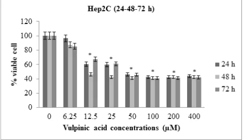
Seven vulpinic acid concentrations were used to treat CaCo2, HepG2, Hep2C, RD, Wehi, Vero and L929 cell lines for 24, 48 and 72 h. Compared with time-dependent manner of vulpinic acid effects, vulpinic acid signifi -cantly inhibited the growth ofall examined cells in a 48 h timeand dosedependent manner (Figure 1). As shown in Figure 2, vulpinic acid showed the best effects on CaCo2 cancer cell line from all studied cells (Figure 2). The highest rates of cell death were observed in approx -imately 12.5 μM concentration vulpinic acid for 48 h on CaCo2cancer cell line ranged from 87.4% (p<0.05) (Figure 2). The value of the IC50 of vulpinic acid for CaCo2, HepG2, Hep2C, RD, Wehicancer cell lines was determined to be 13.7, 23.8, 25.3, 34.4 and 38.6μM, respectively. Vulpinic acid was, for the first time,
Figure 1: Comparative representation of Hep2C cell line (values are statistically significant at * p < 0.05 respective
exposed to CaCo2, HepG2, Hep2C, RD, Wehi cancer cells, Vero and L929 normal cell lines in our study. This result suggests that vulpinic acid significantly inhibited the proliferation of cancer cell lines (p<0.05).As a more interesting result, the cytotoxic effect of vulpinic acid was on cancer cells and it had little cytotoxic effect on normal cells (p<0.05). The application of vulpinic acid did not have a significant decrease in cell viability on L929 and Vero normal cell lines at all increasing concen-tration/time options (p<0.05) (Figure 3). The viability of Vero and L929 normal cells only decreased 12% and 17%, respectively (p<0.05) (Figure 3).One of the most valuable results of this study is that vulpinic acid inhib-ited cancer lines but it had very little inhibiting effect on normal cells.
Effects of vulpinic acid on Bcl2 family members Previous studies have indicated that lichen secondary metabolites are induced by apoptosis of cancer cells.19 Firstly, in this study, the effects of vulpinic acid on molecular mechanisms associated with apoptosis in examined cancer cells and the expression of apoptosis related proteins were analyzed by western blot assay. The value of the IC50 of vulpinic acid demonstrated
increasing Bcl2, P53 and Bax protein levels in examined cancer cells in a 48 h concentrationdependent manner (p<0.05) (Figure 4). Bax expression was 12 fold higher in CaCO2 cells (p<0.05), 10 fold higher in HepG2 (p<0.05), 9 fold higher in Hep2C (p<0.05), 6 fold higher in RD (p<0.05), 5 fold higher in Wehi (p<0.05) than in the
Figure 2: Dose response curves for the effect of vulpinic acid on CaCo2 cell line for 48 h (values are statistically significant
at * p < 0.05 respective control).
Figure 4: Effects of vulpinic acid on apoptosis-related p53 protein in examined cell.
Figure 3: Dose response curves for the effect of vulpinic acid on Vero cell line for 48 h (values are statistically significant at
corresponding normal cells (Vero and L929) (p<0.05). Similarly, p53 expression was 5.2 fold higher in CaCO2 cells (p<0.05), 4 fold higher in HepG2 and Hep2C (p<0.05), approximately 3 fold higher in RD and Wehi cells (p<0.05) than in the VA non-exposed cells (Vero and L929) (p<0.05) (Figure 4). In contrast, Bcl-2 expres -sion was 2.3 fold lower in CaCO2 exposed vulpinic acid (p<0.05), approximately from 0.3 fold to 0.5 fold range on lower in examined all cancer cell lines (p<0.05). Secondly, the value of the IC50 of vulpinic acid was exposed to all examined cells for 48 h and total RNAs were isolated from cell lines. The RNA concentrations were in the range of 137 to 378ng/μL and 230/260 nm ratios were between 0.47 and 0.99. The integrity of the extracted RNAs was also evaluated by gel electrophoresis. The expression of apoptosis related genes (Bax, Bcl-2
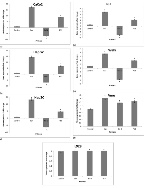
and P53) on cancer and normal cell lines was analyzed by qRT-PCR for the first time in this study. There are no available data in the literature on the effect of vulpinic acid on pro-apoptotic and anti-apoptotic gene expression in different cancer and normal cell lines. Therefore, qRT-PCR assay is demonstrated as one of the most important data explaining the exposure of apoptosis related molecular mechanisms to vulpinic acid on cancer and normal cell lines. Figure 5a-5g summarizes gene expression changes of Bax, Bcl-2 and P53. CaCo2, HepG2, Hep2C, RD and Wehi cancer cell lines exposed to vulpinic acid and the expression of Bax gene was increased by 17.2, 14.3, 13.2, 9.8 and 7.4 fold, respec -tively (p<0.05) (Figure 5a-5e). The expression levels of
Bax gene in Vero and L929 normal cell lines treated with vulpinic acid for 48 h were increased by 1.6 and 1.1 fold, respectively (p<0.05) (Figure 5f-5g). When cancer cell lines and normal cell lines exposed to vulpinic acid in respect to Bax gene expression were compared, an approximately from 7 and 17fold change was determined in Bax gene expression value (p<0.05) (Figure 5a-5g). The P53gene expression levels also increased in examined cell lines treated with vulpinic acid secondary metabolite. It is markedly inreased by 8.7 and 6.8fold on CaCo2 and HepG2 cancer cell line, respectively when compared with vulpinic acid untreated cells(p<0.05)(Figure 5a-5b). The transcriptome levels of P53 genes in the CaCo2 (8.7 fold change) were obviously higher than Vero and L929 normal cell lines (1.4 and 1.9 fold change) (p<0.05). In all cancer cell lines, few significant differences in the expression profiles of Bcl-2 were observed between the other apoptosis related genes (Bax and P53 genes). However Bcl-2 anti-apoptotic gene was 7 fold decreased in CaCo2 and HepG2 cell lines (p<0.05). Bcl-2was more highly expressed in the CaCo2 cancer cell line (7.3 fold change). There was a six and five fold decrease in the
expression of Bcl-2 gene in Hep2C, RD and Wehi cell lines, respectively (p<0.05).
The current study had similar observations to transcrip-tome changes of genes and protein levels. It is obvious that both mRNA and protein levels significantly increased in
Bax and P53 levels and decreased in Bcl-2 level (p<0.05) (Figure 4 and Figure 5a-5g).
DISCUSSION
In this study, vulpinic acid secondary metabolite had more significant cytotoxic effects on CaCo2, HepG2, Hep2C, RD, Wehi cancer cell lines (p<0.05). The effects of vulpinic acid on Bcl2 family members, mRNA and protein levels significantly increased in Bax and P53 levels (p<0.05). This is the first paper on the expression value of apoptosis related genes (Bax, Bcl-2 and P53) on cancer and normal cell lines exposed to vulpinic acid analyzed by qRT-PCR assay.
Karaosmanoğlu and Sivas (2015) investigated the inhibi -tory effect of vulpinic acid on the proliferation of HepG2 hepatocellular carcinoma cell line and F2408 mouse embryonic fibroblasts cell line by MTT assay. Although IC50 value of vulpinic acid was determined from 450 to 500 μM on F2408 normal cell line, the value of the IC50 of vulpinic acid showed a range of 300-400 μM on HepG2 cancer cell line for 48 h.23 In our study, we observed 23.8 µM value of the IC50 of vulpinic acid on HepG2 cancer cell line for 48 h. When the results of both studies were compared, we obtained lower amounts of the IC50 value in our study. As a result of the study, vulpinic acid will be used as a novel drug source in the pharmaceu-tical industry. Furthermore, Karaosmanoğlu and Sivas (2015) evaluated the apoptotic effect of vulpinic acid using AO/EB dye with fluorescence microscopy.23 In this study, we evaluated apoptotic effect of vulpinic acid a wider research at molecular level. Although the most gene expression fold change on CaCo2, HepG2, Hep2C, RD, Wehi cancer cells were shown in the Bax
gene, the lowest gene expression level was on L929 normal cell line. This study confirmed that vulpinic acid plays a major role in the apoptosis of cancer cells. Therefore, studies focusing on specific mechanisms at a molecular level have shown promising results for lichen secondary metabolite based drug therapies.
and western blot assay were applied for the determination of apoptotic effect on cancer cell lines. According to DNA fragment analysis, the intensity of DNA bands in gel decreased on cancer cell lines exposed to CFP-2. It was found that although Bcl-2 expression was at the same level withthe unexposed to CFL-2, Bax expression was increased in exposure to 300 mg/L CFP-2 applica -tion of the western blot assay.24 We also demonstrated the effect of vulpinic acid on cancer and normal cell with apoptosis related protein based data. Similar obser-vation with Lin et al. (2003) was obtained on increase of Bax protein by western blot assay for vulpinic acid (p<0.05). According to our qRT-PCR results, we also found that vulpinic acid was significantly increased in
Bax compared with P53 and Bcl-2 levels (p<0.05). The study of Lin et al. and our study showed comparable results regarding vulpinic acid as an alternative source by inducing apoptosis of cancer cells.
Liu et al. (2010) and Ren et al. (2009) also identified a mitochondria mediated apoptosis pathway by showing changes in ratio of Bax/Bcl-2 proteins after treatment with retigeric acid.25,26 They found retigeric acid was significantly changed in Bax and Bcl-2 levels. The studies by Liu et al., Ren et al. and our study provide similar Bax/Bcl-2 proteins results for an alternative source inducing apoptosis of cancer cells.
Hsu et al. (2015) emphasized several triterpenoids including botulin (K02) and its derivatives (K03, K04 and K06) extracted from Hibiscus syriacus inhibited breast cancer cell and migration.27 Moreover, they investigated cyto-toxic effects on normal mammalian epithelial cells and these molecules inhibited very little on normal cells. This study also showed that betulin and its derivatives induced apoptosis by activating apoptosis-related genes. These results indicate that in the MDA-MB-231 cells, K02 and K06 induced apoptotic gene expression in a Tap63 associated manner. In H184B5F5/M10 normal breast cell, the expression of Bax, Noxa, Puma and
Perp showed no change with K02, K03, K04 and K06 treatment. In this study, the examined cancer cell lines had the highest expression exposed to vulpinic acid, while the lowest expression of normal cell lines was observed. Our study revealed similar results to their studies. Similar observation was made by Zu et al. by qRT PCR for emodin.28 Zu et al. havefound emodin significantly increases in Bax levels and decreases in P53
and Bcl-2 (p<0.05). In our studies, vulpinic acid exposed cancer cells showed significantly higher Bax expression than normal cells. The study by Zu et al. and our study provide similar results for alternative candidate drug molecules on the industrial area. Alhazmi et al. (2014) examined molecular mechanism of NS induced apop
-tosis in MCF-7cell line.29Casp-3, Casp-8,Casp-9 and P53 gene expression changes were determined by dose-and time dependent NS. The expression levels of Casp-3, Casp-8, Casp-9 and P53 in MCF-7 cells treated with 100 μL/mL NS for 48 h markedly increased by 17.37, 4.82, 10.06 and 6.48 fold when compared with control untreated cells, respectively. In our study, Bax gene was the top significantly up-regulated gene at 17.2 fold in CaCo2 cancer cell line while Bcl-2 was the most signi-ficantly down-regulated gene at 7 fold in response to vulpinic acid on CaCo2 and Bcl-2 cell lines, respectively.
CONCLUSION
The present study demonstrates that vulpinic acid signi-ficantly inhibits the growth of cancer cells and provides underlying apoptosis mechanism as an anticancer activity. Vulpinic acid could potentially be an alternative source for cancer therapy in the pharmaceutical industry. Our study determined the expression changes of Bax, Bcl-2
and P53 genes and proteins on exposed cancer and normal cells after vulpinic acid application. The molecular mechanism of vulpinic acid secondary metabolite needs to be investigated in greater detail. Further molecular based research is required for vulpinic acid before devel-oping a promising chemotherapeutic agent for cancer treatment.
ACKNOWLEDGEMENT
We thank Ankara University Project Offices, Project no. 16H0415002 and Project no. 15B0415001 for the partly
financial support.
CONFLICT OF INTEREST
The authors have no conflict of interest to declare.
ABBREVIATIONS
VA: Vulpinic acid; DMSO: Dimethyl Sulfoxide;
qRT-PCR: Quantitative Real Time PCR.
REFERENCES
1. Arslan S, Bölükbaş N. Kanserli hastalarda yaşam kalitesinin değerlendirilmesi. Atatürk Üniv Hemş Yüks Derg. 2003;6(3):38-47.
2. Hanahan D, Weinberg RA. The hallmarks of cancer. Cell. 2000;100(1):57-70.
3. Hanahan D, Weinberg RA. Hallmarks of cancer. The Next Gen Cell. 2011;144(5):646-74.
4. Müller K. Pharmaceutically relevant metabolites from lichens. App Microbiol Biotechnol. 2001;56(1-2):9-16.
5. Singh N, Nambiar D, Kale RK, Singh RP. Usnic acid inhibits growth and induces cell cycle arrest and apoptosis in human lung carcinoma A549 cells. Nut Cancer. 2013;65(1):36-43.
7. Chen Z, Zhang L, Xia L, Jin Y, Wub Q, Guo H, et al. Genomic analysis of drug resistant gastric cancer cell lines by combining mRNA and microRNA expression profiling. Cancer Lett. 2014;350(1):43-51.
8. Huneck S, Yoshimura I. Identification of lichen substances. Springer, Philadelphia, 1996.
9. Mazza F. Constitution and physical properties of vulpinic acid. Zentralblatt Math. 1925;31:182-90.
10. Ren MR, Hur JS, Seo KI. Anti-proliferative effects of Lethariellazahlbruckneri
extracts in HT-29 human colon cancer cells. Food Chem. 2009;47(9):2157-62.
11. Rankovic BR, Kosanic MM, Stanojkovic TP. Antioxidant, antimicrobial and anticancer activity of the lichens Cladonia furcata, Lecanora atra and
Lecanora muralis. BMC Complement Altern Med. 2011;11(1):97.
12. Kosanic MM, Rankovic BR, Stanojkovic TP. Antioxidant, antimicrobial and anti cancer activities of three Parmelia species. J Sci Food Agri. 2012a;92(9):1909-16.
13. Kosanic M, Manojlovic N, Jankovic S. Everniaprunastri and Pseudoevernia furfuracea lichens and their major metabolites as antioxidant, antimicrobial and anticancer agents. FoodChem Toxicol. 2012b;53:112-8.
14. Rankovic B, Kosanic M, Manojlovic N. Chemical composition of Hypogymnia physodes lichen and biological activities of some its major metabolites. Med Chem Res. 2014;23(1):408-16.
15. Özenoğlu S, Aydoğdu G, Dinçsoy AB, Taghidizaj AA, Derici K, Yılmaz E,
et al. Evaluation of the impact on different types of human cancer cell of lichen secondary compounds. Turk Hij Den Biyol Derg. 2013;70(4):215-26. 16. Şekerli M, Kılıç N, Cansaran-Duman D. The Molecular Mechanisms of the
effect of anticancer activity on lichen metabolites. Turk Hij Den Biyol Derg. 2017;74(1):95-102.
17. Dinçsoy AB, Cansaran-Duman D. Changes in apoptosis-related gene expression profiles in cancer cell lines exposed to usnic acid lichen secondary metabolite. Turk J Biol. 2017;41:484-493.
18. Mayer M, O’Neill MA, Murray KE. Usnicacid: a non-genotoxic compound with anticancer properties. Antican Drugs. 2005;16(8):805-9.
19. Backorova M, Backor M, Mikes J. Variable responses of different human cancer cells to the lichen compounds parietin, atranorin, usnic acid and gyrophoric acid. Toxicol in vitro. 2011;25(1):37-44.
20. Burlando B, Ranzato E, Volante A, Appendino G, Pollastro F, Verotta L. Antiproliferative effects on tumour cells and promotion of keratinocyte wound healing by different lichen compounds. Planta Med. 2009;75(6):607-13.
21. Varol M, Türk A, Candan M, Tay T, Koparal T. Photoprotective activity of vulpinic and gyrophic acid toward ultraviolet B-induced damage in human keratinocytes. Phytoter Res. 2016;30(1):9-15.
22. Koparal AT. Anti-angiogenic and antiproliferative properties of the lichen substances (-) - usnic acid and vulpinic acid. Zeitschrfür Naturfor C. 2015;70(5-6):159-64.
23. Karaosmanoğlu O, Sivas H. The in vitro investigation of cytotoxic and apoptotic effects vulpinic acid on normal and cancer cell. J Biotech. 2015;208:S95.
24. Lin X, Cai YJ, Li ZX, Chen Q, Liu ZL. Wang R. Structure determination, apoptosis induction, and telomerase inhibition of CFP-2, a novel lichenin from Cladoniafurcate. Biochim Biophys Acta. 2003;1622(2):99-108. 25. Liu H, Liu YQ, Xu AH, Young CY, Yuan HQ, Lou HX. A novelanticanceragent,
retigeric acid B, displays proliferation inhibition, S phase arrest and apoptosis activation in human prostate cancer cells. Chem Biol Interact. 2010;188(3):598-606.
26. Ren MR, Hur JS, Kim JY, Park KW, Park SC, Seong CN, et al. Antiproliferative effects of Lethariella zahlbruckneri extracts in human HT-29 human colon cancer cells. Food Chem Toxicol. 2009;47(9):2157-62.
27. Hsu RJ, Hsu YC, Chen SP, Fu CL, Yu JC, Chang FW, Chen YH, et al. The
triterpenoids of Hibiscus syriacus induce apoptosis and inhibit cell migration in breast cancer cells. BMC Comp Altern Med. 2015;15(1):65.
28. Zu C, Zhang M, Xue H, Xiaopeng C. Emodin induces apoptosis of human breast cancer cells by modulating the expression of apoptosis-related genes. Oncol Lett. 2015;10(5):2919-24.
29. Alhazmi MI, Hasan TN, Shafi G, Al-Assaf AH, Alfawaz M.A, Alshatwi AA. Roles of p53 and caspases in induction of apoptosis in MCF-7 breast cancer cells treated with a methanolic extract of Nigella sativa seeds. Asi Pas J Canc Preven. 2014;15(22):9655-60.
SUMMARY
Researchers are increasingly studied on new effective candidate drug molecules from biological organisms. Lichens are known to produce secondary metabolites
that find uses as natural drug molecule. Lichen sec
-onder metabolites could play a significantly role in
especially cancer treatment because they are usually less toxic than traditional chemotherapy agents effec-tive, inexpensive and easily available. In this study, we evaluated cytotoxic effect of vulpinic acid and their apoptotic effect. As a result of this study, vulpinic acid could be play an important role in cancer treatment.
Nil Kılıç is PhD student at the Ankara University, Biotechnology Institute, Ankara-Turkey. Areas of interest: Development of the drug candidate molecules for treatment of cancer.
About Authors
PICTORIAL ABSTRACT
Cite this article: Kılıç N, Derici MK, Büyük İ, Aydın SS, Aras S, Cansaran-Duman D. Evaluation of in vitro Anticancer Activity of Vulpinic Acid and its Apoptotic Potential Using Gene Expression and Protein Analysis. Indian J of Pharmaceutical Education and Research. 2018;52(4):626-34.
İlker Büyük is Associate Professor at the Ankara University, Faculty of Science, Department of Biology, Ankara-Turkey. Areas of interest: Gene expression. Areas of interest: Gene expression.
Semra Soydam Aydın is Associate Professor at the Turkish Medicineand Medical Device Agency, Ministiry of Health, Ankara-Turkey.
Sümer Aras is Professor at the Ankara University, Faculty of Science, Department of Biology, Ankara-Turkey. Areas of interest: Gene expression.