1
Manuscript
1 2
Title:
3
Investigation of Brain Vascular Territories in Stroke Patients Detected Non-Valve Atrial 4
Fibrillation as an Etiological Factor 5
Mustafa Karaoglan1, Serkan Demir2 6
1Artvin State Hospital, Artvin-Turkey
7
2Sultan Abdulahid Han Research and Education Hospital, Department of Neurology,
İstanbul-8
Turkey 9
10
Abstract
11
Objective: It was aimed to investigate the cerebral vascular territories in stroke patients with 12
NVAF as an etiologic factor. 13
Material and Methods: A total of 104 patients who were referred to our hospital between 14
January 2015 and September 2016, who were over 55 years of age, identified or documented 15
as having a standard ECG or Holter ECG record on their medical history, and diagnosed with 16
stroke were included. Our study was designed as a retrospective analysis of prospective data. 17
Detailed history, physical examination and electrocardiography (ECG) evaluations of the 18
patients were performed. Descriptive statistics were used in the detection of findings, and t-19
test, Pearson-square test and Fisher's exact test were used for differences analysis. 20
Results: 53.8% (N = 56) of the patients were male and 46.2% (N = 48) were female. The 21
mean age was 73.5. MCA was the most common site of vascular involvement in NVAF-22
dependent strokes. In MCA vascular territory, ischemic infarcts were detected most frequently 23
in the upper and lower divisions. SCA and PCA followed MCA. Approximately 64% of the 24
NVAF-related strokes were anterior circulation infarction (ASE) and 22% were posterior 25
circulation infarct (PSE). There was a significant difference in age and past stroke history 26
factors in favor of ASE (p<0.05). There was no significant difference between ASE and PSE 27
in HT, cardiac history and DM factors (p>0.05). 28
Conclusion: It was emphasized that the area of the vessel that underwent ischemia in the 29
acutely displayed infarcts and the etiological factor for this vessel area could be predicted. 30
31
Key words: Brain vessel, ischemic stroke, non-valvular atrial fibrillation 32
33
2
Introduction
34
Non-valvular atrial fibrillation (NVAF) is an independent risk factor for ischemic stroke and 35
cardioembolic causes account for close to 20% of all ischemic stroke (1). Studies indicate that 36
the first stroke in the presence of atrial fibrillation (AF) is twice as fatal as in the absence of 37
AF, and the risk of recurrent stroke is higher in survivors. The prevalence of AF in people 38
over 65 years old is 5%. The annual risk of stroke in patients with AF is determined by the 39
CAHDVAS2C score. This rate is significantly increased with age, accompanying comorbid 40
diseases and especially stroke history. 41
Heart-borne emboli is the reason in two-thirds of patients with AF and ischemic stroke. When 42
the etiologic factor is NVAF in ischemic stroke, the superiority of anticoagulant therapy is 43
demonstrated in the preservation. Therefore, NVAF is an important etiologic factor for 44
ischemic stroke in terms of treatment and prognosis (2). In this study, it was aimed to 45
investigate the cerebral vascular territories in stroke patients who have non-valvular atrial 46
fibrillation as an etiologic factor and it is aimed to emphasize the importance of stroke in 47
patients with AF. 48
49
Material and Methods
50
Our study was designed as a retrospective analysis of prospective data. The patient population 51
was determined to be over 55 years old, who applied to [the name of the hospital will be 52
indicated after the referee evaluations] Education Research Hospital Neurology Department 53
between January 2015 and September 2016. A detailed history, physical examination and 54
electrocardiography (ECG) evaluations of 104 patients were performed. 55
Patients who were identified or documented as having AF case in their medical history, 56
standard ECG or Holter ECG record and who received a stroke diagnosis were included in the 57
study. Stroke diagnosis were admitted with clinical evaluations in patients whose symptoms 58
lasted longer than 24 hours. It was also accepted that only the ischemic region formed in the 59
brain by MRI diffusion was shown. The demographic characteristics of all patients were 60
adjusted with the CHADS VASC scores recommended for use in the European Society of 61
Cardiology (ESC) guidelines for atrial fibrillation published in 2010. 62
Risk factors for CHADS VASC score were age, gender, past infarction, diabetes mellitus 63
(DM), hypertension (HT) and cardiac history. As cardiac history; coronary artery disease, past 64
myocardial infarction, coronary artery bypass graft, and congestive heart failure were 65
3
once. Fetal posterior cerebral arteries were excluded from the study and vessel areas were 67
classified based on the relevant literature (3). 68
These vessels were defined as anterior cerebral artery (ACA), middle cerebral artery (MCA), 69
lenticulostriate artery (LSA), anterior choroidal artery (AchA), posterior cerebral artery 70
(PCA), vertebral artery (VA), posterior inferior cerebellar artery (PICA) and superior 71
cerebellar artery (SCA). MCA was classified as total MCA, MCA upper division, MCA lower 72
division, total MCA with deep branches and malign MCA with deep branches affected by 73
infarct areas. Border zone infarcts and subcortical lacunar infarcts were classified separately. 74
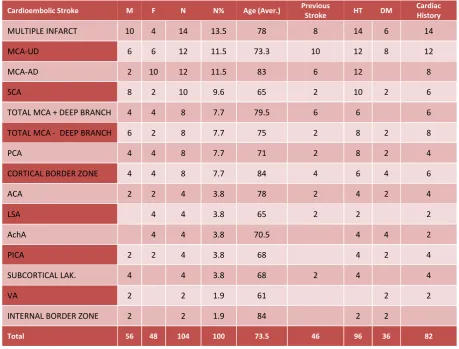
Patients with infarcts in more than one vascular territory at the same time were identified as 75
multiple infarcts (Figure 1). Descriptive statistics were used in the detection of findings, and t-76
test, Pearson-square test and Fisher's exact test were used for differences analysis. 77
This study was conducted with the ethical approval of HNEH-KAEK-2017/406 number and 78
dated 24.04.2017 issued by the Haydarpasa Numune Research And Education Hospital 79
Ethical committee. 80
81
Results
82
A total of 104 patients participated in our study. Of these patients, 53.8% (N = 56) were male 83
and 46.2% (N = 48) female. The mean age of the patients was 73.5. In addition, 44.2% (N = 84
46) of patients had previous stroke, 92.3% had HT, 34.6% had DM, and 78.8% had a cardiac 85
history (Figure 2). 86
As a result of the detailed evaluations made, the most common venous occlusion area was 87
determined as MCA in the strokes due to NVAF. In MCA vascular territory, ischemic infarcts 88
were detected most commonly in upper and lower divisions. SCA and PCA followed MCA. 89
The most important risk factor for multiple infarcts was age and the rate was 13.5%. All of the 90
seven patients had HT and cardiac history, and 5 patients had prior strokes. 91
A total of 10 patients with border zone infarcts had an average age of 84 years and 8 patients 92
had infarct cortical localization. Four of the 4 patients with subcortical lacunar infarcts had 93
HT history. Findings obtained as a result of our study are given in Table-1. 94
Approximately 64% of the NVAF-related strokes were anterior circulation infarction (ASE) 95
and 22% were posterior circulation infarction (PSE). The average age at ASE was 75 and 66 96
at PSE. The ratio of female to male in ASE was found to be 1.6, and this ratio was found to be 97
0.5 in PSE, and the difference was in favor of males. The stroke history in ASE was found to 98
4
While the rate of all patients with HT risk factor in their background was 92% in ASE and 100
91% in PSE, these rates for DM were identified 30% for ASE and 33% for PSE. The rate of 101
all patients with cardiac disease history was found 80% in ASE and 66% in PSE. While 102
female gender is preliminary for strokes formed in the ASE areas, there is male dominance in 103
PSE. While previous stroke history was frequent in ASE strokes, mean age in PSE strokes 104
was found younger than ASE (Table-2). 105
When Table-2 is examined; the factors of age and past stroke history were significantly 106
different in favor of ASE (p<0.05). However, there was no significant difference between 107
ASE and PSE in HT, cardiac history and DM factors (p>0.05). 108
109
Discussion
110
Atrial fibrillation is a known cardiac risk factor (2). Studies have shown that strokes which 111
develop in AF patients may be 2 times more mortal than non-AF patients (14,15). Especially 112
with the increase of the CHADVASC score, the risk of stroke in patients with AF is 113
increasing. It is suggested to fight with AF in all diagnosis and treatment guidelines for the 114
prevention of major strokes and mortality and morbidity that may develop. New-generation 115
anticoagulant agents have also been used in combination with warfarin, a vitamin K 116
antagonist, to combat AF (16). 117
Stroke may develop in patients with NWAF despite proper antithrombotic treatment. Emboli 118
originating from the left appendage is most often responsible for these patients’ infarctions 119
(10). There are larger particulate emboli in patients with AF compared to emboli that develop 120
secondary to carotid disease and are more prevalent as transient ischemic attack (TIA). They 121
cause large ischemic strokes (11). In addition to causing massive strokes, silent cerebral 122
infarction and TIA can also be seen (12,13). 123
In a study of Chung and colleagues with 2702 stroke patients, 15.6% of all strokes were 124
associated with AF and the most common vessel area was MCA. AF was detected as the 125
reason for 50% of SCA infarcts (5 of 10 patients) (1). In the evaluation of 1000 patients who 126
underwent their first stroke in the Lausanne stroke registry study, MCA was the most 127
common vessel of heart embolization (5). In the Besancon stroke registry, prospective 128
recordings of 2500 disease were also recorded, MCA is the most common embolization vessel 129
(6). Rovira et al. [7] also found similar findings in their studies of stroke involvement and 130
stroke mechanisms. The work of Stecco et al. (8) and Paciaroni (9) confirm all these studies. 131
5
Conclusion
133
We carried out this study with the aim of emphasizing the particular artery bed-related stroke 134
that the NWAF, which can cause great strokes, could develop. We also noted that stroke is a 135
disease that needs to be taken precautions. 136
As a result of our study, MCA was found as the most frequent vessel involvement area in 137
NVAF-related strokes, and SCA and PCA followed. In addition, about 64% of strokes with 138
NVAF are anterior circulation infarction (ASE) and 22% are posterior circulation infarction 139
(PSE) in our study. 140
Considering that some of the developed strokes are disabling strokes, the importance of 141
prognosis in AF treatment is once again revealed in our study. In particular, left hemisphere 142
MCA infarcts lead to right hemiparesis and limits the quality of life due to motor and sensory 143
aphasia. The most common vessel associated with NWAF is MCA, which is consistent with 144
the literature. However, the restrictive factors of our study were the lack of comparable 145
studies, being single-centered and retrospective, having a small number of patients and 146
reflecting a certain population. 147
In our study, it was predicted to determine the area of the vessel that underwent ischemia in 148
the acutely displayed infarcts. It was also predicted that the etiological factor for this area of 149
the vessel can be estimated. We think that further elaboration of these vascular territories and 150
a more detailed examination of risk factors may give us more information about the etiology. 151
Currently available classifications do not fully demonstrate the etiology of stroke and cause 152
recurrent strokes (4). Therefore, the vascular area and proper treatment options need to be 153
improved. 154
155
Acknowledgments: 156
The acknowledgment will be made after the referee evaluations. 157
158
Conflict of Interest:
159
There is no conflict of interest in the study. 160
161
References (Vancouver Style)
162
1- Chung JW, Park SH, Kim N, Kim WJ, Park JH, Ko Y, et al. Trial of ORG 10172 in Acute 163
Stroke Treatment (TOAST) classification and vascular territory of ischemic stroke lesions 164
6
2- Fuster V, Rydén LE, Cannom DS, Crijns HJ, Curtis AB, Ellenbogen KA, et al. 166
ACC/AHA/ESC 2006 guidelines for the management of patients with atrial fibrillation. 167
Circulation. 2006;114(7):e257-e354. 168
3- Tatu L, Moulin T, Vuillier F, Bogousslavsky J. Arterial territories of the human brain. 169
Manifestations of Stroke. 2012;30:99-110. 170
4- Gladstone DJ, Spring, M, Dorian P, Panzov V, Thorpe KE, Hall J, et al. Atrial fibrillation 171
in patients with cryptogenic stroke. N Eng J Med. 2014;370(26):2467-2477. 172
5- Bogousslavsky J, Van Melle G, Regli F. The Lausanne Stroke Registry: analysis of 1,000 173
consecutive patients with first stroke. Stroke. 1988;19:1083–1092. 174
6- Moulin T, Tatu L, Crépin-Leblond T, Chavot D, Bergès S, Rumbach L. The Besancon 175
Stroke Registry: An acute stroke registry of 2,500 consecutive patients. Eur Neurol 176
1997;38(1):10-20. 177
7- Rovira A, Grive E, Alvarez-Sabin J. Distribution territories and causative mechanisms of 178
ischemic stroke. Eur Radiol. 2005;15(3):416-426. 179
8- Stecco A, Quagliozzi M, Soligo E, Naldi A, Cassarà A, Coppo L, et al. Can neuroimaging 180
differentiate PFO and AF-related cardioembolic stroke from the other embolic sources? 181
Clinical-radiological correlation on a retrospective study. La radiologia medica. 182
2017;122(6):412-418. 183
9- Paciaroni M., Silvestrelli G, Caso V, Corea F, Venti M, Milia P, et al. Neurovascular 184
territory involved in different etiological subtypes of ischemic stroke in the Perugia Stroke 185
Registry. Eur Neurol. 2003;10(4):361-365. 186
10- Anderson DC, Kappelle LJ, Eliasziw M, Babikian VL, Pearce LA, Barnett HJM. 187
Occurrence of hemispheric and retinal ischemia in atrial fibrillation compared with carotid 188
stenosis. Stroke. 2002;33:1963. 189
11- Harrison MJ, Marshall J. Atrial fibrillation, TIAs and completed strokes. Stroke. 1984; 190
15:441. 191
12- Ezekowitz MD, James KE, Nazarian SM, Davenport J, Broderick JP, Gupta SR, et al. 192
Silent cerebral infarction in patients with nonrheumatic atrial fibrillation. The Veterans 193
Affairs Stroke Prevention in Nonrheumatic Atrial Fibrillation Investigators. Circulation. 1995; 194
92:2178. 195
13- Demir S, Ozdag MF, Kendirli MT, Togrol RE. What do anticoagulants say about 196
7
14- Lin HJ, Wolf PA, Kelly-Hayes M, Beiser AS, Kase CS, Benjamin EJ, et al. Stroke 198
severity in atrial fibrillation. The Framingham Study. Stroke. 1996;27(10):1760. 199
15- Lamassa M, Di Carlo A, Pracucci G, Basile AM, Trefoloni G, Vanni P, et al. 200
Characteristics, outcome, and care of stroke associated with atrial fibrillation in Europe: data 201
from a multicenter multinational hospital-based registry (The European Community Stroke 202
Project). Stroke. 2001;32(2):392. 203
16- Cohen AT, Hamilton M, Mitchell SA, Phatak H, Liu X, Bird A, et al. Comparison of the 204
novel oral anticoagulants apixaban, dabigatran, edoxaban, and rivaroxaban in the initial and 205
long-term treatment and prevention of venous thromboembolism: systematic review and 206
network meta-analysis. PLoS One. 2015;10(12):e0144856 207
208 209 210 211 212 213 214 215 216 217 218 219 220
8 231
232 233 234 235
236
Table and Legends
237
Table 1. Vascular fields and demographic data 238
M: Male, F: Female, N: Number of Patients, N%: Percentage of Patients 239
240 241
Table 2. ASE and PSE Comparison 242
Cardioembolic Stroke M F N N% Age (Aver.) Previous
Stroke HT DM
Cardiac History
MULTIPLE INFARCT 10 4 14 13.5 78 8 14 6 14
MCA-UD 6 6 12 11.5 73.3 10 12 8 12
MCA-AD 2 10 12 11.5 83 6 12 8
SCA 8 2 10 9.6 65 2 10 2 6
TOTAL MCA + DEEP BRANCH 4 4 8 7.7 79.5 6 6 6
TOTAL MCA - DEEP BRANCH 6 2 8 7.7 75 2 8 2 8
PCA 4 4 8 7.7 71 2 8 2 4
CORTICAL BORDER ZONE 4 4 8 7.7 84 4 6 4 6
ACA 2 2 4 3.8 78 2 4 2 4
LSA 4 4 3.8 65 2 2 2
AchA 4 4 3.8 70.5 4 4 2
PICA 2 2 4 3.8 68 4 2 4
SUBCORTICAL LAK. 4 4 3.8 68 2 4 4
VA 2 2 1.9 61 2 2
INTERNAL BORDER ZONE 2 2 1.9 84 2 2
Total 56 48 104 100 73.5 46 96 36 82
ASE (N=67) PSE (N=23) p
9
243
244
245
246
247 248
†: t-test result, ˟: chi-square result, ‡: Fisher’s Exact Test result 249
Figures and Legends
250 251
Female / Male Ratio 1.6 0.5 -
Previous Stroke History %53 %16 0.002˟
HT %92 %91 0.668‡
Cardiac Disease History %80 %66 0.181˟
10 252
11
a-Acute phase MCA Upper and Lower Division involvement (Deep Branch Protection) MRI 254
Diffusion Sequence 255
b-Subacute period total MCA involvement (Upper + Lower and Deep Branches) MRI 256
Diffusion Sequence 257
c-MRI Diffusion Sequence compatible with acute multiple infarct 258
d-MRI Diffusion Sequence Consistent with Acute Phase Subcortical Lacunar Infarct 259
e- MRI Diffusion Sequence compatible with Acute-Subacute period MCA-PCA common 260
irrigation area 261
f- MRI Diffusion Sequence compatible with Acute-Subacute period MCA-MCA Deep branch 262
common irrigation area 263
g-MRI Diffusion Sequence compatible with acute-subacute period PCA irrigation area 264
h- MRI Diffusion Sequence (including vertebral irrigation area) compatible with Acute-265
Subacute period PICA irrigation area 266
i- MRI Diffusion Sequence compatible with Acute-Subacute period MCA upper division 267
irrigation area 268
j- MRI Diffusion Sequence compatible with Acute-Subacute period MCA subdivision 269
irrigation area 270
271
272
12 274
Figure 2. Ratio of Risk Factors to Total Number of Patients (%) 275
53.8
46.2 44.2
92.8
34.6
78.8
0 25 50 75 100