Stromal-derived factor-1 (SDF-1) expression
during early chick development
RIZWAN REHIMI
1,2, NARGIS KHALIDA
1, FAISAL YUSUF
1, FANGPING DAI
1,
GABRIELA MOROSAN-PUOPOLO
1,2and BEATE BRAND-SABERI
1,*
1 Institute of Anatomy and Cell Biology, Department of Molecular Embryology and 2 Faculty of Biology, University of Freiburg, Freiburg, Germany
ABSTRACT Cell migration plays a fundamental role in a wide variety of biological processes including development, tissue repair and disease. These processes depend on directed cell migration along and through cell layers. Chemokines are small secretory proteins that exert their effects by activating a family of G-protein coupled receptors and have been shown to play numerous fundamental roles in the control of physiological and pathological processes during development and in adult tissues, respectively. Stromal-derived factor-1 (SDF-1/CXCL12), a ligand of the chemokine receptor, CXCR4, is involved in providing cells with directional cues as well as in controlling their proliferation and differentiation. Here we studied the expression pattern of SDF-1 in the developing chick embryo. We could detect a specific expression of SDF-1 in the ectoderm, the sclerotome, the intersomitic spaces and the developing limbs. The expression domains of SDF-1 reflect its role in somitic precursor migration and vessel formation in the limbs.
KEY WORDS:
SDF-1, CXCR4, cell migration, chick embryo, differentiation
Introduction
The role of cell migration is of paramount importance in numerous biological processes occuring during development and disease. During development, many cells actively migrate from one place to another where they differentiate into mature tissues and organs. Cell migration is a complex process involving dy-namic interactions between migrating cells and tissues through which they migrate. In order to migrate and invade a target tissue, cells change shape and adhesion properties. Cells are able to read the guidance cues provided by target tissues that tell them where to go and when to stop. There are some complex groups of regulators that direct cell movement by modulating adhesion, attraction and repulsion. The foremost regulators of chemo-attraction are members of the chemokine family (Luster 1998, Kim and Broxmeyer 1999). Chemokines are small secretory proteins that exert their effects by activating a family of seven-pass transmembrane G-protein coupled receptors that typically recog-nize a wide variety of ligands and have been shown to play numerous fundamental roles in the control of physiological and pathological processes. Due to the large number of chemokines and rapid progress within the field, many chemokines have been reported by different research groups and have been given
*Address correspondence to: Prof. Dr. Beate Brand-Saberi. Institute of Anatomy and Cell Biology, Department of Molecular Embryology, Albert-Ludwig University, Albertstrasse 17, 79104 Freiburg, Germany. Fax: +49-761-2035-387. e-mail: [email protected]
Accepted: 13th June 2007. Published online: 8th November 2007. Edited by: Patrick Tam.
0214-6282/2008/$35.00
© UBC Press Printed in Spain
multiple names. Chemokines are able to bind to multiple chemok-ine receptors, but until recently, stromal derived factor (SDF-1) was known to bind exclusively to chemokine receptor CXCR4 and
that in turn also binds solely to SDF-1. This however has changed after finding from the work of groups that could show that SDF-1 can also bind and signal through a previously known orphan receptor RDC1/CXCR7 (Balabanian et al., 2005;
Dambly-Chaudiere et al., 2007). These findings open up more possibilities
for SDF-1 signalling which as yet was only thought to occur via CXCR4.
The chemokine and its receptor pair, CXCR4/SDF-1 are well investigated in developmental process such as in vascularization, embryogenesis, T-cell activation and migration at sites of inflam-mation and T-Cell homing, hematopoiesis and HIV pathogenesis. (Murdoch 2000; Balkwill 2004). During brain development, SDF-1
plays a very critical role in several aspects such as cell migration and axon pathfinding, but recently its role to stimulate axonal branching and to regulate axonal patterning has been described (Pujol et al., 2005).
Stromal cell-derived factor (SDF-1) is a member of the CXC
Abbreviations used in this paper: SDF, stromal cell-derived factor.
subfamily of chemokines. SDF-1 is expressed
in many organs including bone, lung, liver, brain, thymus and lymph nodes (Shirozu et al.,
1995, Nagasawa et al., 1998), but it is mainly
produced by stromal cells such as osteo-blasts, fibroblasts and endothelial cells in the bone marrow (Yun et al., 2003). Initially, SDF-1 was cloned from murine bone marrow and
characterized as pre-B cells growth stimulat-ing factor (Nagasawa et al., 1994, Shirozu et al., 1995). Physiological functions of
chemokines have been described as SDF-1
plays a very important role in the migration of germ cells. During development these cells undergo a very specialized process of migra-tion from their site of origin to the future gonad. Chemokine CXCR4/SDF-1 mutant germ cells
in zebrafish are unable to migrate directly towards their target tissue from their site of origin, this result indicating that ligand SDF-1 gives the guiding cues to migratory germ cells at all stages of migration towards their target (Knaut et al., 2003). In SDF-1(-/-) mice, PGCs
migrate normally through tissues of embryos, but the numbers of PGCs in the gonads are remarkably reduced. These findings revealed an aberrant colonization of the gonads by germ cells lacking a functional chemokine ligand and receptor (Ara et al., 2003). Further
evidence for the role of this pair during germ cell migration in mice and avian comes from the work of Stebler and collegues, who could show the relevance of the SDF-1 expression
to the primodial germ cells migratory pathway (Stebler et al., 2004). The SDF-1\CXCR4 axis
is also required for normal myelopoiesis and lymphopoiesis (Murdoch 2000). Recently, we have reported the first use of a peptidic inhibi-tor for CXCR4 in the developing chick embryo
and observed that cells migrating from the dermomyotome into the limb bud acquire an angiogenic fate, whereas the myogenic fate is repressed, implying that SDF-1 signalling is
directly or indirectly involved in the myogenic determination of the CXCR4 expressing cells
in the limb (Yusuf et al., 2006).
Many severe defects have been observed during development in knockout studies of
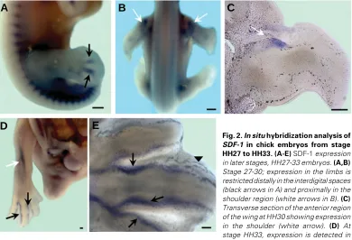
Fig. 1. In situ hybridization analysis of SDF-1 in chick embryo from stage HH3 to HH25. (A-O) Expression of SDF-1 in developing chick embryos. At stages HH3-5, strong expression is noted at the level of the primitive streak (white arrowheads in A and B). (C) Section of (A) at the level of the primitive streak, showing expression in the epiblast (black arrows). (E,F) Stages HH12-14. Prominent ectodermal expression can be observed in the caudal region of the embryo (black arrows), whereas more cranially, SDF-1 is not expressed. Also note the expression domains in the head region (white arrows). (D,G) Sections of stage HH12 and HH14 embryos respectively, showing SDF-1 signal overlying the paraxial mesoderm, intermediate mesoderm and medial parts of lateral plate (black arrows). However, the signal is missing in the ectoderm overlying the neural tube (red arrows). (J,K) Stages HH18+-20+; expression in the intersomitic spaces (green arrowhead) as well as in the branchial arches can be seen (white arrowhead in K). (H) Cross section of interlimb region of HH20+ showing a prominent expression domain around the dorsal aortic wall (green arrow) and sclerotome (white arrow). Sclerotomal staining also observed at HH25 (white arrowhead in N). (I,L,M,N) Stages 20+-25. Transcripts of SDF-1 are observable in the limbs (white arrow). (M) Expression domain in the cloacal region (green arrow). (O) Transverse section of limb, displaying restricted expression domains in the limb and mesenchyme (white arrows). The scale bar represents 100 µm in each photo.
O
B
C
D
E
F
H
I
J
K
L
A
M
N
CXCR4 or its ligand SDF-1,
such as cardiac ventricular defects, aberrations of intesti-nal vasculature and several affected hemopoietic compart-ments (Zou et al., 1998).
Ani-mals, deficient for either
CXCL12 or CXCR4, exhibit
severe aberration of cerebel-lar and hippocampal morphol-ogy, defects currently believed to result from the disconcerted migration of neuronal precur-sors (Zou et al., 1998, Zhu et al., 2002; Bagri et al., 2002; Lu et al., 2002). CXCL12 is also
known to modulate axonal pathfinding (Xiang et al., 2002;
Chalasani et al., 2003).
Interactions between SDF-1 and its receptor CXCR4 are
involved in cell progression and metastasis of several cers, including pancreatic can-cer (Koshiba et al., 2000),
kid-ney cancer (Schrader et al.,
2002), prostate cancer
B
C
D
E
A
(Taichman et al., 2002) lung and ovarian cancer (Scotton et al.,
2001, 2002; Kijima et al., 2002). Muller reported that CXCL12\CXCR4 have been associated with metastasis of breast
cancer (Muller and Neville 2001). In mice, the expression patterns of stromal cell-derived factor-1 and its receptor are also particu-larly noticeable in the developing cardiac, vascular and craniofa-cial system during organogenesis (McGrath et al., 1999). CXCR4
receptor is widely expressed in various tissues of the developing chick embryo such as in the ventral forgut portal, developing somites, tailbud, Wolffian duct, the lateral plate mesoderm and developing blood vessels (Yusuf et al., 2005). In our study we
documented the normal expression pattern of CXCL12 (SDF-1) in
chick embryos and we observed that SDF-1 is widely expressed
in most of the embryonic tissues during development such as in the ectoderm, the sclerotome, the intersomitic spaces and the mesenchyme of the developing limbs. Although the expression profile in SDF-1 has been reported in context of primordial germ
cell migration (Stebler et al., 2004), we present the expression
analysis of this important developmental gene in a broader context especially, highlighting its role in limb myogenesis and vasculogenesis and somitogenesis.
Results
Transcripts of SDF-1 can be detected as early as stage
HH3-5 in the chick embryos (Fig. 1A, B). The gastrulating embryo (Fig. 1A, B) stained for SDF-1 intensely in the epiblast
layer (Fig. 1C). At the level of the primitive streak, where the epiblast cells invaginate, staining is intense in the epiblast and mild in the underlying newly formed mesoderm (Fig. 1C), whereas more posteriorly, where gastrulation has not ad-vanced yet, the epiblast stains faintly for SDF-1 (not shown).
At stage HH12-15, the SDF-1 transcripts are to be found
predominantly in the ectoderm. Whole mounts for stages HH12 and HH14 show a prominent ectodermal staining in the caudal parts of the embryos and a faint signal in the head region is also detectable (Fig. 1 E, F). Sections revealed the ectodermal presence of SDF-1 signal overlying the paraxial mesoderm,
intermediate mesoderm and medial parts of the lateral plate mesoderm at stage HH12-14, however, the signal is missing in the ectoderm above the neural tube (Fig. 1D, G). Interestingly, the ectodermal presence of SDF-1 seems to follow a gradient
pattern along the antero-posterior axis. The unsegmented paraxial mesoderm is seen to be always covered by an SDF-1 expressing ectoderm, but more cranially, the ectoderm
over-lying mature epithelial somites is devoid of SDF-1 transcripts.
No SDF-1 expression can be seen in the underlying neural
tissue or mesodermal tissue at these stages (Fig. 1 D, G). A radical shift of SDF-1 expression pattern is observed from
stage HH18 onwards, where the transcripts are no longer detectable in the ectoderm, but are now predominantly seen in the mesodermal derivatives. At stage HH18+ whole mounts, one can detect a symmetrically arranged expression pattern in the intersomitic spaces and in the sclerotome, which is also visible on sections (Fig. 1J, H). High amounts of SDF-1
tran-scripts are also visible around the dorsal aorta wall (Fig. 1H). At HH20+, the intersomitic and sclerotomal expression persists (Fig. 1K), in addition, an expression domain in the branchial arches (Fig. 1K), in the limb bud (Fig. 1I, L, M) and the cloacal region is visible (Fig. 1M). The intersomitic expression domain persists at HH25 (Fig. 1N). The SDF-1 transcripts at HH25
of the distal 2/3 region of the limb (Fig. 1O).
As the limbs develop, the SDF-1 expression domain gets
restricted to the interdigital zones at HH27 (Fig.2 A) distally and proximally in the shoulder region at HH30 (Fig. 2B). Transverse section at the anterior end of the wing bud reveals the expression zone in the shoulder region (Fig. 2C). The interdigital expression domains at HH33 get limited to the perichondral tissue (Fig. 2 D, E). Control embryos hybridized using the sense probe showed no staining (data not shown).
Discussion
SDF-1 is highly conserved in the entire animal kingdom (Bleul et al., 1996). SDF-1 was till recently known to bind exclusively to
chemokine receptor, CXCR4, as its sole ligand (McGrath et al.,
1999, Murdoch 2000), however, finding have now shown that it also can bind and signal through RDC1/CXCR7. Stebler and
group have previously reported the functionally relevant expres-sion of SDF-1 and its receptor CXCR4 during primordial germ cell
migration in the chick embryo (Stebler et al., 2004). We now report
a more general expression analysis of SDF-1 during chick embryo
development, pointing towards its functional relevance to migra-tory cell populations. Our SDF-1 RNA probe is designed to bind
with SDF-1α mRNA. However as there is considerable homology between the 5’end of the SDF-1α and SDF-1β, it is difficult to discriminate between the expression of SDF-1α and SDF-1β due to the small splice specific probe lengths. Our probe therefore was able to detect the expression of both SDF-1α and SDF-1β.
In the developing embryo, CXCR4 has been described as the
most abundantly expressed chemokine receptor, the expression starting as early as gastrulation stages (McGrath et al., 1999). We
have previously reported the expression pattern of CXCR4 in the
developing chick embryo (Yusuf et al., 2005). In our present work,
we noticed that complementarity exists between the expression pattern of SDF-1 and CXCR4 during several stages of embryonic
development. This complementarity has also been reported by others in mouse and zebrafish embryos (McGrath et al., 1999;
Doitsidou et al., 2002; Vasyutina et al., 2005).
As in the case of the mouse embryos, the expression domains of SDF-1 and CXCR4 in the chick embryos also undergo a
dramatic profile change along the antero-posterior axis as devel-opment proceeds. At the start of gastrulation, SDF-1 transcripts
can be detected in the epiblast, whereas at these stages, the expression of CXCR4 although also present in the epiblast, is
mainly restricted to the invaginating mesoderm and endoderm at the level of the primitive streak. At around stage HH6, the mesodermal signal is more prominent as reported previously (Stebler et al., 2004). The expression domains of CXCR4 and SDF-1 in the gastrulating tissue point towards a probable autocrine
interaction between the ligand and receptor that guide the migrat-ing epiblast cells. Similar autocrine interactions between the two have been described during endothelial cell branching (Salvucci
et al., 2002). Once the three germ layers have been established
and the mesoderm starts to organize itself into paraxial, interme-diate and lateral plate mesoderm at HH14-16, the ectodermal expression of SDF-1 is maintained above the differentiating
mesoderm, which at this stage is positive for CXCR4 (McGrath et al., 1999; Yusuf et al., 2005). However, as the paraxial mesoderm
organizes to form epithelial somites, there is a progressive
de-crease of SDF-1 transcripts cranially in the overlying ectoderm.
As the CXCR4/SDF-1 axis has been implicated in cell migration,
it is probable that receptor-ligand interactions are needed for the cell movements that are occurring as the paraxial mesoderm arranges to give rise to epithelial somites. Similar interactions may also be active in the maturation of the kidney apparatus which develops from the intermediate mesoderm that expresses
CXCR4 (Yusuf et al., 2005).
A shift of expression domain from ectodermal to mesenchymal tissue similar to the one described in murine embryos was also observed in chick embryos (McGrath et al., 1999). From stage
HH18 onwards, there are no SDF-1 transcripts to be seen in the
overlying ectoderm and its derivative, the neural tube. SDF-1
expression is now evident in the sclerotome and mesenchymal tissue surrounding the aorta. At similar stages the expression profile of CXCR4 has now shifted to the neural tube as described
before (Yusuf et al., 2005; McGrath et al., 1999). The role of SDF-1 has been well investigated in endothelial cell organization and
vessel formation (Chen et al., 2007). Recently SDF-1 has also
been advocated as a probable candidate for therapeutic neovascularization (Zhou et al., 2007). SDF-1 transcripts could
also be detected in the lateral plate mesoderm and the genital ridge (data not shown) as published earlier (Stebler et al., 2004). SDF-1 was detected at sites of active vessel formation in the
intersomitic regions and the interdigital zones. We had earlier described streaks of CXCR4 positive cells moving into the limbs
from the intersomitic regions at stage HH19-20 (Yusuf et al.,
2005), at similar stages we also observe a faint SDF-1 staining in
the limb mesenchyme in Fig1 I and L. The SDF-1/CXCR4 axis has
been shown to induce chemotaxis and cell migration in several physiological and pathological situations during development and disease (Murdoch 2000). In context of the embryo, SDF-1
prob-ably functions in hand with other chemoattractants like Scatter Factor/Hepatocyte Growth Factor (SF/HGF) to make possible the
migration of the myogenic and angiogenic precursors into the limb mesenchyme. Recently, work from the lab of Carmen Birchmeier and from our lab has implicated SDF-1 signalling in muscle
patterning (Vasyutina et al., 2005; Yusuf et al., 2006).
SDF-1 expression was also observed in the dorsal aspect of the forelimb and cloacal region (data not shown) at HH 26-27, pointing towards a possible role in pectoral gridle and cloacal muscle formation.
Our expression analysis shows that SDF-1 has considerable
complementarity to the expression pattern of CXCR4 in chick
embryos. Furthermore, its expression pattern is highly suggestive for its role in cell migration and vasculogenesis during embryo-genesis and organoembryo-genesis.
Materials and Methods
Preparation of probe and in situ hybridization
Fertilized chicken eggs obtained from a local breeder were incubated at 38°C and 80% humidity. Embryos were staged according to Ham-burger and Hamilton (1995). After removing the extra-embryonic mem-brane, embryos were fixed overnight in 4% PFA at 4°C. For dehydration, embryos were entered into a series of different graded of methanol and were subsequently stored at -20°C or directly entered into the procedure of in situ hybridization (Nieto, et al., 1996) with SDF-1 antisense RNA
pair of SDF-1α gene specific primers (sense primer: 5’ GCCTGCACCGTCGCCAGAATG 3’; and antisense primer: 5’ AGGCCAACTCCAAACCCATCTTCA 3’). The RT-PCR product (425bp) was cloned into pDrive vector and confirmed by sequencing. For prepa-ration of the RNA antisense probe to detect SDF-1 gene expression
patterns in this study, the plasmid was linearized with Not I and synthe-sized with T7 RNA polymerase. The sense RNA probe for SDF-1 was synthesized as control. After the in situ hybridization experiment, the
embryos were analyzed and photographed. The embryos were further sectioned by using a Leica vibratome at a thickness of 35-60µm. For permanent slides, sections were embedded in Aquatex from Merck.
Acknowledgements
All authors would like to extend their thanks to Ellen Gimbel, Susanna Konradi and Ulrike Pein for their excellent technical assistances. We acknowledge the support of “molecular mechanisms of migration, inva-sion and metastasis”, a Baden-Württemberg grant, the Myores Project (511978) funded by the EU’s sixth framework Programme and Grako 1104.
References
ARA, T., NAKAMURA, Y., EGAWA, T., SUGIYAMA, T., ABE, K., KISHIMOTO, T., MATSUI, Y. and NAGASAWA, T. (2003). Impaired colonization of the gonads by primordial germ cells in mice lacking a chemokine, stromal cell-derived factor-1 (sdf-1). Proc Natl Acad Sci USA 100: 5319-23.
BAGRI, A., GURNEY, T., HE, X., ZOU, Y.R., LITTMAN, D.R., TESSIER-LAVIGNE, M. and PLEASURE, S.J. (2002). The chemokine sdf1 regulates migration of dentate granule cells. Development 129: 4249-60.
BALABANIAN, K., LAGANE, B., INFANTINO, S., CHOW, K.Y., HARRIAGUE, J., MOEPPS, B., ARENZANA-SEISDEDOS, F., THELEN, M. and BACHELERIE, F. (2005). The chemokine sdf-1/cxcl12 binds to and signals through the orphan receptor rdc1 in t lymphocytes. J Biol Chem 280: 35760-6.
BALKWILL, F. (2004). The significance of cancer cell expression of the chemokine receptor cxcr4. Semin Cancer Biol 14: 171-9.
BLEUL, C.C., FUHLBRIGGE, R.C., CASASNOVAS, J.M., AIUTI, A. and SPRINGER, T.A. (1996). A highly efficacious lymphocyte chemoattractant, stromal cell-derived factor 1 (sdf-1). J Exp Med 184: 1101-9.
CHALASANI, S.H., SABELKO, K.A., SUNSHINE, M.J., LITTMAN, D.R. and RAPER, J.A. (2003). A chemokine, sdf-1, reduces the effectiveness of multiple axonal repellents and is required for normal axon pathfinding. J Neurosci 23: 1360-71.
CHEN, T., BAI, H., SHAO, Y., ARZIGIAN, M., JANZEN, V., ATTAR, E., XIE, Y., SCADDEN, D.T. and WANG, Z.Z. (2007). Stromal cell-derived factor-1/cxcr4 signaling modifies the capillary-like organization of human embryonic stem cell-derived endothelium in vitro. Stem Cells 25: 392-401.
DAMBLY-CHAUDIERE, C., CUBEDO, N. and GHYSEN, A. (2007). Control of cell migration in the development of the posterior lateral line: Antagonistic interac-tions between the chemokine receptors cxcr4 and cxcr7/rdc1. BMC Dev Biol 7: 23.
DOITSIDOU, M., REICHMAN-FRIED, M., STEBLER, J., KOPRUNNER, M., DORRIES, J., MEYER, D., ESGUERRA, C.V., LEUNG, T. and RAZ, E. (2002). Guidance of primordial germ cell migration by the chemokine sdf-1. Cell 111: 647-59.
DUNUSSI-JOANNOPOULOS, K., ZUBEREK, K., RUNYON, K., HAWLEY, R.G., WONG, A., ERICKSON, J., HERRMANN, S. and LEONARD, J.P. (2002). Efficacious immunomodulatory activity of the chemokine stromal cell-derived factor 1 (sdf-1): Local secretion of sdf-1 at the tumor site serves as t-cell chemoattractant and mediates t-cell-dependent antitumor responses. Blood 100: 1551-8.
HONCZARENKO, M., LE, Y., GLODEK, A.M., MAJKA, M., CAMPBELL, J.J., RATAJCZAK, M.Z. and SILBERSTEIN, L.E. (2002). Ccr5-binding chemokines modulate cxcl12 (sdf-1)-induced responses of progenitor b cells in human bone marrow through heterologous desensitization of the cxcr4 chemokine receptor. Blood 100: 2321-9.
HORI, M., MITSUHASHI, S., KOBAYASHI, T., NAGASAWA, T., MORI, N., MIWA, M. and ABE, T. (1994). Cd30-positive lymphoma in human t-cell leukaemia virus
type 1 carrier without monoclonal integration of htlv-1. Br J Haematol 88: 419-20.
HOSHI, M., NAGASAWA, T., ABE, T., SATOH, T. and MITSUI, Y. (1994). Matura-tion of human megakaryocytic cell line (cmk-7) on the human umbilical vein endothelial cell (ec) monolayer. Int J Hematol 59: 191-9.
IWASAKI, Y., NIWA, S., NAKANO, H., NAGASAWA, T. and YAMANE, T. (1994). Purification and properties of phosphatidylinositol-specific phospholipase c from streptomyces antibioticus. Biochim Biophys Acta 1214: 221-8.
KIJIMA, T., MAULIK, G., MA, P.C., TIBALDI, E.V., TURNER, R.E., ROLLINS, B., SATTLER, M., JOHNSON, B.E. and SALGIA, R. (2002). Regulation of cellular proliferation, cytoskeletal function and signal transduction through cxcr4 and c-kit in small cell lung cancer cells. Cancer Res 62: 6304-11.
KIM, C.H. and BROXMEYER, H.E. (1999). Chemokines: Signal lamps for trafficking of t and b cells for development and effector function. J Leukoc Biol 65: 6-15.
KNAUT, H., WERZ, C., GEISLER, R. and NUSSLEIN-VOLHARD, C. (2003). A zebrafish homologue of the chemokine receptor cxcr4 is a germ-cell guidance receptor. Nature 421: 279-82.
KOJIMA, H., SUZUKAWA, K., YATABE, Y., HORI, M., NAGASAWA, T. and ABE, T. (1994). Establishment of a new natural killer (nk) cell line, tks-1, from a patient with aggressive type of large granular lymphocyte (lgl) leukemia. Leukemia 8: 1999-2004.
KOSHIBA, T., HOSOTANI, R., MIYAMOTO, Y., IDA, J., TSUJI, S., NAKAJIMA, S., KAWAGUCHI, M., KOBAYASHI, H., DOI, R., HORI, T. et al. (2000). Expression of stromal cell-derived factor 1 and cxcr4 ligand receptor system in pancreatic cancer: A possible role for tumor progression. Clin Cancer Res 6: 3530-5.
LAPIDOT, T. and KOLLET, O. (2002). The essential roles of the chemokine sdf-1 and its receptor cxcr4 in human stem cell homing and repopulation of trans-planted immune-deficient nod/scid and nod/scid/b2m(null) mice. Leukemia 16: 1992-2003.
LU, M., GROVE, E.A. and MILLER, R.J. (2002). Abnormal development of the hippocampal dentate gyrus in mice lacking the cxcr4 chemokine receptor. Proc Natl Acad Sci USA 99: 7090-5.
LUSTER, A.D. (1998). Chemokines—chemotactic cytokines that mediate inflam-mation. N Engl J Med 338: 436-45.
MCGRATH, K.E., KONISKI, A.D., MALTBY, K.M., MCGANN, J.K. and PALIS, J. (1999). Embryonic expression and function of the chemokine sdf-1 and its receptor, cxcr4. Dev Biol 213: 442-56.
MULLER, W.J. and NEVILLE, M.C. (2001). Introduction: Signaling in mammary development and tumorigenesis. J Mammary Gland Biol Neoplasia 6: 1-5.
MURDOCH, C. (2000). Cxcr4: Chemokine receptor extraordinaire. Immunol Rev 177: 175-84.
NAGASAWA, T., KIKUTANI, H. and KISHIMOTO, T. (1994). Molecular cloning and structure of a pre-b-cell growth-stimulating factor. Proc Natl Acad Sci USA 91: 2305-9.
NAGASAWA, T., TACHIBANA, K. and KISHIMOTO, T. (1998). A novel cxc chemokine pbsf/sdf-1 and its receptor cxcr4: Their functions in development, hematopoiesis and hiv infection. Semin Immunol 10: 179-85.
NIETO, M.A., PATEL, K. and WILKINSON, D.G. (1996). In situ hybridization analysis of chick embryos in whole mount and tissue sections. Methods Cell Biol 51: 219-35.
PUJOL, F., KITABGI, P. and BOUDIN, H. (2005). The chemokine sdf-1 differentially regulates axonal elongation and branching in hippocampal neurons. J Cell Sci 118: 1071-80.
RATAJCZAK, M.Z., ZUBA-SURMA, E., KUCIA, M., RECA, R., WOJAKOWSKI, W. and RATAJCZAK, J. (2006). The pleiotropic effects of the sdf-1-cxcr4 axis in organogenesis, regeneration and tumorigenesis. Leukemia 20: 1915-24.
SALVUCCI, O., YAO, L., VILLALBA, S., SAJEWICZ, A., PITTALUGA, S. and TOSATO, G. (2002). Regulation of endothelial cell branching morphogenesis by endogenous chemokine stromal-derived factor-1. Blood 99: 2703-11.
SCHRADER, A.J., LECHNER, O., TEMPLIN, M., DITTMAR, K.E., MACHTENS, S., MENGEL, M., PROBST-KEPPER, M., FRANZKE, A., WOLLENSAK, T., GATZLAFF, P. et al. (2002). Cxcr4/cxcl12 expression and signalling in kidney cancer. Br J Cancer 86: 1250-6.
tumours. Br J Cancer 85: 891-7.
SCOTTON, C.J., WILSON, J.L., MILLIKEN, D., STAMP, G. and BALKWILL, F.R. (2001). Epithelial cancer cell migration: A role for chemokine receptors? Cancer Res 61: 4961-5.
SCOTTON, C.J., WILSON, J.L., SCOTT, K., STAMP, G., WILBANKS, G.D., FRICKER, S., BRIDGER, G. and BALKWILL, F.R. (2002). Multiple actions of the chemokine cxcl12 on epithelial tumor cells in human ovarian cancer. Cancer Res 62: 5930-8.
SHIROZU, M., NAKANO, T., INAZAWA, J., TASHIRO, K., TADA, H., SHINOHARA, T. and HONJO, T. (1995). Structure and chromosomal localization of the human stromal cell-derived factor 1 (sdf1) gene. Genomics 28: 495-500.
STEBLER, J., SPIELER, D., SLANCHEV, K., MOLYNEAUX, K.A., RICHTER, U., COJOCARU, V., TARABYKIN, V., WYLIE, C., KESSEL, M. and RAZ, E. (2004). Primordial germ cell migration in the chick and mouse embryo: The role of the chemokine sdf-1/cxcl12. Dev Biol 272: 351-61.
STUMM, R.K., RUMMEL, J., JUNKER, V., CULMSEE, C., PFEIFFER, M., KRIEGLSTEIN, J., HOLLT, V. and SCHULZ, S. (2002). A dual role for the sdf-1/cxcr4 chemokine receptor system in adult brain: Isoform-selective regulation of sdf-1 expression modulates cxcr4-dependent neuronal plasticity and cere-bral leukocyte recruitment after focal ischemia. J Neurosci 22: 5865-78.
TAICHMAN, R.S., COOPER, C., KELLER, E.T., PIENTA, K.J., TAICHMAN, N.S. and MCCAULEY, L.K. (2002). Use of the stromal cell-derived factor-1/cxcr4 pathway in prostate cancer metastasis to bone. Cancer Res 62: 1832-7.
VASYUTINA, E., STEBLER, J., BRAND-SABERI, B., SCHULZ, S., RAZ, E. and BIRCHMEIER, C. (2005). Cxcr4 and gab1 cooperate to control the development
of migrating muscle progenitor cells. Genes Dev 19: 2187-98.
XIANG, Y., LI, Y., ZHANG, Z., CUI, K., WANG, S., YUAN, X.B., WU, C.P., POO, M.M. and DUAN, S. (2002). Nerve growth cone guidance mediated by g protein-coupled receptors. Nat Neurosci 5: 843-8.
YUN, H.J. and JO, D.Y. (2003). Production of stromal cell-derived factor-1 (sdf-1)and expression of cxcr4 in human bone marrow endothelial cells. J Korean Med Sci 18: 679-85.
YUSUF, F., REHIMI, R., DAI, F. and BRAND-SABERI, B. (2005). Expression of chemokine receptor cxcr4 during chick embryo development. Anat Embryol (Berl) 210: 35-41.
YUSUF, F., REHIMI, R., MOROSAN-PUOPOLO, G., DAI, F., ZHANG, X. and BRAND-SABERI, B. (2006). Inhibitors of cxcr4 affect the migration and fate of cxcr4+ progenitors in the developing limb of chick embryos. Dev Dyn 235: 3007-15.
ZHOU, B., HAN, Z.C., POON, M.C. and PU, W. (2007). Mesenchymal stem/stromal cells (msc) transfected with stromal derived factor 1 (sdf-1) for therapeutic neovascularization: Enhancement of cell recruitment and entrapment. Med Hypotheses 68: 1268-71.
ZHU, Y., YU, T., ZHANG, X.C., NAGASAWA, T., WU, J.Y. and RAO, Y. (2002). Role of the chemokine sdf-1 as the meningeal attractant for embryonic cerebellar neurons. Nat Neurosci 5: 719-20.
ZOU, Y.R., KOTTMANN, A.H., KURODA, M., TANIUCHI, I. and LITTMAN, D.R. (1998). Function of the chemokine receptor cxcr4 in haematopoiesis and in cerebellar development. Nature 393: 595-9.
Related, previously published Int. J. Dev. Biol. articles
See our Special Issue Invasion in Cancer and Embryonic Development edited by Marc Mareel and Juan Aréchaga at: http://www.ijdb.ehu.es/web/contents.php?vol=48&issue=5-6
See our Special Issue Mammalian Reproduction & Development in honor of Anne McLaren and edited by Brigid Hogan at: http://www.ijdb.ehu.es/web/contents.php?vol=45&issue=3
Primordial germ cell migration
Kathleen Molyneaux and Christopher Wylie Int. J. Dev. Biol. (2004) 48: 537-543
The chemokine network in cancer - much more than directing cell movement
Hagen Kulbe, Neil R. Levinson, Fran Balkwill and Julia L. Wilson
Int. J. Dev. Biol. (2004) 48: 489-496
Collective cell migration in morphogenesis and cancer
Peter Friedl, Yael Hegerfeldt and Miriam Tusch Int. J. Dev. Biol. (2004) 48: 441-449