Arve Opheim, PT, PhD
Anna Danielsson, PT,
PhD
Margit Alt Murphy, PT,
PhD
Hanna C. Persson, PT,
MSc
Katharina Stibrant
Sunnerhagen, MD,
PhD
Correspondence to Dr. Opheim: [email protected]
Supplemental data at Neurology.org
Early prediction of long-term upper limb
spasticity after stroke
Part of the SALGOT study
ABSTRACT
Objective:
To identify predictors and the optimal time point for the early prediction of the presence
and severity of spasticity in the upper limb 12 months poststroke.
Methods:
In total, 117 patients in the Gothenburg area who had experienced a stroke for the first
time and with documented arm paresis day 3 poststroke were consecutively included. Assessments
were made at admission and at 3 and 10 days, 4 weeks, and 12 months poststroke. Upper limb
spasticity in elbow flexion/extension and wrist flexion/extension was assessed with the modified
Ashworth Scale (MAS). Any spasticity was regarded as MAS
$
1, and severe spasticity was
re-garded as MAS
$
2 in any of the muscles. Sensorimotor function, sensation, pain, and joint range
of motion in the upper limb were assessed with the Fugl-Meyer assessment scale, and, together with
demographic and diagnostic information, were included in both univariate and multivariate logistic
regression analysis models. Seventy-six patients were included in the logistic regression analysis.
Results:
Sensorimotor function was the most important predictor both for any and severe
spastic-ity 12 months poststroke. In addition, spasticspastic-ity 4 weeks poststroke was a significant predictor
for severe spasticity. The best prediction model for any spasticity was observed 10 days
post-stroke (85% sensitivity, 90% specificity). The best prediction model for severe spasticity was
observed 4 weeks poststroke (91% sensitivity, 92% specificity).
Conclusions:
Reduced sensorimotor function was the most important predictor both for any and
severe spasticity, and spasticity could be predicted with high sensitivity and specificity 10 days
poststroke.
Neurology®2015;85:873–880GLOSSARY
ADL5activities of daily living;ARAT5Action Research Arm Test;CI5confidence interval;FMA-UE5Fugl-Meyer Assess-ment Upper Extremity Scale;MAS5modified Ashworth Scale;NIHSS5NIH Stroke Scale;NLR5negative likelihood ratio;
PLR5positive likelihood ratio;ROM5range of motion;SALGOT5Stroke Arm Longitudinal Study at the University of Gothenburg.
Upper limb spasticity has been found to be associated with reduced arm function and low levels of
independence, and with a 4-fold increase in direct care costs during the first year poststroke.
1–5The
prevalence of upper limb spasticity in all patients 12 months poststroke varies from 17% to 38%
6–10and was found to be 46% in patients with initial impaired arm function.
5It has been found that 4%
–
13% of patients need treatment for spasticity 6
–
12 months poststroke.
6,9Previous studies during the
first 10 days poststroke have identified several predictors for spasticity 3
–
12 months poststroke, e.g.,
reduced sensorimotor function and activities of daily living (ADL), muscle weakness, left-sided paresis,
and smoking.
8,9,11,12These studies were relatively small (n
5
47),
9assessed patients several days after
stroke onset,
11or assessed spasticity in both upper and lower limbs. Whether early assessments of
upper limb function and impairments can predict the occurrence and degree of upper limb spasticity
12 months poststroke with good accuracy is uncertain. The optimal time for early prediction of upper
limb spasticity 12 months poststroke is also unknown.
12This information is of clinical relevance, as
patients with an increased risk of developing spasticity-related impairments, complications, and
From the Institute of Neuroscience and Physiology (A.O., A.D., M.A.M., H.C.P., K.S.S.), Rehabilitation Medicine, Sahlgrenska Academy, University of Gothenburg, Sweden; and Sunnaas Rehabilitation Hospital (A.O.), Nesoddtangen, Norway.
Go to Neurology.org for full disclosures. Funding information and disclosures deemed relevant by the authors, if any, are provided at the end of the article. The Article Processing Charge was paid by the authors.
increased disability may be identified.
1,13The
study aims were to identify predictor variables
and the optimal time for early prediction of
any spasticity and severe spasticity in the upper
limb 1 year poststroke.
METHODS All patients with first-ever stroke in an 18-month period in 2009–2010 who were admitted to the largest of 3 acute stroke units at the Sahlgrenska University Hospital, Gothenburg, Sweden, within 3 days after stroke onset were eligible for consecutive screening for inclusion in the present study, which was a part of the Stroke Arm Longitudinal Study at the University of Gothenburg (SALGOT). In the SALGOT study, the recovery of upper extremity function was investigated in a nonselected sample during the first year poststroke.14All included patients
had ischemic or hemorrhagic stroke15for the first time, were
over 18 years old, and had impaired upper extremity function, which was assessed at day 3 with the Action Research Arm Test (ARAT) (0–57)16and defined as,57 points. The study sample
size estimation (n 5 88) for SALGOT was to determine a medium change of 6 points (10%) on ARAT, with a power of 0.8 and a significance level of 0.05. With an expected dropout rate of 30%, the aim was to include 120 patients.14
Standard protocol approvals, registrations, and patient consents.Study approval was provided by the Regional Ethics Committee of the Western region of Sweden (Registration num-ber 225/08), and written informed consent was obtained. The study is registered at www.clinicaltrials.gov (NCT 01115348).
Assessment procedure. In SALGOT, the patients were as-sessed 9 times during the first year: at admission; at 3 and 10 days; at 3, 4, and 6 weeks; and at 3, 6 and 12 months poststroke. In the current study, data from admission, 3 and 10 days, 4 weeks, and 12 months were used. Predominantly, the assessments were car-ried out by 3 physiotherapists and were performed according to a standardized protocol.14A majority of the assessments were
per-formed at the university hospital. If traveling was not possible for the patient, the assessments were conducted in the patient’s home, nursing home, or rehabilitation unit.
Variables.Predictor variables collected at admission (day 0).
Clinical characteristics and assessments routinely registered at admission during the acute stage of stroke were selected as poten-tial predictor variables (age, sex, ischemic or hemorrhagic stroke, side of stroke, and smoking in the last 3 months). Stroke locali-zation was classified using the Oxfordshire Classification17and
ischemic stroke was classified after cause of lesion using the Trial of Org 10172 in Acute Stroke Treatment criteria.18The initial
severity of stroke and arm paresis was assessed with the 0- to 42-point ordinal NIH Stroke Scale (NIHSS) and the NIHSS arm subscale (0–4), respectively.19NIHSS arm was treated as a
cate-gorical variable with 0 as the reference category.
Predictor variables collected at 3 and 10 days and 4 weeks poststroke.Common clinical assessment scales of sensorimotor impairments assessed at 3 and 10 days and 4 weeks poststroke were selected as potential predictors. Sensorimotor function in the upper limb was assessed with the motor function part (sec-tions A–D) of the Fugl-Meyer Assessment Upper Extremity Scale (FMA-UE).20The FMA-UE (sections A–D) includes 33
active motor function tests, where a higher score indicates a better performance (0–66). The nonmotor domains of the same scale (sections H–J) were used to assess sensation (0–12), joint pain (0– 24), and range of motion (ROM) during passive joint motions
(0–24); lower scores indicate reduced sensation, more pain, and reduced ROM, respectively. Spasticity in elbow flexors, elbow extensors, wrist flexors, and wrist extensors were assessed with the 6-level modified Ashworth Scale (MAS).21The original MAS
categories were reordered into integers between 0 and 5, to incorporate the score 11. MAS were dichotomized, and spasticity was considered to be present if the MAS score was$1 in any of these muscle groups.
Dependent variables.At 12 months poststroke, spasticity was reassessed in a similar way as previously. Any spasticity was considered if the MAS score was$1, and severe spasticity was considered if the MAS score was $2 in any of the muscle groups.22
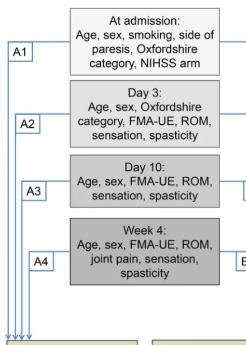
Statistical methods.Continuous and normally distributed var-iables are presented with means and SDs. Ordinal and non-normally distributed variables are presented with medians and 1st and 3rd quartile (Q1–Q3). Univariate logistic regression analyses were used to assess the relationship between the potential predictor variables and the outcome variable. The predictor variables were tested for correlations, and when 2 variables had a high correlation (r . 0.8), one was omitted before multivariate logistic regression analysis. The multivariate logistic regression analyses were used to predict the presence of (A) any spasticity and (B) severe spasticity at 12 months poststroke. In both (A) and (B), 4 models (A1–A4 and B1–B4) were found and compared (figure 1). The criteria for including a potential predictor variable in the multivariate logistic regression analyses were as follows: (1) significant predictor identified in previous studies, with the condition that if these variables were not found predictive in models A/B1, they were not included in models A/ B2–4, and (2) univariate logistic regressionpvalue,0.20. Age and sex were included in all models. The variables in the multivariate logistic regression analysis are shown in figure 1.
In the multivariate logistic regression analysis, the enter method was used stepwise and nonsignificant variables were removed manually one at the time, to ensure that only significant variables (p,0.05) were included in the final model. To control for possible nonlinearity between summed ordinal predictors and the dependent variable, a multivariate model with the squared predictor variables (FMA-UE2and Sensation2) was tested.
Non-significant associations were interpreted as nonlinearity not shown.23The results are presented with unstandardized
coeffi-cients,pvalues, and odds ratios with 95% confidence intervals (95% CIs).24The sensitivity (%), specificity (%), positive
likeli-hood ratio (PLR), negative likelilikeli-hood ratio (NLR), and the cor-responding 95% CI for each of the models was calculated using MedCalc for Windows, version 12.7.7.0 (MedCalc Software, Ostend, Belgium). All other statistical calculations were per-formed using IBM SPSS statistics 21.0 (IBM, Armonk, NY). The Strengthening the Reporting of Observational Studies in Epidemiology (STROBE) guidelines were followed.25,26
In total, 76 patients were assessed 12 months
post-stroke (35% dropout) and included in the logistic
regression analysis. The 2 main causes for dropout
were death (n
5
14) and study withdrawal (n
5
7).
The clinical characteristics of the 76 patients included
in the logistic regression analysis are shown in table 1.
At 3 and 10 days and 4 weeks, 24%, 43%, and 46%
of the 76 patients were assessed with any spasticity,
respectively. At 12 months, 46% and 29% were assessed
as having any spasticity and severe spasticity, respectively.
Prediction of any spasticity 12 months poststroke.
The
univariate logistic regression analysis for all potential
predictor variables for any spasticity and severe
spas-ticity is presented in tables e-1 and e-2 on the
Neu-rology
®Web site at Neurology.org. The multivariate
logistic regression analysis results are presented in
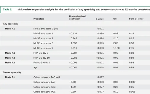
table 2. In models A2 and A3, the FMA-UE was a
significant predictor. In model A4, age at stroke onset
was a significant predictor in addition to FMA-UE,
and higher FMA-UE scores and higher age were
associated with reduced probability for spasticity.
The sensitivity, specificity, PLR, and NLR of model
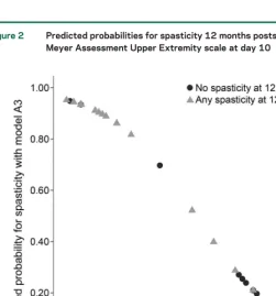
A3 indicated that this model had the highest
predictive value (table 3). The fit of prediction
model A3 was assessed in a scatterplot of the
predicted probabilities in relation to FMA-UE at
day 10 (figure 2). The squared FMA-UE variable
(FMA-UE
2) was not significant in models A2
–
A4,
and nonlinearity could not be shown.
Prediction of severe spasticity 12 months poststroke.
The
results from multivariate logistic regression models
B1
–
B4 are shown in table 2. Sensorimotor function
(FMA-UE) was a significant predictor in all models,
except B1. Lower FMA-UE scores (models B2
–
B4)
were associated with an increased predicted
probability for severe spasticity. In model B2
reduced sensation and in model B4 spasticity at 4
weeks poststroke were additional significant predictors.
The sensitivity, specificity, PLR, and NLR of model
B4 indicated that this model had the highest
predictive value (table 3). The squared variables
(FMA-UE
2and Sensation
2) were not significant
predictors in any of the models B2
–
B4, thus
nonlinearity could not be shown.
DISCUSSION
The present study demonstrated that
any spasticity was best predicted with variables
col-lected at day 10 poststroke and severe spasticity was
best predicted with variables collected 4 weeks
post-stroke. Lower sensorimotor function score, as
identi-fied with the FMA-UE, consistently and significantly
predicted both any spasticity and severe spasticity at
12 months poststroke. Age at stroke onset was a
significant predictor 4 weeks poststroke, with higher
age
associated
with
reduced
probability
for
spasticity. The presence of upper limb spasticity 4
weeks poststroke was a significant predictor for
severe spasticity.
These findings support previous studies reporting
paresis and reduced ADL function to be significant
predictors for spasticity poststroke.
8,11,12However,
none of the previous studies used the FMA-UE to
assess sensorimotor function, and thus, a direct
com-parison cannot be made with those studies.
Addition-ally, none of the former studies compared different
prediction models for spasticity at 12 months
post-stroke based on the assessments at different time
points relatively early after stroke onset, as was
per-formed in the present study. Predicting upper limb
spasticity with a relatively high accuracy based on
early assessments may have high clinical relevance,
as patients with an increased risk of developing
spasticity-related impairments may be identified early
and monitored more closely in order to implement
appropriate interventions.
In the present study, patients with higher age were
predicted to have reduced probability for spasticity.
This finding may be in accordance with a previous
Figure 1 The multivariate logistic regression analyses models
At day 3, Oxfordshire category and spasticity was included only in model B2. FMA-UE5
study
22where more severe spasticity was found in
younger patients 3 months poststroke, but not 18
months poststroke. Muscle force generation from
tendon reflexes has been found to be slower and
weaker with increasing age
27and if this also is the case
for tonic reflexes associated with spasticity, spastic
responses may be weaker in older patients.
In the present study, there was no association
between the side of weakness and spasticity 1 year after
stroke, as described previously.
12There may be several
reasons for this discrepancy; for instance, there were
methodologic differences between the studies, as the
Tone Assessment Scale was used to assess spasticity,
and spasticity and weakness were assessed in both arms
and legs
12as opposed to only upper limb spasticity and
sensorimotor function in the present study. An
associ-ation between smoking and severe spasticity has been
found previously,
12which could not be confirmed
here. The authors discussed that their model, which
included smoking, had an event per variable of 7,
com-pared to the recommended minimum of 10.
12,28Model B1 showed that patients with lacunar
stroke had a lower OR for severe spasticity than those
with other stroke locations. Therefore, the Oxford
Table 1 Demographic and clinical data of the patients included in the logistic regression analysis (n576)
Characteristics Values
M/F, % 60/40
Age, y, mean (SD) 67.2 (12.0)
Ischemic/hemorrhagic stroke, % 82/18
Paretic arm, R/L, % 43/57
Side of lesion, R/L/bilateral/cerebellum, % 54/41/4/1
Stroke localization, Oxfordshire classification, TAC/LAC/PAC/POC, % 12/29/51/8
Ischemic stroke subtypes, TOAST (n562), LAA/CE/SAO/OC/UND, % 16/31/29/13/11
NIHSS, arm, median (Q1–Q3)a 2 (1–4)
NIHSS, total, median (Q1–Q3)a 7 (3–12)
Smoking, no/yes, %a 77/23
Days at stroke unit, mean (SD) 12.4 (7.3)
Admitted to inpatient rehabilitation, n 51
Botulinum toxin treatment for spasticity, n 9
Abbreviations: CE5cardioembolism; LAA5large artery atherosclerosis; LAC5lacunar anterior circulation; OC 5 other determined cause; PAC 5partial anterior circulation; POC5posterior circulation; Q1-Q351st and 3rd quartile; SAO5small vessel occlusion; TAC5total anterior circulation; TOAST5Trial of Org 10172 in Acute Stroke Treatment; UND5undetermined cause.
aAt admission.
Table 2 Multivariate regression analysis for the prediction of any spasticity and severe spasticity at 12 months poststroke
Predictors
Unstandardized
coefficient pValue OR 95% CI lower 95% CI upper Any spasticity
Model A1 NIHSS arm, score 0 (ref) 0.001
NIHSS arm, score 1 20.134 0.888 0.88 0.14 5.58
NIHSS arm, score 2 0.742 0.494 2.10 0.25 17.59
NIHSS arm, score 3 1.030 0.325 2.80 0.36 21.73
NIHSS arm, score 4 2.911 0.003 18.38 2.75 122.94
Model A2 FMA-UE day 3 20.087 ,0.001 0.92 0.89 0.95
Model A3 FMA-UE day 10 20.083 ,0.001 0.92 0.89 0.95
Model A4 FMA-UE week 4 20.092 ,0.001 0.91 0.88 0.95
Age 20.061 0.044 0.94 0.89 0.99
Severe spasticity
Model B1 Oxford category, TAC (ref) 0.027
Oxford category, LAC 23.00 0.003 0.05 0.007 0.37
Oxford category, PAC 21.39 0.077 0.25 0.05 1.16
Oxford category, POC 22.30 0.077 0.10 0.008 1.29
Model B2 FMA-UE day 3 20.128 0.005 0.88 0.81 0.96
Sensation day 3 20.164 0.045 0.85 0.72 0.99
Model B3 FMA-UE day 10 20.164 0.024 0.85 0.74 0.98
Model B4 FMA-UE week 4 20.111 0.001 0.90 0.84 0.96
Spasticity week 4 3.42 0.009 30.62 2.34 401.5
Abbreviations: CI5confidence interval; FMA-UE5Fugl-Meyer Assessment Upper Extremity scale; LAC5lacunar anterior circulation; NIHSS5NIH Stroke Scale; OR5odds ratio; Oxford5Oxfordshire Stroke Classification; PAC5partial anterior circulation; POC5posterior circulation; ref5reference category; TAC5total anterior circulation.
categories were included in model B2 to check for
stroke location as a possible predictive factor. No
association between stroke location and spasticity
was observed in model B2.
Presence of upper limb spasticity in the
assess-ments during the first month was not a significant
predictor for any spasticity at 12 months poststroke.
Spasticity at 4 weeks poststroke was a significant
pre-dictor only for severe spasticity at 12 months. There
may be both neurologic and muscular causes for this
observation, as the tonic stretch reflexes may increase
during the first 3 months, and intrinsic muscle
changes may occur later.
29Therefore, spasticity may
be an unstable impairment during the first months,
before a more stable and manifest impairment is
observed. A recent study based on the same study
population supported this finding,
5as the authors
found that patients changed both from no spasticity
to any spasticity and vice versa during the first months
poststroke.
Figure 2 shows the predicted probabilities in model
A3 and the sensorimotor function at day 10, and
in-dicates a fairly good fit of the model. A perfect
agree-ment would have resulted in a straight, negative line,
with FMA-UE
5
0 equivalent to the highest
probabil-ity (1.00), and FMA-UE
5
66 equivalent to the lowest
probability. Those scoring
.
40 points on the
FMA-UE at day 10 had less than 20% probability for
spas-ticity and those scoring
,
15 points had more than
80% probability for spasticity 12 months poststroke.
There were relatively few patients scoring in the middle
range (20
–
40 points) on the FMA-UE; therefore, the
predictions of spasticity may be more uncertain in this
range. The fit of model A3 was also confirmed by the
relatively high sensitivity and specificity.
The clinical implications of the current study are
mainly within 2 areas. First, the assessment of motor
function at an early stage, either with the NIHSS at
admission or with the FMA-UE, may give a good
indication of the probability of a patient developing
spasticity 12 months poststroke. At 3 days poststroke,
the sensitivity and specificity of the prediction models
were 85% and increased further at day 10. From a
clinical perspective, the assessment of sensorimotor
function and the early identification of patients at risk
of developing spasticity and in particular severe
Table 3 Predictive properties of the different models to predict any spasticity and severe spasticity 12 months poststroke (n576)
Any spasticity 12 mo poststroke (95% CI)
Model A1 admission Model A2 day 3 Model A3 day 10 Model A4 week 4 Sensitivity, % 60.0 (42.1–76.1) 85.7 (69.7–95.1) 84.4 (67.2–94.7) 74.3 (56.7–87.5)
Specificity, % 90.2 (76.9–97.3) 85.4 (70.8–94.4) 90.6 (75.0–97.9) 90.0 (76.3–97.2)
PLR 6.15 (2.33–16.22) 5.86 (2.76–12.42) 9.00 (3.03–26.70) 7.43 (2.87–19.21)
NLR 0.44 (0.29–0.67) 0.17 (0.07–0.38) 0.17 (0.08–0.39) 0.29 (0.16–0.51)
Severe spasticity 12 months poststroke (95% CI)
Model B1 admission Model B2 day 3 Model B3 day 10 Model B4 week 4 Sensitivity, % 27.3 (10.7–50.2) 90.5 (69.6–97.9) 94.7 (73.9–99.1) 90.9 (70.8–98.6)
Specificity, % 94.4 (84.6–98.8) 92.5 (81.8–97.9) 86.7 (73.2–94.9) 92.4 (81.8–97.9)
PLR 4.91 (1.35–17.91) 11.99 (4.62–31.07) 7.11 (3.35–15.08) 12.05 (4.65–31.19)
NLR 0.77 (0.59–1.00) 0.10 (0.03–0.39) 0.06 (0.01–0.41) 0.10 (0.03–0.37)
Abbreviations: CI5confidence interval; NLR5negative likelihood ratio; PLR5positive likelihood ratio.
Figure 2 Predicted probabilities for spasticity 12 months poststroke and Fugl-Meyer Assessment Upper Extremity scale at day 10
spasticity may be important. Spasticity has been
found to be associated with pain, reduced range of
motion, and reduced motor function, which can have
a negative impact on the functional ability of the
patient.
1,5,13Patients at risk can be followed more
closely, and spasticity may be treated both
pharma-cologically and non-pharmapharma-cologically.
30It is
uncer-tain whether early treatment reduces spasticity in the
long term. However, it has been well-established that
such treatments have led to significant improvements
in spasticity-related impairments, motor function,
and quality of life among patients poststroke.
10,30–33The second clinical implication may come from
the finding that the assessment of spasticity at day 3
or 10 could not predict spasticity after 1 year.
Although the univariate logistic regression showed a
significant association between spasticity at day 10
and spasticity 12 months poststroke, this association
was lost in the multivariate regression analysis, as
the FMA-UE was a much stronger predictor. Only
after 4 weeks was the presence of spasticity a
predic-tor, as patients with spasticity had a 30 times higher
OR for severe spasticity 12 months poststroke than
those without spasticity. Additionally, the chance of
developing severe spasticity 1 year after stroke was
very low if the assessment of spasticity at week 4
showed no spasticity. Consequently, there was a time
point between 10 and 28 days poststroke at which
spasticity became a predictor for severe spasticity,
indicating that spasticity predominantly emerged
and became manifest during this period. Therefore,
the clinical value of assessing spasticity in patients
much earlier than 4 weeks poststroke to predict
long-term severe spasticity may be limited. However,
the assessment of spasticity at this time may be
impor-tant for other purposes. Both of these clinical
implica-tions indicate a need for regular and structured
follow-ups for patients poststroke
34as spasticity and
related impairments may develop over months.
The assessment of spasticity may be a limitation as
the MAS is an ordinal, clinical assessment scale and
not a metric measure of spasticity. However, it does
not require any equipment, is easy to apply in
differ-ent settings, is frequdiffer-ently used, and has relatively
good intrarater reliability.
21In the present study,
MAS was dichotomized for both any spasticity and
severe spasticity, which may be claimed to be arbitrary
and not necessarily coincide with important clinical
divisions. The dichotomization for any spasticity has
shown that patients with spasticity had poorer
senso-rimotor function, more pain, and reduced ROM than
those without spasticity.
5In the current study, the summed score of the
ordinal FMA-UE scale was used in the prediction
models, which can be a limitation. However, the
FMA-UE has been shown to have excellent
psychomet-ric properties, to be a valid indicator of motor recovery,
and is widely used to indicate stroke severity.
35–37The
unidimensional hierarchy of the FMA-UE has been
demonstrated both in acute and chronic stroke
37,38and as nonlinearity of the FMA-UE could not be
shown, we chose to use the FMA-UE in the analysis.
The patients in the present study may be regarded
as fairly representative for patients with first stroke,
with reduced arm function at day 3, living in a
west-ern European country, and receiving modwest-ern stroke
care according to evidence-based practice. The
pa-tients in the present study may not be representative
of the global population of persons poststroke.
AUTHOR CONTRIBUTIONS
Dr. Opheim conducted the analyses and wrote the drafts and the revi-sions of the manuscript. Dr. Danielsson developed the study design
Comment:
How and why to predict spasticity after stroke?
Although many stroke patients present with spasticity, this impairment re-mains a riddle for physicians. Why, when, and how does a patient develop spas-ticity, whereas another patient with a similar cerebral lesion does not? Moreover, the evolution of spasticity among these chronic patients and its relation to functional activity are not straightforward. Thus, the assessment and treatment of spasticity remain a challenge in neurorehabilitation.
Opheim et al.1identify the early predictors of spasticity among stroke patients:
age, sex, and neurologic impairments assessed with the Fugl-Meyer scale. Assessing the patient 10 days and 4 weeks after stroke allows the prediction, respectively, of the presence of spasticity and its severity at 1 year poststroke. Interestingly, stroke severity assessed by the NIH Stroke Scale at admission was not a predictor. This emphasizes the importance of assessing patients regularly and accurately during rehabilitation. Ideally, this assessment should not focus only on neurologic impairments. Following the WHO International Classification of Functioning, Disability, and Health (www.who.int/ classifications/icf), the activities that the patient performs in his or her environment and his or her social participation should also be assessed.
This study also underlines the usefulness of the Fugl-Meyer scale. However, whereas the authors used the original ordinal scale, they submitted the results to complex statistical methods. The Fugl-Meyer scale, as many other scales used in neurorehabilitation,2has been transformed to a linear scale through Rasch
anal-ysis. In clinical practice and future research, it would be preferable to use these improved versions to optimize the quality of assessment and to gather continuous data suitable to powerful parametric statistics.3,4
Early identification of patients at risk of developing spasticity should improve the quality of care. They should be regularly assessed and would benefit from early treatment to avoid long-term complications (e.g., contractures), espe-cially for the most impaired patients or those with reduced access to specialists.
1. Opheim A, Danielsson A, Alt Murphy M, Persson HC, Sunnerhagen KS. Early prediction of long-term upper limb spasticity after stroke: part of the SALGOT study. Neurology 2015;85:873–880.
2. Belvedere SL, Morton NA. Application of Rasch analysis in health care is increasing and is applied for variable reasons in mobility instruments. J Clin Epidemiol 2010;63:1–11. 3. Grimby G, Tennant A, Tesio L. The use of raw scores from ordinal scales: time to end
malpractice? J Rehabil Med 2012;44:97–98.
4. Smith AG, Burns TM. Clinical measurement tools in therapeutic trials: time to make a Rasch decision? Neurology 2014;83:2104–2105.
Thierry M. Lejeune, MD, PhD
Gaëtan Stoquart, MD, PhD
From the Physical Medicine and Rehabilitation Department, Cliniques Universitaires Saint-Luc, Université Catholique de Louvain, Brussels, Belgium.
Study funding: No targeted funding reported.
and commented on and critically reviewed manuscript drafts and revi-sions. Dr. Alt Murphy developed the study design, collected the data, and commented on and critically reviewed manuscript drafts and revi-sions. H.C. Persson developed the study design, collected the data, and commented on and critically reviewed manuscript drafts and revisions. Dr. Sunnerhagen developed the study design and commented on and critically reviewed manuscript drafts and revisions.
ACKNOWLEDGMENT
The authors thank the patients for participation, Eva-Lena Bustrén for helping with data collection and entry, and The Riks-Stroke Collaboration for data on smoking.
STUDY FUNDING
Funded in part by the Swedish Research Council (VR K 2012-70X-22122-01-3), the Foundation of the Swedish National Stroke Association, the Health & Medical Care Committee of the Regional Executive Board, Region Västra Götaland, an unconditional grant from Allergan, the Local Research and Development Board for Gothenburg, Södra Bohuslän, the Norrbacka-Eugenia Foundation, and the Promobilia Foundation.
DISCLOSURE
A. Opheim reports no disclosures relevant to the manuscript. A. Daniels-son received payment from Allergan Norden AB for a lecture on physi-otherapy in spasticity and from Camp Scandinavia for travel costs to a rehabilitation meeting as payment for a lecture on physiotherapy. M. Murphy and H. Persson report no disclosures relevant to the manu-script. K. Sunnerhagen works for the National Board of Health and Wel-fare regarding guidelines forStroke. She has talked about these guidelines at a meeting sponsored by Pfizer. She has been invited to a meeting by Allergan Europe to discuss outcome measures; poststroke checklist. Go to Neurology.org for full disclosures.
Received November 6, 2014. Accepted in final form April 9, 2015.
REFERENCES
1. Brainin M, Norrving B, Sunnerhagen KS, et al. Poststroke chronic disease management: towards improved identifica-tion and intervenidentifica-tions for poststroke spasticity-related complications. Int J Stroke 2011;6:42–46.
2. Zorowitz RD, Gillard PJ, Brainin M. Poststroke spasticity: sequelae and burden on stroke survivors and caregivers. Neurology 2013;80:S45–S52.
3. Lundström E, Smits A, Borg J, Terént A. Four-fold increase in direct costs of stroke survivors with spasticity compared with stroke survivors without spasticity: the first year after the event. Stroke 2010;41:319–324.
4. Brainin M. Poststroke spasticity: treating to the disability. Neurology 2013;80:S1–S4.
5. Opheim A, Danielsson A, Alt Murphy M, Persson HC, Sunnerhagen KS. Upper limb spasticity during the first year after stroke: a longitudinal study at the University of Gothenburg (SALGOT). Am J Phys Med Rehabil 2014;93:884–896.
6. Lundström E, Terént A, Borg J. Prevalence of disabling spasticity 1 year after first-ever stroke. Eur J Neurol 2008; 15:533–539.
7. Watkins CL, Leathley MJ, Gregson JM, Moore AP, Smith TL, Sharma AK. Prevalence of spasticity post stroke. Clin Rehabil 2002;16:515–522.
8. Urban PP, Wolf T, Uebele M, et al. Occurrence and clin-ical predictors of spasticity after ischemic stroke. Stroke 2010;41:2016–2020.
9. Lundstrom E, Smits A, Terent A, Borg J. Time-course and determinants of spasticity during the first six months fol-lowing first-ever stroke. J Rehabil Med 2010;42:296–301.
10. Wissel J, Manack A, Brainin M. Toward an epidemiology of poststroke spasticity. Neurology 2013;80:S13–S19. 11. Kong KH, Lee J, Chua KS. Occurrence and temporal
evolu-tion of upper limb spasticity in stroke patients admitted to a rehabilitation unit. Arch Phys Med Rehabil 2012;93:143–148. 12. Leathley MJ, Gregson JM, Moore AP, Smith TL, Sharma AK, Watkins CL. Predicting spasticity after stroke in those surviving to 12 months. Clin Rehabil 2004;18:438–443.
13. Sunnerhagen KS, Olver J, Francisco GE. Assessing and treating functional impairment in poststroke spasticity. Neurology 2013;80:S35–S44.
14. Alt Murphy M, Persson HC, Danielsson A, Broeren J, Lundgren-Nilsson A, Sunnerhagen KS. SALGOT: Stroke Arm Longitudinal study at the University of Gothenburg, prospective cohort study protocol. BMC Neurol 2011;11:56. 15. Tunstall-Pedoe H; for the WHO MONICA Project. The World Health Organization MONICA Project (monitoring trends and determinants in cardiovascular disease): a major international collaboration. J Clin Epi-demiol 1988;41:105–114.
16. Nordin A, Alt Murphy M, Danielsson A. Intra-rater and inter-rater reliability at the item level of the Action Research Arm Test for patients with stroke. J Rehabil Med 2014;46:738–745.
17. Bamford J, Sandercock P, Dennis M, Burn J, Warlow C. Classification and natural history of clinically identifiable sub-types of cerebral infarction. Lancet 1991;337:1521–1526. 18. Adams HP Jr, Bendixen BH, Kappelle LJ, et al.
Classifi-cation of subtype of acute ischemic stroke: definitions for use in a multicenter clinical trial: TOAST: Trial of Org 10172 in Acute Stroke Treatment. Stroke 1993;24:35–41. 19. Brott T, Adams HP, Olinger CP, et al. Measurements of acute cerebral infarction: a clinical examination scale. Stroke 1989;20:864–870.
20. Fugl-Meyer AR, Jaasko L, Leyman I, Olsson S, Steglind S. The post-stroke hemiplegic patient: 1: a method for eval-uation of physical performance. Scand J Rehabil Med 1975;7:13–31.
21. Bohannon RW, Smith MB. Interrater reliability of a mod-ified Ashworth Scale of muscle spasticity. Phys Ther 1987; 67:206–207.
22. Welmer AK, Widen Holmqvist L, Sommerfeld DK. Location and severity of spasticity in the first 1–2 weeks and at 3 and 18 months after stroke. Eur J Neurol 2010;17:720–725. 23. Cox DR, Wermuth N. Tests of linearity, multivariate
nor-mality and the adequacy of Linear scores. J R Stat Soc Ser C Appl Stat 1994;43:347–355.
24. Pallant J. SPSS Survival Manual, 2nd ed. Berkshire, UK: Open University Press; 2005.
25. Vandenbroucke JP, von Elm E, Altman DG, et al. Strengthening the Reporting of Observational Studies in Epidemiology (STROBE): explanation and elaboration. Epidemiology 2007;18:805–835.
26. von Elm E, Altman DG, Egger M, et al. The Strengthen-ing the ReportStrengthen-ing of Observational Studies in Epidemiol-ogy (STROBE) statement: guidelines for reporting observational studies. Epidemiology 2007;18:800–804. 27. Chung SG, Van Rey EM, Bai Z, Rogers MW, Roth EJ,
Zhang LQ. Aging-related neuromuscular changes charac-terized by tendon reflex system properties. Arch Phys Med Rehabil 2005;86:318–327.
29. Thilmann AF, Fellows SJ, Garms E. The mechanism of spastic muscle hypertonus: variation in reflex gain over the time course of spasticity. Brain 1991;114:233–244. 30. Thibaut A, Chatelle C, Ziegler E, Bruno MA, Laureys S,
Gosseries O. Spasticity after stroke: physiology, assessment and treatment. Brain Inj 2013;27:1093–1105.
31. Ward AB. A summary of spasticity management: a treat-ment algorithm. Eur J Neurol 2002;9:48–52.
32. Cousins E, Ward A, Roffe C, Rimington L, Pandyan A. Does low-dose botulinum toxin help the recovery of arm function when given early after stroke? A phase II random-ized controlled pilot study to estimate effect size. Clin Rehabil 2010;24:501–513.
33. Ward AB. A literature review of the pathophysiology and onset of post-stroke spasticity. Eur J Neurol 2012;19:21–27. 34. Philp I, Brainin M, Walker MF, et al. Development of a poststroke checklist to standardize follow-up care for stroke survivors. J Stroke Cerebrovasc Dis 2013;22:e173–e180.
35. Gor-García-Fogeda MD, Molina-Rueda F, Cuesta-Gómez A, Carratalá-Tejada M, Alguacil-Diego IM, Mian-golarra-Page JC. Scales to assess gross motor function in stroke patients: a systematic review. Arch Phys Med Rehabil 2014;95:1174–1183.
36. Salter K, Campbell N, Richardson M, et al. Outcome Meas-ures in Stroke Rehabilitation: Evidence Reviews [Serial Online]. 2013:1–144. Available at: http://www.ebrsr.com/ sites/default/files/Chapter21_Outcome-Measures_FINAL_ 16ed.pdf. Accessed April 3, 2015.
37. Crow JL, Kwakkel G, Bussmann JB, Goos JA, Harmeling-van der Wel BC. Are the hierarchical properties of the Fugl-Meyer assessment scale the same in acute stroke and chronic stroke? Phys Ther 2014;94:977–986. 38. Crow JL, Harmeling-van der Wel BC. Hierarchical
prop-erties of the motor function sections of the Fugl-Meyer assessment scale for people after stroke: a retrospective study. Phys Ther 2008;88:1554–1567.