1
A Retrospective Analysis of the Cartilage and Intervertebral Disc Kunitz ProteaseInhibitory Proteins Identifies These as Members of The Inter--Trypsin Inhibitor Superfamily with potential roles in the protection of the articulatory surface
Susan M Smith1, James Melrose1, 2, 3, 4
1Raymond Purves Bone and Joint Research Laboratory, Kolling Institute, Northern Sydney
Local Health District, St. Leonards, NSW 2065, Australia. 2Graduate School of Biomedical
Engineering, University of New South Wales, Sydney, NSW 2052, Australia. 3Sydney Medical
School, Northern, The University of Sydney. 4Faculty of Medicine and Health, University of
Sydney, Royal North Shore Hospital, St. Leonards, NSW 2065.
¶Address correspondence to: - Prof. J. Melrose,
Raymond Purves Bone and Joint Research Laboratories, Level 10, Kolling Institute of Medical Research B6, The Royal North Shore Hospital,
St. Leonards, NSW 2065, Australia. Ph +61 2 9926-4806,
Fax +61 2 9926-5266
Email: [email protected]
Keywords: serine proteinase inhibitor; Kunitz; bikunin; inter--trypsin inhibitor; pre--trypsin inhibitor.
Abbreviations used.
AC, articular cartilage; ITI, inter--trypsin inhibitor; Pre--TI, pre--trypsin inhibitor; SPI, serine proteinase inhibitor; KPI. Kunitz protease inhibitor; CS, chondroitin sulphate; HC, heavy chain (ITI); OA, osteoarthritis; RA, rheumatoid arthritis; HA, hyaluronan; SLPI, secretory leucocyte proteinase inhibitor; TOM, Trappin ovine molecule; bT, biotinylated trypsin; TSG-6, tumour necrosis factor-stimulated gene-6; GAG, glycosaminoglycan; KS, Kaposi sarcoma; UTS, urinary trypsin inhibitor; BPTI, basic pancreatic trypsin inhibitor; IVD, intervertebral disc; ChD, chondrodystrophoid; Non-ChD, non-chondrodystrophoid.
Preprints (www.preprints.org) | NOT PEER-REVIEWED | Posted: 31 December 2018
© 2018 by the author(s). Distributed under a Creative Commons CC BY license.
Preprints (www.preprints.org) | NOT PEER-REVIEWED | Posted: 31 December 2018 doi:10.20944/preprints201812.0369.v1
© 2018 by the author(s). Distributed under a Creative Commons CC BY license.
Abstract
3
IntroductionInter--trypsin inhibitor (ITI) was first discovered as a serum proteinase inhibitor by Steinbuch and Loeb in 1961 (1). A series of studies subsequently demonstrated ITI in the sera of a number of animals including, bovine, cervine, equine, porcine, donkey, lapine, canine, rat and ovine sources and its susceptibility to limited proteolysis by a trypsin-like serine proteinase yielding a number of characteristic trypsin inhibitory species (2-5). Further studies led to the identification of two Kunitz protease inhibitor domains in the ITI light chain which was subsequently named bikunin (2, 5). A further related large ITI related serum inhibitor of 120kDa was also identified which could be cleaved by limited digestion with kallikrein into 100 and 35 kDa fragments (6) and was named pre--trypsin inhibitor (pre--TI) (7). Bikunin was subsequently shown to be a proteoglycan with the identification of a chondroitin sulphate (CS) side chain (8-12). Bikunin is unique among the proteoglycans in that this CS chain does not merely decorate the proteoglycan core protein but it acts as a linkage module for the attachment of a number of heavy chain proteins in ITI (5, 13). The mature ITI/pre--TI molecule was thus shown to contain three heavy and one light chain in a molecule of 120-250 kDa in size which was synthesized in the liver and secreted into serum (5). Further studies have demonstrated up to six heavy chains attached to ITI in a number of tissues (14, 15) and truncated heavy chains in fibrillated regions of osteoarthritic human articular cartilage (OA AC) (16).
ITI has interactive properties with hyaluronan (HA) which are promoted by the small glycoprotein TSG-6 particularly under inflammatory conditions. This may protect the HA through transfer of ITI heavy chains cross-linking the HA stabilising and countering its depolymerisation by free radicals released by inflammatory cells during OA and rheumatoid arthritis (RA)(17). The ITI Kunitz serine protease inhibitor (SPI) proteins detected in AC are biosynthetic products of the chondrocytes within AC (18). Previous studies with isolated chondrocytes and intervertebral disc (IVD) cells have demonstrated the synthesis of 6, 12, 26, 36, and 58 kDa SPI species in alginate bead culture (18, 19). How intact ITI is degraded into its Kunitz SPI domains (KPIs) in AC has not been determined but by analogy to in-vitro studies on serum ITI this is likely to involve a serine proteinase, chondrocytes synthesise a Preprints (www.preprints.org) | NOT PEER-REVIEWED | Posted: 31 December 2018
Preprints (www.preprints.org) | NOT PEER-REVIEWED | Posted: 31 December 2018 doi:10.20944/preprints201812.0369.v1
chymotrypsin-like serine protease which is a likely candidate (20). Chymotrypsin affinity chromatography of cartilage SPI-120 and SPI-58 converts these to 36, 26, 12 and 6 kDa KPI species confirming earlier ovine studies (18, 21) similar KPIs have been generated from sheep serum ITI (22). The ITI SPIs are normally identified using antibodies however this does not give any information on their biological status. In the course of our studies on the SPIs of AC and IVD we have developed a sensitive Affinity blotting procedure using biotinylated trypsin (bT) which can be used alongside conventional antibody blots. This is a useful sensitive technique capable of detecting low ng-pg levels of SPI and also demonstrates that the SPI is in a biologically active form (21, 23, 24). In the present study we have used both of these techniques which complement one another to demonstrate that many of the SPIs previously identified in AC and IVD are in fact members of the ITI superfamily (18, 19, 21, 25-31).
5
promote wound repair (51). KPI domains have also been shown to be bi-functional in a number of marine, parasite, snake, scorpion and tick venom KPI proteins providing voltage gated ion blocking properties as well as protease inhibition (52-58). This provides clues as to the full functional capability of ITI in mammalian tissues. The present study isolated and characterised cartilage KPIs generated in-situ from an 1-microglobulin-bikunin precursor protein which are related to ITI and pre--TI. These should not be confused with two additional 6 and 12 kDa cationic SPIs found in human AC (secretory leucocyte proteinase inhibitor, SLPI; and elafin) which are also Kunitz domain SPIs but derived from different precursor proteins to the ITI SPIs (43, 51, 59, 60). Ovine AC contains two homologues of SLPI and elafin (ovine SLPI and Trappin ovine molecule, TOM) which share moderate levels of sequence homology (~60%) (61-63). Preprints (www.preprints.org) | NOT PEER-REVIEWED | Posted: 31 December 2018Preprints (www.preprints.org) | NOT PEER-REVIEWED | Posted: 31 December 2018 doi:10.20944/preprints201812.0369.v1
Materials and Methods
The sources and quality of reagents used in this study were as noted earlier (18, 20, 21). Chondroitinase-ABC from Proteus vulgaris (Catalogue # C3667), bovine pancreatic trypsin type XI (Catalogue # T1005), Porcine pancreatic trypsin and chymotrypsin, porcine kallikrein and porcine plasmin, human plasma 1-proteinase inhibitor were purchased from Sigma-Aldrich, Sydney, Australia. 1,9-dimethyl methylene blue (Catalogue # 341088) were obtained from Sigma-Aldrich, Sydney, Australia. Nitroanilide chromogenic protease substrates were purchased from Bachem Feinchernikalien AG, Bubendorf, Switzerland. BPTI as Trasylol ® (Aprotinin) was obtained from Bayer Healthcare through our in-house pharmacy. Human leucocyte elastase and cathepsin G were isolated from out of date neutrophil buffy coat preparations obtained from our blood bank as specified earlier (64, 65). Anti-BPTI antibodies were raised in chickens using trasylol as antigen, the anti-BPTI IgY was harvested from egg-yolks as specified earlier (29). Antibodies to 1-proteinase inhibitor (ab9373) and inter- -trypsin inhibitor (ab204513), rabbit polyclonal anti-bikunin antibody (ab43073) were purchased from Abcam (Cambridge, MA). Affinity purified mouse monoclonal antibody to TSG-6 (Catalogue # AF2326) and mouse anti-human 1-microglobulin (Catalogue # MAB7724) were obtained from R & D systems, In-vitro Technologies Pty Ltd, Noble Park, Vic, Australia. 2-B-6 Hybridoma conditioned medium and anti-lubricin (MAb 3A4) were gifts from Prof B. Caterson, University of Cardiff, UK. MAb 12C5 to versican G1 domain, was purchased from the Developmental Studies Hybridoma Bank, University of Iowa, USA.
Tissues
Ovine stifle joints were harvested from 2-year-old control merino wethers from other projects currently being undertaken in our laboratory. All ethical permissions in these projects covered the use of ovine tissues in other projects such as the one covered in the present study. Bovine knees were obtained from a local abattoir.
Visualisation of cartilage surface components
7
earlier to visualise surface HA deposition (133) and lubricin identified using antibody 3A4.Preparation of biotinylated trypsin
Biotinylated trypsin (bT) was prepared as noted earlier (24) and subjected to a final clean up step on a SBTI Affinity column to ensure a fully active enzyme preparation of high specific activity, this was aliquoted and stored frozen and its activity determined by active site titration (-20° C).
Preparation of immobilised hyaluronan.
A column of immobilised HA was prepared as outlined earlier (66, 67).
Determination of trypsin inhibitory activity
Aliquots (50 l) of chromatographic fractions were added to individual wells of a flat bottom 96 well microtitre plate followed by assay buffer (50mM Tris-HCl pH 8.2 containing 10mM CaCl2).
Trypsin (20-50ng) in assay buffer (50 l) was then added with mixing. The assay was initiated by addition of substrate (50 l of CBZ-Arg-4-nitroanilide in 100% DMSO) and the plates incubated at 37° C for 3-5 h. The plates were then read at A405nm using a plate reader. Trypsin inhibitory activity was determined from the drop in A405nm compared to control wells containing trypsin but no chromatographic fraction.
Measurement of the Relative Protease Inhibitory Activity of Bikunin KPI-1, KPI-2 and BPTI Trypsin (EC 3.4.21.4, Sigma type XIII from bovine pancreas) treated with N-tosyl-phenylalanine chloromethyl ketone to inactivate any contaminating chymotrypsin activity was assayed with the substrate N--carbobenzoxyL-arginine-p-nitroanilide (ZAPNA) (68). The level of active trypsin was measured using the active site titrant N carbobenzoxy-L-arginine p-nitrophenyl ester (69) and used in turn to standardise the KPI-1, KPI-2 and BPTI samples. Chymotrypsin (EC 3.4.21.1, type II from bovine pancreas) was assayed using the substrate Ala-Ala-Val-Ala 4-NA (AAVANA), human neutrophil cathepsin G (EC 3.4.21.20) activity was assayed with N-Succinyl-Ala-Ala-Pro-Phe 4-NA (SAAPPNA). Plasma kallikrein (EC 3.4.21.34) was assayed using D-Val-Leu-Arg 4NA and plasmin (EC 3.4.21.7) was assayed using D-Val-Leu-Lys 4NA. Preprints (www.preprints.org) | NOT PEER-REVIEWED | Posted: 31 December 2018
Preprints (www.preprints.org) | NOT PEER-REVIEWED | Posted: 31 December 2018 doi:10.20944/preprints201812.0369.v1
Human leucocyte elastase (HLE) (EC 3.4.21.37) was assayed with N- -succinyl-alanyl-alanyl-valine p-nitroanilide (SAAVNA) (70). These assays were performed in 50 mM Tris HCI, 100 mM NaCI, 20 mM CaCI2, 0.1 mg/ml of bovine serum albumin (BSA), and 0.02% (w/v) NaN3, pH
8.2. Cathepsin G (EC 3.4.21.20) was isolated aspreviously described (64, 65), and along with commercial chymotrypsin (EC 3.4.21.1, type II from bovine pancreas) was assayed in 100 mM Tris HCI, 100 mM NaCI, 10 mM CaCI2, 0.1 mg/ml of BSA, and 0.02% NaN3 pH 7.5, using N-
-succinyl-alanyl-alanyl-prolylphenylalanine-p-nitroanilide (SAPA) as substrate. Assays were performed with substrate concentrations of 0.2 mM in 4% (v/v) DMSO at 37°C in 96-well microplates in a final reaction volume of 250 l. Absorbance values at 405 nm were recorded, using the absorbance at 690 nm as a zero reference value.
Extraction of serine proteinase inhibitory proteins from articular cartilage.
Finely diced articular cartilage from eight stifle joints of 2 year old control merino wethers (18g wet weight) was extracted with 200ml of extraction buffer (6M Urea 50mM Tris-HCl pH 7.2) with constant end-over-end mixing at 4° C for 72h. The tissue residue was then separated by filtration through muslin and the extract clarified by centrifugation (16,000 x g for 20 min).
DEAE Sepharose 4B Anion Exchange chromatography.
Aliquots of the tissue extract (50ml) were applied batch-wise to a 60ml bed volume column of DEAE Sepharose at a flow rate of 10ml/h and the column eluted with starting buffer 50mM Tris-HCl pH 7.2 (150ml). Bound material was eluted with a gradient of 2M NaCl in starting buffer as follows, (i) a linear gradient from 0-0.4 M NaCl (50ml), (ii) stepwise change to 2M NaCl (100ml). Eluant fractions of 10ml were collected; aliquots of fractions (50 l) were monitored for relative trypsin inhibitory activity and sulphated GAG content by reaction with 1,9-dimethyl methylene blue. Protein was monitored by the A280nm of the fractions. SPI containing fractions 14-18 were subsequently pooled and further purified by HA affinity chromatography.
HA Affinity chromatography of the DEAE SPI pools.
9
was eluted with 2M NaCl in starting buffer. Eluant fractions (5ml) were collected at a flow rate of 10ml/h. Aliquots (50l) of fractions were monitored for trypsin inhibitory activity. Bound fractions containing SPI activity were subsequently pooled as shown in Fig 3.Sephadex G100 gel permeation chromatography of HA affinity purified SPI samples.
A XK16/100 (1.6 x 98cm) column of Sephadex G100 was equilibrated in 50mM Tris-HCl 150mM NaCl pH 7.2 and the void volume and total volume of the column determined using Dextran blue and D-glucose respectively. Samples of the HA affinity purified SPI pool (2.0ml) were applied to the Sephadex G100 column at a flow rate of 8ml/h. Eluent fractions (2ml) were monitored for protein (A280nm) and trypsin inhibitory activity. Two SPI pools were subsequently collected by pooling fractions 4-9 (SPI pool 1), and fractions 18-25 (SPI pool 2). After one month storage at 4° C these samples were re-chromatographed.
Chymotrypsin Affinity Chromatography
SPI samples from HA affinity or Sephadex G100 gel permeation chromatography were applied to a column of immobilized chymotrypsin (1 ml bed volume). The column was washed with 1 M NaCl in 0.5 M sodium acetate, buffer pH 4.0 starting buffer, to remove non-bound material. Bound SPIs were eluted from the column with 3 mM HCl, pH 2.0, into aliquots of 1M Tris free base to adjust the pH of the collected fraction to 7.4.
Concanavalin A Lectin Affinity chromatography of SPI samples.
SPI samples from HA affinity or Sephadex G100 gel permeation chromatography were applied to a Concanavalin A Sepharose 4B column (3.5 X 1.5 cm) in 50 mM Tris, 200 mM NaCl buffer pH 7.2 starting buffer containing 1 mM CaCl2, 1 mM MgCl2, and 1 mM MnCl2, at room
temperature. The column was washed with 10 bed volumes of starting buffer to remove non-bound material. Bound SPIs were eluted with a linear gradient of 0-0.2 M methyl- -D-glucopyranoside in starting buffer. The ovine AC SPIs eluted at ~0.07 M methyl -D-glucopyranoside.
Detection of active SPIs using biotinylated trypsin and Affinity blotting. Preprints (www.preprints.org) | NOT PEER-REVIEWED | Posted: 31 December 2018
Preprints (www.preprints.org) | NOT PEER-REVIEWED | Posted: 31 December 2018 doi:10.20944/preprints201812.0369.v1
SPI samples were electrophoresed on Novex 4-20% SDS-PAGE gradient gels then transferred to 0.45 m nitrocellulose membranes in 12 mM Tris, 96 mM glycine, pH8.3, 20% v/v methanol transfer buffer for 2 h at 200 mA (24). Nitrocellulose membranes were blocked overnight in 20 mM Tris-HCl 500 mM NaCl pH 7.2 containing 0.1%v/v Tween 20 (TBS-Tween) then rinsed in TBS-Tween prior to incubation with biotinylated trypsin (bT, 0.5 g/ml) in TBS-Tween for 1h at room temperature. The membranes were then washed again in TBS-Tween then incubated for 1h with avidin-alkaline phosphatase 1/1000 dilution. After a final wash the membranes were incubated in NBT/BCIP substrate in 0.1M Tris-HCl pH 9.5 colour development buffer for 20 minutes.
Development of SPI Western blots with antibodies to chondroitin-4 sulphate stub epitope, bikunin, TSG-6 and 1-microglobulin.
SPI samples which had been subjected to electrophoresis on 4-20% PAG gradient gels in 0.1M Tris 0.1M Tricine pH 8.4 and transferred to nitrocellulose membranes in 12 mM Tris, 96 mM glycine, pH8.3, 20% v/v methanol transfer buffer for 2 h at 200 mA were blocked overnight in 2% w/v BSA in 50mM Tris-HCl pH 7.2 containing 0.15 M NaCl (Tris-saline).
Chondroitinase ABC 0.05 U/ml was added to the 2-B-6 (+) blots and these were incubated at 37° C to generate the 2-B-6 (+) stub epitopes from the CS linkage regions during the membrane blocking step. All other blots were blocked overnight in 2% BSA in Tris-saline at 4° C. The blocked blots were incubated with a 1/10 dilution of 2-B-6 hybridoma medium, anti-bikunin (10
g/ml), anti- 1-microglobulin (5 g/ml) or anti-TSG-6 antibody (5 g/ml). After incubation
overnight at 4° C the blots were washed in Tris-saline then anti-rabbit or anti-mouse IgG alkaline phosphatase conjugated secondary antibodies (1/1000 dilution) were added at room temperature for 2h. After washing again, the blots were incubated with NBT/BCIP in alkaline phosphatase development buffer. Colour development was terminated after 45 minutes.
Sulphated GAG analysis.
11
Preprints (www.preprints.org) | NOT PEER-REVIEWED | Posted: 31 December 2018Preprints (www.preprints.org) | NOT PEER-REVIEWED | Posted: 31 December 2018 doi:10.20944/preprints201812.0369.v1
Results
Anion exchange chromatography of the 6M urea extract of pooled ovine stifle joint cartilage resulted in ~60% of the extracted protein unbound and ~8% of the SPI activity (Fig 1). The SPI of interest was bound to the DEAE support matrix and was eluted at between 0.1 to 0.3M NaCl in fractions 13-19. These SPI containing fractions were pooled for further analysis. A minor proportion of the total sulphated GAG of the extract was also eluted in the pooled SPI fractions, however the majority of the sulphated GAG eluted at 2M NaCl in the step change of the elution profile. This represented aggrecan, the major AC CS-proteoglycan (Fig 1).
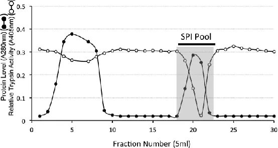
Figure 1. DEAE Anion exchange chromatography of a 6M urea extract of ovine articular cartilage showing protein (A280nm), sulphated glycosaminoglycan (A535nm), and relative trypsin inhibitory activity of the fractions (A405nm). The column was eluted with a gradient of NaCl (dotted line). Fractions displaying serine proteinase inhibitor (SPI) activity were pooled as shown for further purification.
13
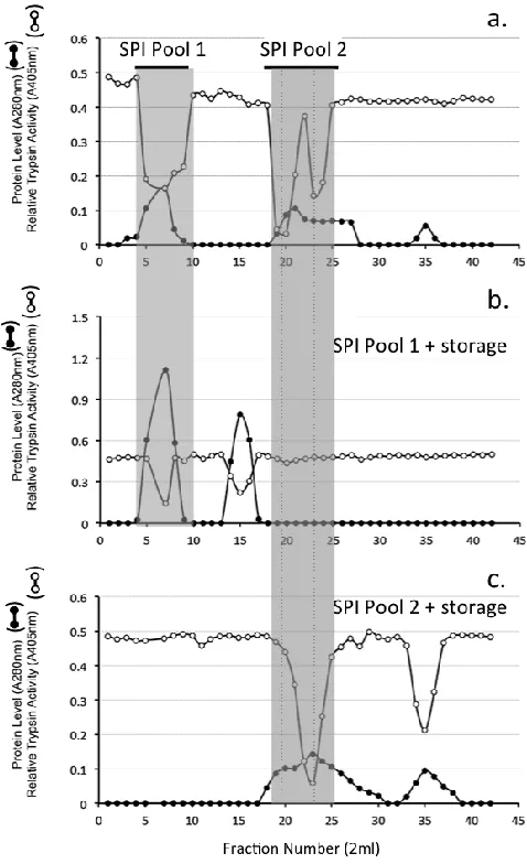
Figure 2. HA Affinity chromatography of the SPI pool from DEAE Anion exchange chromatography of the ovine AC SPIs. The column was eluted with 2 bed volumes of starting buffer 50mM Tri-HCl pH 7.2 and the bound SPI eluted by a step change elution with 2M NaCl in starting buffer. Fractions containing trypsin inhibitory activity were pooled (SPI pool) for further analysis.Sephadex G100 gel permeation chromatography of the HA affinity bound SPI resulted in the elution of ~ 30% of the SPI in the void volume of the column (Fraction 4-8) while a larger proportion of the SPI eluted well included into the column over fractions 18-26 (Fig 3A). These SPI containing fractions were separately pooled (SPI pool 1 and SPI pool 2) and stored at 4°C for one month prior to re-chromatography on the same column (Fig 3B, C). The void volume SPI pool sample eluted as two discernable SPI peaks at the void volume and also in fractions 12-16 (Fig 3B). The SPI pool 2 sample from the initial separation on Sephadex G100 (Fig 3A) eluted over a similar size range after storage however the SPI activity profile of this peak was skewed towards a smaller molecular weight range and an additional SPI peak was evident in fractions 32-38 (Fig 3C).
Preprints (www.preprints.org) | NOT PEER-REVIEWED | Posted: 31 December 2018
Preprints (www.preprints.org) | NOT PEER-REVIEWED | Posted: 31 December 2018 doi:10.20944/preprints201812.0369.v1
Figure 3 Sephadex G100 gel permeation chromatography of the ovine AC SPI sample from HA affinity chromatography (a). Fractions were monitored for protein (A280nm) and relative trypsin inhibitory activity (A405nm). Fractions containing trypsin inhibitory activity were pooled. Two SPI pools were collected as shown and separately re-chromatographed on the same column after 2 weeks storage at 4C (b, c).
15
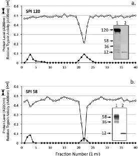
Figure 4. Chymotrypsin affinity chromatography of the SPI pools 1 and 2 from Fig 3a. The insets depict bT affinity blots, which identify active SPIs in the sample prior to (1) and after (2) chymotrypsin affinity chromatography.Concanavalin A lectin affinity chromatography of the SPI58 sample from Sephadex G100 chromatography (SPI Pool 2, Fig 3A) resulted in the isolation of a bound SPI pool containing a closely spaced SPI doublet (45-58 kDa) and minor 36kDa species (Fig 5).
Preprints (www.preprints.org) | NOT PEER-REVIEWED | Posted: 31 December 2018
Preprints (www.preprints.org) | NOT PEER-REVIEWED | Posted: 31 December 2018 doi:10.20944/preprints201812.0369.v1
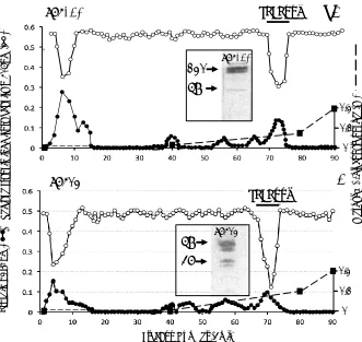
Figure 5. Concanavalin A affinity chromatography of SPI pool 2 Fig 3a. The SPI58 pool was applied and the column eluted with 10 bed volumes of starting buffer (50 mM Tris, 200 mM NaCl buffer pH 7.2 starting buffer containing 1 mM CaCl2, 1 mM MgCl2, and 1 mM MnCl2,).
Bound material was eluted with a linear 0-0.2 M gradient of methyl -glucopyranoside as shown. The inset shows active 58 and 36 kDa SPIs on bT affinity blots of the SPI58 sample prior to and after Concanavalin A affinity chromatography.
Affinity blotting of SPI samples from various stages of the isolation procedure, 1. anion exchange, 2. HA affinity, 3-6. Sephadex G100 chromatography SPI pools 1 and 2 ± storage at 4°C and 7, 8 after chymotrypsin affinity identified a range of SPI species of molecular weight 120, 86, 58, 36, 26 and 12 kDa (Fig 6a). Western blotting using antibodies to the 2-B-6 stub epitope of the CS side chain of bikunin. 1-microglobulin, bikunin and TSG-6 identified the molecular organization of these SPIs. The 36, 58, 86 and 120 kDa SPIs were
Diagrammatic representations of the structural organisation of bikunin/ITI and the deduced inter-relationships between the KPIs identified in the present study are presented in Figures 7 and 8. M e th yl a D -g lu co p yr an o si d e (M ) SPI 58
58
36
Frac; on Number (3ml)
SPI Pool 0 0.1 0.2
a.
P ro te in A 2 8 0 n m(
)
R el a; ve tr yp si n in h ib it o ry ac ; vi ty A 4 0 5 n m(
)
SPI 120
SPI Pool
17
Figure 6.Identification of serine proteinase inhibitor (SPI) species using biotinylated trypsin and avidin alkaline phosphatase conjugate by affinity blotting (a). SPIs are identified in samples from the various isolation procedures used in this study.Selected regions of replicate blot lanes from anion exchange isolated ovine cartilage SPIs were cut into regions corresponding to the 120, 58, 36 and 12 kDa SPI bands and examined using antibodies to the chondroitin-4 sulphate stub epitope of CS chains (2B6+), a1-microglobulin (a1-M), bikunin and TSG-6. The blot segments for the 2B6 (+) determinations were digested overnight with chondroitinase ABC during the blocking step. This removes the CS chains generating a 2B6(+) stub epitope which identifies CS substitution on that SPI species which migrates with the position of the intact CS-SPI during SDS PAGE. A summary of the antibody stainings for each SPI band is provided in Table C. Comparison of a bikunin blot (lanes 1-3) and bT affinity blot (lanes (4-6) of serum, plasma and urine samples (d).The smaller SPI species in lane 5 not detected in lane 2 is due to a1-proteinase inhibitor which binds to the HA affinity column during the isolation steps.
58
36
12 120
a1M Bikunin TSG-6
SPI Pool kDa SPI Pool 120 58 36 12
2B6(+) a1M Bikunin TSG-6
+++ ++++ ++ ++ + +++ ++ +++ ++ Summary of Ab staining
1 2
3 4
5
6
460 268 238 171 117 71 55 41 31 100 70 56 40 140 260 HiMark Std (kDa) Novex Std (kDa)
b.
c.
d.
2-B-6(+)Pool 1 Pool 2 Preprints (www.preprints.org) | NOT PEER-REVIEWED | Posted: 31 December 2018
Preprints (www.preprints.org) | NOT PEER-REVIEWED | Posted: 31 December 2018 doi:10.20944/preprints201812.0369.v1
Figure 7. Sequence of the CS chain of bikunin as proposed by Ly et al 2011(a) and Lord et al 2013 (b). The KPI SPI domains are shown in b. and the attached N-glycan chain of KPI-1. The oversulphated CS-D disaccharide is shown which forms part of the MO-225 epitope embedded in the CS-A chain of bikunin. Arrangement of sulphated, disulphated and non-sulphated regions of the CS chain of bikunin are shown.
N
b
4
b
3
b
4
b
3
4S
6S
2S
n
n
CS-A
CS-D
b b a
Asn 1 4b 1 4b
1 4b 1 4 1 2
1 2b 1
6
3 1 2
2 6
6 a
Kunitz domain 1 8.46 kDa Kunitz domain 2 5.9 kDa
N-linked Oligosaccharide
2.4 kDa O-linked CS chain 11.7 kDa
a. Li et al 2011 b. Lord et al 2013 c. CS Disaccharides
1-B-5
MO-225
2-B-6 (+)
Intact
19
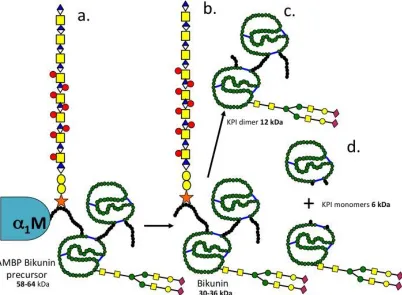
Figure 8. Schematic representations and proposed inter relationship between SPI species seen in this study. a. 1 microglobulin-bikunin precursor (58-64 kDa), b. bikunin 30-36 kDa, c. bikunin KPI dimer (12-16 kDa) after removal of the 11.7 kDa CS chain from bikunin, d.bikunin KPI monomers (6-9 kDa) with the N-linked 2-4 kDa glycan oligosaccharide attached to the KPI-1 domain which facilitates its isolation by ConA affinity chromatography and the 6 kDa KPI-2 domain which does contain oligosaccharide substitution.Assessment of the inhibitory activity of the isolated KP1 and KP2 ITI SPI domains against a range of serine proteinases using specific anilide substrates demonstrated these had potent inhibitory activity against porcine pancreatic trypsin and chymotrypsin, human leucocyte elastase and cathepsin G, porcine kallikrein and plasmin comparable to BPTI (Table 1A). KPI-1 was somewhat less inhibitory against human leucocyte cathepsin G, porcine kallikrein and plasmin compared to BPTI however still displayed appreciable inhibitory activity against these proteinases (Table 1B).
Preprints (www.preprints.org) | NOT PEER-REVIEWED | Posted: 31 December 2018
Preprints (www.preprints.org) | NOT PEER-REVIEWED | Posted: 31 December 2018 doi:10.20944/preprints201812.0369.v1
Table I
Serine proteinase inhibitory (SPI) activities of the Bikunin Kunitz
protease inhibitor (KPI) domains and BPTI A.
Proteinase
Substrate*
Mean Relative inhibitory activity by 1 Unit of
SPI** (% Inhibition) ± SD (n=6)
Sheep Kunitz
domain 1
Sheep Kunitz
domain 2
BPTI
Porcine pancreatic
trypsin
ZAPNA
95 ± 2.61
97 ± 1.47
96 ± 3.54
Porcine Pancreatic
chymotrypsin
AAVANA
52 ± 4.05
55 ± 2.86
53 ± 3.73
Human Leucocyte
elastase
SAAVNA
67 ± 4.79
69 ± 3.06
78 ± 4.07
Human Leucocyte
cathepsin G
SAAPPNA
18 ± 7.09
25 ± 5.01
28 ± 4.03
Porcine Kallikrein
VLANA
55 ± 8.84
86 ± 4.41
92 ± 5.68
Porcine Plasmin
VLLNA
51 ±4.84
85 ± 1.94
88 ± 8.84
**One unit of inhibitory activity was defined as the amount of SPI required to give
50% inhibition of 0.2
g of active site titrated trypsin. Each proteinase (0.2
g) was
incubated with one unit of SPI and residual enzyme activity assessed relative to
enzyme samples incubated in the absence of inhibitor using the indicated substrates.
*Abbreviations:
ZAPNA: CBZ-Arg-4 NA; AAVANA:
Ala-Ala-Val-Ala 4-NA
; SAAVNA:
N-Succinyl-Ala-Ala-Val-4NA, SAAPPNA: N-Succinyl-Ala-Ala-Pro-Phe 4-NA; VLANA:
D-Val-Leu-Arg
4NA
; VLLNA: D-Val-Leu-Lys 4NA; SPI: Serine proteinase inhibitor; KPI: Kunitz
protease inhibitor
B.
Proteinase
Statistical significance
KPI-1 vs BPTI
KPI-2 vs BPTI
Porcine pancreatic trypsin
NSD
NSD
Porcine Pancreatic
chymotrypsin
NSD
NSD
Human Leucocyte elastase
NSD
NSD
Human Leucocyte
cathepsin G
KPI-1 < BPTI
P
<0.05
NSD
Porcine Kallikrein
KPI-1 < BPTI
P
<0.05
NSD
Porcine Plasmin
KPI-1 < BPTI
P
<0.05
NSD
NSD-not significantly different
21
was used to localise HA, this was visualised using avidin-FITC (Fig 9b). Lubricin was immunolocalised using MAb 3A4 (Fig 9c).Figure 9. Immunolocalisation of versican in the surface regions of bovine articular cartilage (a) which immobilize HA at the cell surface (b). This is visualized using biotinylated HABP and avidin-FITC. Lubricin is also a component of the surface lamina of articular cartilage (c) and has roles in joint lubrication acting synergistically with HA and other proteins such as fibronectin and pentraxin-3 which aid in joint lubrication. ITI SPIs are also attached to HA and these protect the cell surface lubricin. HA is also visualized intra- and pericellularly in the articular chondrocytes (b). Cell nuclei were stained with propidium iodide in b and c. HA was visualized using bHABP/avidin-FITC as indicated earlier (133).
12C5
3A4
bHABP
a.
b.
c.
100mm100mm
100mm
Preprints (www.preprints.org) | NOT PEER-REVIEWED | Posted: 31 December 2018
Preprints (www.preprints.org) | NOT PEER-REVIEWED | Posted: 31 December 2018 doi:10.20944/preprints201812.0369.v1
Discussion
Identity of the ovine cartilage SPIs
The present study has identified a 58 kDa 1-microglobulin-bikunin precursor protein (SPI 58) which was converted to a number of smaller SPIs either by prolonged storage or by chymotrypsin affinity chromatography (18, 21). A 120kDa SPI was also detected which had the CS attachment stub epitope identified by MAb 2-B-6 (+) and was also reactive with antibodies to bikunin and TSG-6 consistent with its identity as pre--TI. All of the SPIs generated from SPI 58 were reactive with an antibody to bikunin. A canine IVD study has previously identified 120-250 kDa SPIs cross-reactive with an ITI antibody (31). Pre--TI is susceptible to cleavage by kallikrein into 100 and 35 kDa fragments (6) and trypsin also degrades ITI into a number of characteristic fragments of similar size to those seen in the present study (2, 3, 72). The ovine cartilage SPI 58 and 120 was also isolated by concanavalin A lectin affinity chromatography confirming N-glycosylation known to be present on bikunin. SPI 26 was also isolated using concanavalin A lectin affinity chromatography, SPI-26 was also reactive with anti-bikunin but not antibodies to 2-B-6(+), 1-M or TSG-6 thus identifying this as a KPI dimer devoid of the CS chain of bikunin.
The structural organisation of bikunin
CS-23
A chain using monoclonal antibody MO-225, these are considered important for the interactive properties of the bikunin CS chain (10). The general depiction of the bikunin molecule by Fries and Blom (32) shows the O- linked CS chain in the N-terminal extension peptide of KPI domain 1 and N-glycosylation site at Asn-45 (10, 11).Inter-relationships between ovine articular cartilage SPI species and bikunin/ITI
Comparison of data generated in the present study with published data on ITI fragments generated by trypsin or kallikrein digestion allowed us to interpret inter-relationships between the ovine AC SPIs observed in the present study. A diagram of the deduced structures of these SPIs is presented in Fig 8. SPI58 was detected using antibodies to 1-microglobulin, bikunin and 2-B-6 (+) CS stub epitope thus represented the CS substituted AMBP-bikunin precursor protein. Chymotrypsin affinity chromatography generated 12 and 6 kDa SPIs from SPI58. This protein appears quite labile and prolonged storage of SPI58 at 4°C also generated 36, 26 and 12 kDa KPI species, these were identified by a polyclonal anti-bikunin antibody, SPI36 contained a 2-B-6 (+) reactive CS epitope demonstrating the presence of a CS side chain while SPI26 did not, but contained variable portions of the N and C terminal peptide extensions attached to the KPI dimer. We also deduced that SPI12 represented the double headed KPI devoid of these peptide extensions. Furthermore, SPI6 could be generated by chymotrypsin affinity representing individual KPI domains 1 and 2, these were separable from one another using concanavalin-A lectin chromatography on the basis of the absence or presence of N-glycosylation.
ITI/bikunin is a multifunctional protein
Despite its discovery almost six decades ago the primary role of ITI has been elusive. Subsequent studies have established functional roles for the ITI HC and KPI domains in their own right. While the KPI domains of the bikunin light chain of ITI confer protease-inhibitory properties they also provide anti-tumour and anti-viral properties. Furthermore, the HCs attached to the CS chain of bikunin mediate ITI’s protein-protein interactions with many ECM components. TSG6 is one such interactive protein, which promotes the transfer of the ITI HCs to HA, cross-linking and stabilising HA through a unique trans-esterification reaction (74). In Preprints (www.preprints.org) | NOT PEER-REVIEWED | Posted: 31 December 2018
Preprints (www.preprints.org) | NOT PEER-REVIEWED | Posted: 31 December 2018 doi:10.20944/preprints201812.0369.v1
some cases the accumulation of HA-HCs in tissues can be beneficial such as in the ripening and fertilization of the oocyte or the maturation of the growth plate cartilages during endochondral ossification(39-41, 75). In other cases HC-HA accumulation in a condensed form in tissues hampers tissue function. Besides AC, ITI is also expressed in brain, placenta, liver, heart, lung, kidney and IVD and may be of importance in organ development (19, 38). Several observations suggest that like other members of the KPI superfamily, bikunin has anti-tumour
and anti-viral activities, expression of bikunin by human glioblastoma cells suppresses tumour
invasion (76) and addition of bikunin to human chondrosarcoma cell cultures blocks cell
spreading (77). Prior to this, low molecular weight factors had been detected in pregnant urine
and pharmaceutical grade preparations of human chorionic gonadotrophin (hCG) which
actively inhibited Karposi Sarcoma (KS) lesion development in HIV infections. These factors
were initially termed antiviral lysozyme-C or antiviral RNases (78-82). The appreciation that
bikunin possessed anti-viral activity became apparent when a 15.8 kDa fragment of bikunin
was identified as a contaminant in hCG preparations which inhibited the spread of KS lesions
in HIV infections (83).
Detrimental aspects of HC-HA complex transfer in tissues.
During inflammation and developmental processes, HCs from ITI are covalently transferred to HA via the enzyme TSG-6 to form an HC-HA complex. This is a significantly more adhesive substrate for leukocytes than non-cross-linked HA, and can enhance inflammation in pathological conditions. The accumulation of pro-inflammatory HC-HA in lung tissue in cystic fibrosis exacerbates chronic inflammation in airway disease and increases mucus viscosity making it difficult to eliminate from the bronchioles reducing O2 transfer and impairing lung
function (84). Hyperglycaemia induces accumulation of HA around vascular smooth muscle cells, increases aortic stiffness and strength, and primes the vascular wall for the deposition
of cholesterol, accumulation of leucocytes and accelerated development of atherosclerosis
in ApoE -/- mice (85). Accumulation of HA and HC-HA in lung tissue correlates with impaired
25
and are implicated in their pathogenesis (88, 89). Renal epithelial cells produce bikunin when stimulated by nephrotoxic agents such as oxalate (90), HA also inhibits calcium oxalate crystallization in-vitro (91). Urinary trypsin inhibitor (UTI) has a regulatory effect on local vascular tone by regulation of Ca2+ influx suppressing smooth muscle contraction (92) and also prevents lipopolysaccharide-induced increases in cytosolic free Ca2+ in human neutrophils and human umbilical vein endothelial cells (93).The KPI proteins are a diverse group of proteins occurring in all phyla in nature. Many invertebrate KPIs display blocking properties for voltage gated ion-channels. The parasitic worm Echinococcus granulosus synthesises a KPI peptide that blocks cation channels (94). ShPI-1 and APEKTx1, BPTI-like KPIs from the sea anemones Stichodactyla helianthus (53) and Anthopleura elegantissima (55)and the anemone toxins kalicludines and kaliseptine are homologous to snake venom dendrotoxins and also display ion-blocking properties (56). Calcicludine, a venom peptide of Dendroaspis angusticeps, homologous to snake dendrotoxins and the KPI APP/protease nexin-2 domain of the brain proteoglycan appican (54, 95) blocks high-threshold Ca2+ channels in cerebellar granule neurons (57) and homologous to LmKTT-1a a bifunctional KPI and potassium channel blocking scorpion peptide toxin (96). The snake toxin BF9 also displays KPI activity and potassium channel inhibitory activity (58). These ion-blocking properties are related to the neuroregulatory properties displayed by bikunin and appican in the CNS.
Tissue isoforms of ITI
Serum ITI has historically been depicted as a molecule containing two heavy chains (HC1 and HC2) while pre--TI has another HC (HC3) however up to six HCs can be attached to the CS chain of bikunin/ITI with tissue development and pathology (97). A systematic review of gene and transcript expression profiles using microarray and sequencing based functional genomics and antibody based profiling (98-102) is working towards a full description of a tissue based map of the human proteome (103). Thus a comparison of the expression profiles of the ITIH1-ITIH5 genes in tissues demonstrates that ITIH1-ITIH4 are predominantly expressed in liver while ITIH5 is expressed in breast, skin, adipose tissue and placenta (15, 97). ITIH5 is Preprints (www.preprints.org) | NOT PEER-REVIEWED | Posted: 31 December 2018
Preprints (www.preprints.org) | NOT PEER-REVIEWED | Posted: 31 December 2018 doi:10.20944/preprints201812.0369.v1
over-expressed in inflammatory skin diseases such as psoriasis, atopic dermatitis and allergic contact dermatitis (15) and specifically in the suprabasal layers of the epidermis. ITIH5 is also expressed by normal skin fibroblasts but not by epidermal keratinocytes (97) and is a novel putative tumour suppressor gene in colon cancer (104). Renal ITIH3 expression may regulate oxalate kidney stone formation (91).HA also inhibits calcium oxalate crystallization in-vitro (91). Novel truncated 50 kDa forms of HC1 and HC2 have been detected in OA AC (16), full length 90 kDa HCs attached to HA were also observed in the synovial fluids of OA patients. Bikunin and ITI are abundant in regions of surface fibrillation in OA AC.
Beneficial aspects of HC-HA transfer in connective tissues
Mesenchymal stem cells (MSCs) are pluripotent, differentiating into osteoblasts, chondrocytes, and adipocytes in-vitro and in-vivo. Umbilical MSCs (UMSCs) exposed to inflammatory cells synthesize an extracellular glycocalyx rich in HA bound to ITI HCs and the enzyme TSG6 which catalyzes the transfer of HCs to HA, versican, and pentraxin-3 (105). This glycocalyx regulates inflammatory cells and allows UMSCs to survive host immune rejection. Furthermore, the focal up-regulation of HA and ITI in areas of muscle damage and the temporal acute expression of TSG6 by MSCs is conducive to the creation of a microenvironment favouring the engraftment of MSCs in areas of damaged muscle promoting tissue repair (106).
Protective roles for ITI KPIs in connective tissues
MMPs have important roles in tissue remodelling in physiological and pathological
conditions in tissue development, morphogenesis, angiogenesis, tissue repair, arthritis and in
tumour development. AC SPIs have roles in the prevention of excessive degradation of ECM
components following traumatic overload in post-traumatic OA or in the inflammatory conditions of RA. Kunitz domain 2 of ITI (trypstatin) is taken up by mast cells and is found complexed with serine proteases in intracellular granules (107).
27
The human, canine and ovine IVD contain both low molecular weight (12 kDa) SPIs and 120 and 250 kDa ITI-like SPIs (28, 31) active against human leucocyte elastase (HLE), cathepsin-G, chymotrypsin and trypsin, urokinase, plasmin, kallikrein (108), similar SPIs have been identified in costal and AC and fibrocartilaginous meniscus (25). N-terminal sequencing demonstrated identical amino acid sequences for the cartilage and IVD SPI with SLPI from parotid and seminal plasma secretions (25). IVD cells and articular chondrocytes also synthesized mRNA to SLPI (59). Affinity blotting with bT (24), a solid phase enzyme linked immunofiltration assay to SLPI (109) and competitive inhibition assay developed to quantitate SPIs in normal and degenerate human IVDs (30), demonstrated depleted levels of active IVD SPI with advancing IVD pathology. Studies on canine IVD SPIs in chondrodystrophic (ChD) and non-chondrodystrophic (non-ChD) breeds (31) have differing rates of disc degeneration and an age dependant decline in active IVD SPI levels in the ChD (but not the non-ChD) breeds. BPTI has similar electrophoretic properties to the 6kDa ovine AC SPI (21). A chicken anti-BPTI IgY demonstrated homologies between the ovine SPIs and BPTI (28) and immunolocalised these in mast cells and chondrocytes in ovine and bovine lung and AC (29). BPTI cross-reactive SPIs were synthesized by ovine AF and NP cells in alginate bead culture with a 6 kDa SPI secreted into the culture media and a 34-36 kDa SPI was retained within alginate beads (19). Ovine chondrocytes also synthesised 14C-lysine- 6 and 58kDa SPIs in alginate bead culture(18). A biotinylated potato chymotrypsin inhibitor affinity probe demonstrated that chondrocytes synthesised an active 14C-chymotrypsin-like serine proteinase in alginate bead culture (20)
which may generate the 6kDa ovine Kunitz SPI from the 58 kDa SPI precursor.
Bikunin as a cell regulatory proteoglycan.
Oversulphated CS/DS promotes neuritogenesis and regulates CNS development. The disulphated disaccharide CS-D-unit, promotes neurite outgrowth through the DSD-1 epitope embedded in the CS chains of DSD-1-PG/phosphacan (110-115). Over sulphated DS also exhibits neurite outgrowth activity (116), the short isoform variant of phosphacan/receptor protein tyrosine phosphatase-, interacts with neuronal receptors and promotes neurite outgrowth (117). Bikunin is also expressed in brain tissue (118, 119) and accumulates in brain tumours (89). Like phosphacan, bikunin contains embedded CS-D motifs within the repeat Preprints (www.preprints.org) | NOT PEER-REVIEWED | Posted: 31 December 2018
Preprints (www.preprints.org) | NOT PEER-REVIEWED | Posted: 31 December 2018 doi:10.20944/preprints201812.0369.v1
disaccharide region of its CS chain. Such motifs in phosphacan promote neurite outgrowth suggesting that bikunin may also have roles to play in neural development. Appican is another CS brain proteoglycan containing embedded CS-E residues within its CS side chains (95, 120) and is produced by astrocytes (121). These CS-E motifs (122) interact with neuroregulatory factors (123) inducing morphological change in C6 glioma cells and directed adhesion of neural cells to the ECM (124) and also promote the chondrocytic differentiation of ATDC5 cells (125).
HC chain transfer from the bikunin CS chain provides matrix stabilization, oocyte expansion and is essential in fertilization but CS-HCs may effect HA turnover adversely and have deleterious effects on physiological processes in cystic fibrosis, diabetes, asthma, hyperglycemia, tumour development and atherosclerosis. Microarray analysis has identified a number of bikunin target genes in ovarian cancer cells and these have been categorised as transcriptional regulators, oncogenes/tumor suppressor genes, signalling molecules, growth/cell cycle, invasion/metastasis, cytokines, apoptosis, ion channels, extracellular matrix proteins, as well as some proteases (126). This further emphasises bikunin as a multifunctional cell regulatory proteoglycan.
Localisation of HA and ITI SPIs at the articular surface is of physiological significance.
Versican is found localised at the surface of AC where it localises HA. As shown in this study, the ITI SPIs share an affinity for HA thus we may deduce that these would also localise in the surface regions of AC where they would protect the AC from proteolytic damage from serine proteinases. The ITI SPIs have broad inhibitory activity against a range of serine proteinases (Table 1 and 2) some of which have MMP activating activity, which is another class of cartilage degradative proteinase. Thus the ITI SPIs may have a significant protective role to play. The ITI SPIs also display potent ant-bacterial, ant-fungal and anti-viral activities further expanding their prospective biological protection roles.
Lubricin (PRG4) was also shown to be a component of the cartilage surface in the present study (Fig 9c),
lubricin acts synergistically with HA (Fig 9b) to provide elastoviscous properties which are important
29
boundary lubricant. Localisation of the ITI SPIs with HA in the articular surface may therefore also
protect lubricin in the surface lamina and make important contributions to the preservation of joint
function. A number of catabolic enzymes have been observed which digest lubricin (128-130) and can
be inhibited by ITI (Table 1). ITI accumulates in HA depositions in skin in a condition known as lichen
sclerosis (131). HA has been shown to be depleted in AC in human OA and in a mouse model of human
mucopolysaccharidosis IX which displays similar cartilage changes as found in OA (132). In normal AC,
HA is found localised to the articulatory surface regions and in the epiphyseal growth plate where it
also has roles to play in endochondral ossification. HA is also localised pericellularly and intracellularly
in the hypertrophic cells of the vertebral growth plate of the IVD (133).
Umbilical cord derived MSCs interact with inflammatory cells synthesizing a glycocalyx rich in
versican bound HCs linked to HA [BB1]. TSG-6 catalyses the transfer of HCs to HA, HCs are also
transferred to versican. This may explain a mechanism whereby versican in the surface regions of
articular cartilage observed in the present study immobilize HA at the cartilage surface by a means
other than through direct interactions between the versican G1 domain and HA (Fig 9). ITI SPIs are
also components of such glycocalyx formations in cornea which occur on the corneal surface under
inflammatory conditions (134). Thus the ITI isoforms attached to the articular surface previously
described by Yoshihara et al 2008 may also be attached to versican at the articular surface which also
immobilizes HA in the surface lamina.(16). TSG-6 catalyses the transfer of HC chains from ITI to HA in
such formations and this stabilizes the HA. This process also inhibits angiogenesis (135). Pentraxin-3
also binds to these HC-HA complexes further contributing to their anti-angiogenic properties.
Furthermore, pentraxin-3 also has roles in innate immunity and this complements the interaction of
lubricin with TLR-2 and TLR-4 in the surface regions of cartilage to regulate inflammatory processes and
maintain cartilage homeostasis(136, 137). Pentraxin-3 is a soluble pattern recognition receptor with
essential roles to play in the innate immune system (138, 139). High molecular weight HA also has
anti-inflammatory properties (140), its localisation at the cartilage surface by versican also contributes to
joint lubrication. Lubricin in the surface regions of cartilage also binds to a number of cartilage proteins
(141) and along with fibronectin and HA this contributes to joint lubrication, retains the lubricin at the
cartilage surface (142) and may also promote cartilage regeneration (141, 143). Furthermore, ITI SPIs
Preprints (www.preprints.org) | NOT PEER-REVIEWED | Posted: 31 December 2018
Preprints (www.preprints.org) | NOT PEER-REVIEWED | Posted: 31 December 2018 doi:10.20944/preprints201812.0369.v1
also bind to the cell surface HA (16) and this would be expected to afford proteolytic protection from
degradative proteinases known to be present in synovial fluid particularly during OA and under
inflammatory conditions (144, 145). Collectively all of the aforementioned components thus provide
protection to the articulating surface of cartilage and are essential in the preservation of knee joint
31
Conclusions.1. Ovine AC and IVD contain SPIs related to ITI/bikunin which are released from the bikunin precursor protein and display potent inhibitory activity against leucocyte elastase, cathepsin G, trypsin, chymotrypsin, plasmin and kallikrein.
2. ITI/bikunin are multifunctional KPI domain proteins with a diverse range of biological properties beyond protease inhibition and are beneficial to tissues in terms of prevention of bacterial, fungal and viral infection, modulation of innate immunity in host-defense and have cell proliferative and anti-tumour properties. The dual function of some KPI domains in protease inhibition and blocking of ion-fluxes in voltage gated ion-channels indicate KPIs may be of therapeutic value in neural disorders, the embedded CS-D motifs in the CS chains of bikunin may equip it with cell regulatory and neuritogenic properties.
3. HC chain transfer from the bikunin CS chain provides matrix stabilization, oocyte expansion and is essential in fertilization but CS-HCs may effect HA turnover adversely and have deleterious effects on physiological processes in cystic fibrosis, diabetes, asthma, hyperglycemia and atherosclerosis.
4. Localisation of ITI in the surface regions of articular cartilage have important tissue protective roles to play and preservation of joint functional properties.
Acknowledgements
This study was funded by NHMRC Project Grant 1004032. Prof Bruce Caterson is thanked for the gifts of 2-B-6 hybridoma conditioned medium and MAb 3A4 to lubricin.
Disclosures.
The author has no financial disclosures or conflicts to report.
Preprints (www.preprints.org) | NOT PEER-REVIEWED | Posted: 31 December 2018
Preprints (www.preprints.org) | NOT PEER-REVIEWED | Posted: 31 December 2018 doi:10.20944/preprints201812.0369.v1
References
1.
Steinbuch, M., and Loeb, J. (1961) Isolation of an alpha2-globulin from human
plasma,
Nature
192
, 1196.
2.
Hochstrasser, K., Albrecht, G., Schonberger, O. L., and Wachter, E. (1983)
Kunitz-type proteinase inhibitors derived by limited proteolysis of the
inter-alpha-trypsin inhibitor, VII. Characterization of the bovine inhibitor as
double-headed trypsin-elastase inhibitor,
Hoppe Seylers Z Physiol Chem
364
,
1689-1696.
3.
Hochstrasser, K., Wachter, E., Albrecht, G. J., and Reisinger, P. (1985)
Kunitz-type proteinase inhibitors derived by limited proteolysis of the
inter-alpha-trypsin inhibitor, X. The amino-acid sequences of the inter-alpha-trypsin-released inhibitors
from horse and pig inter-alpha-trypsin inhibitors,
Biol Chem Hoppe Seyler
366
,
473-478.
4.
Wachter, E., and Hochstrasser, K. (1979) Kunitz-type proteinase inhibitors
derived by limited proteolysis of the inter-alpha-trypsin inhibitor, III. Sequence
of the two Kunitz-type domains inside the native inter-alpha-trypsin inhibitor, its
biological aspects and also of its cleavage products,
Hoppe Seylers Z Physiol
Chem
360
, 1305-1311.
5.
Salier, J. P. (1990) Inter-alpha-trypsin inhibitor: emergence of a family within
the Kunitz-type protease inhibitor superfamily,
Trends Biochem Sci
15
,
435-439.
6.
Nishimura, H., Kakizaki, I., Muta, T., Sasaki, N., Pu, P. X., Yamashita, T., and
Nagasawa, S. (1995) cDNA and deduced amino acid sequence of human
PK-120, a plasma kallikrein-sensitive glycoprotein,
FEBS Lett
357
, 207-211.
7.
Saguchi, K., Tobe, T., Hashimoto, K., Sano, Y., Nakano, Y., Miura, N. H., and
Tomita, M. (1995) Cloning and characterization of cDNA for inter-alpha-trypsin
inhibitor family heavy chain-related protein (IHRP), a novel human plasma
glycoprotein,
J Biochem
117
, 14-18.
8.
Hochstrasser, K., Schonberger, O. L., Rossmanith, I., and Wachter, E. (1981)
Kunitz-type proteinase inhibitors derived by limited proteolysis of the
inter-alpha-trypsin inhibitor, V. Attachments of carbohydrates in the human urinary
trypsin inhibitor isolated by affinity chromatography,
Hoppe Seylers Z Physiol
Chem
362
, 1357-1362.
9.
Chi, L., Wolff, J. J., Laremore, T. N., Restaino, O. F., Xie, J., Schiraldi, C.,
Toida, T., Amster, I. J., and Linhardt, R. J. (2008) Structural analysis of bikunin
glycosaminoglycan,
J Am Chem Soc
130
, 2617-2625.
10.
Lord, M. S., Day, A. J., Youssef, P., Zhuo, L., Watanabe, H., Caterson, B., and
Whitelock, J. M. (2013) Sulfation of the bikunin chondroitin sulfate chain
determines heavy chain.hyaluronan complex formation,
J Biol Chem
288
,
22930-22941.
11.
Ly, M., Leach, F. E., 3rd, Laremore, T. N., Toida, T., Amster, I. J., and Linhardt,
R. J. (2011) The proteoglycan bikunin has a defined sequence,
Nat Chem Biol
7
, 827-833.
12.
Zhuo, L., Salustri, A., and Kimata, K. (2002) A physiological function of serum
proteoglycan bikunin: the chondroitin sulfate moiety plays a central role,
Glycoconj J
19
, 241-247.
13.
Zhuo, L., Hascall, V. C., and Kimata, K. (2004) Inter-alpha-trypsin inhibitor, a
covalent protein-glycosaminoglycan-protein complex,
J Biol Chem
279
,
38079-38082.
33
chain (ITIH) genes in multiple human solid tumors: a systematic expression
analysis,
BMC Cancer
8
, 25.
15.
Huth, S., Heise, R., Vetter-Kauczok, C. S., Skazik, C., Marquardt, Y., Czaja,
K., Knuchel, R., Merk, H. F., Dahl, E., and Baron, J. M. (2015)
Inter-alpha-trypsin inhibitor heavy chain 5 (ITIH5) is overexpressed in inflammatory skin
diseases and affects epidermal morphology in constitutive knockout mice and
murine 3D skin models,
Exp Dermatol
24
, 663-668.
16.
Yoshihara, Y., Plaas, A., Osborn, B., Margulis, A., Nelson, F., Stewart, M.,
Rugg, M. S., Milner, C. M., Day, A. J., Nemoto, K., and Sandy, J. D. (2008)
Superficial zone chondrocytes in normal and osteoarthritic human articular
cartilages synthesize novel truncated forms of inter-alpha-trypsin inhibitor
heavy chains which are attached to a chondroitin sulfate proteoglycan other
than bikunin,
Osteoarthritis Cartilage
16
, 1343-1355.
17.
Lamkin, E., Cheng, G., Calabro, A., Hascall, V. C., Joo, E. J., Li, L., Linhardt,
R. J., and Lauer, M. E. (2015) Heavy chain transfer by tumor necrosis
factor-stimulated gene 6 to the bikunin proteoglycan,
J Biol Chem
290
, 5156-5166.
18.
Rodgers, K. J., Melrose, J., and Ghosh, P. (1996) Purification and
characterisation of 6 and 58 kDa forms of the endogenous serine proteinase
inhibitory proteins of ovine articular cartilage,
Biol Chem
377
, 837-845.
19.
Melrose, J., Smith, S., and Ghosh, P. (2002) Synthesis of a Kunitz-type serine
proteinase inhibitory protein that shares homology with bovine pancreatic
trypsin inhibitor by ovine intervertebral disc cells in serum-free alginate bead
culture,
J Spinal Disord Tech
15
, 164-171.
20.
Rodgers, K. J., Melrose, J., and Ghosh, P. (1995) Biotin-labeled potato
chymotrypsin inhibitor-1: a useful probe for the detection and quantitation of
chymotrypsin-like serine proteinases on western blots and its application in the
detection of a serine proteinase synthesised by articular chondrocytes,
Anal
Biochem
227
, 129-134.
21.
Rodgers, K. J., Melrose, J., and Ghosh, P. (1996) Biotinylated trypsin and its
application as a sensitive, versatile probe for the detection and characterisation
of an ovine chondrocyte serine proteinase inhibitor using Western blotting,
Electrophoresis
17
, 213-218.
22.
Rasp, G., Hochstrasser, K., Wachter, E., and Reisinger, P. W. (1987) The
amino-acid sequence of the trypsin-released inhibitor from sheep
inter-alpha-trypsin inhibitor,
Biol Chem Hoppe Seyler
368
, 727-731.
23.
Melrose, J., Rodgers, K. (1996) Preparation and use of biotinylated probes for
the detection and characterisation of serine proteinases and serine proteinase
inhibitory proteins, In
A laboratory guide to biotin labelling in Biomolecule
analysis
(F, M. T. a. F., Ed.), Birkhauser publishing, Basel.
24.
Melrose, J., Rodgers, K., and Ghosh, P. (1994) The preparation and use of
biotinylated trypsin in western blotting for the detection of trypsin inhibitory
proteins,
Anal Biochem
222
, 34-43.
25.
Andrews, J. L., Melrose, J., and Ghosh, P. (1992) A comparative study of the
low-molecular mass serine proteinase inhibitors of human connective tissues,
Biol Chem Hoppe Seyler
373
, 111-118.
26.
Melrose, J., and Ghosh, P. (1992) Development of an avidin-biotin competitive
inhibition assay and validation of its use for the quantitation of human
intervertebral disc serine proteinase inhibitory proteins,
Anal Biochem
204
,
372-382.
27.
Melrose, J., Ghosh, P., Taylor, T. K., and Andrews, J. L. (1992) The serine
proteinase inhibitory proteins of the human intervertebral disc: their isolation,
characterization and variation with ageing and degeneration,
Matrix
12
,
456-470.
Preprints (www.preprints.org) | NOT PEER-REVIEWED | Posted: 31 December 2018
Preprints (www.preprints.org) | NOT PEER-REVIEWED | Posted: 31 December 2018 doi:10.20944/preprints201812.0369.v1