Scholarship@Western
Scholarship@Western
Electronic Thesis and Dissertation Repository
7-12-2017 12:00 AM
Mechanical Properties of Retrieved Highly Cross-linked
Mechanical Properties of Retrieved Highly Cross-linked
Polyethylene Acetabular Liners in Total Hip Arthroplasty
Polyethylene Acetabular Liners in Total Hip Arthroplasty
Gurpreet Singh
The University of Western Ontario
Supervisor Brent Lanting
The University of Western Ontario Joint Supervisor Robert Klassen
The University of Western Ontario Joint Supervisor Douglas Naudie
The University of Western Ontario Graduate Program in Surgery
A thesis submitted in partial fulfillment of the requirements for the degree in Master of Science © Gurpreet Singh 2017
Follow this and additional works at: https://ir.lib.uwo.ca/etd
Part of the Musculoskeletal Diseases Commons, Orthopedics Commons, and the Surgical Procedures, Operative Commons
Recommended Citation Recommended Citation
Singh, Gurpreet, "Mechanical Properties of Retrieved Highly Cross-linked Polyethylene Acetabular Liners in Total Hip Arthroplasty" (2017). Electronic Thesis and Dissertation Repository. 4667.
https://ir.lib.uwo.ca/etd/4667
This Dissertation/Thesis is brought to you for free and open access by Scholarship@Western. It has been accepted for inclusion in Electronic Thesis and Dissertation Repository by an authorized administrator of
i
Highly crosslinked polyethylene (HXLPE) has replaced the conventional
ultra-high-molecular-weight-polyethylene (UHMWPE) as the gold standard bearing surface in total hip
arthroplasty (THA) due to superior wear characteristics and survivorship. However, HXLPE
has demonstrated poorer mechanical properties, in vivo oxidation, and concerns of rim
fractures. The purpose of this project was to study the mechanical properties at the rim of
retrieved HXLPE acetabular liners.
We developed a simple technique for measurement of hardness at the rim of irradiated,
remelted, HXLPE liners of a specific design. The effect of shelf time on mechanical
properties of retrieved liners was determined and showed no correlation between hardness
with shelf time. Furthermore, hardness testing of retrieved samples showed no correlation
between hardness and time in vivo. This suggests that rim fractures in this design of liners
are likely not a result of in vivo decline of mechanical properties.
Keywords
Total hip arthroplasty, Highly crosslinked polyethylene, Mechanical properties, Indentation
ii
Co-Authorship Statement
Chapter 1
• Gurpreet Singh – Author
• Brent Lanting – Assistance with Manuscript preparation
• Matthew Teeter - Assistance with Manuscript preparation
• James Howard – Assistance with Manuscript preparation
• Douglas Naudie - Assistance with Manuscript preparation
Chapter 2
• Gurpreet Singh – Author, Study Design
• Brent Lanting – Study Design, Assistance with Manuscript preparation
• Matthew Teeter - Assistance with Manuscript preparation
• James Howard – Assistance with Manuscript preparation
• Douglas Naudie - Assistance with Manuscript preparation
• Robert Klassen - Assistance with Manuscript preparation
Chapter 3
• Gurpreet Singh – Author
Chapter 4
• Gurpreet Singh – Author
• Brent Lanting – Assistance with Manuscript preparation
• James Howard – Assistance with Manuscript preparation
• Robert Klassen - Assistance with Manuscript preparation
Chapter 5
• Gurpreet Singh – Author, Study Design, Analysis, Interpretation
• Brent Lanting – Assistance with Study Design, Interpretation, Manuscript preparation
iii
• James Howard – Assistance with Manuscript preparation
• Robert Klassen - Assistance with Testing of the samples
• Ariful Islam – Testing of the samples, Assistance with Study Design
Chapter 6
• Gurpreet Singh – Author, Study Design, Analysis, Interpretation
• Brent Lanting – Assistance with Study Design, Interpretation, Manuscript preparation
• Matthew Teeter - Assistance with Study Design, Analysis
• James Howard – Assistance with Manuscript preparation
• Robert Klassen - Assistance with Testing of the samples
• Ariful Islam – Testing of the samples, Assistance with Study Design
Chapter 7
• Gurpreet Singh – Author
• Brent Lanting – Assistance with Manuscript preparation
iv
Acknowledgments
First and most importantly, I would like to thank my mentors Dr. Lanting, Dr. Teeter, Dr.
Howard, Dr. Naudie, and Dr. Klassen for their endless support and and guidance throughout
this process. Each mentor had a unique but very important contribution towards this thesis.
Thank You Dr. Lanting for pushing me throughout the process and spending the enormous
amount of time, and always being available. To Dr. Teeter, thank you for your assistance
with the analysis as well as your help with the implant retrieval laboratory. To Dr. Howard
and Dr. Naudie, thank you for your input and feedback with the writings. To Dr. Klassen,
thank you for helping me understand materials testing better and allowing me to use your lab
for testing. Through this thesis, I was hoping to learn ways to develop a foundation to
conduct research as I move forward into my own practice and I certainly achieved that with
all your support.
Secondly, I would like to thank Ariful Islam from Dr. Klassen’s laboratory for his help with
testing of the samples and helping me understand the process better.
To the arthroplasty staff at UH, thank you for a great year of training. Thank you for
allowing me to use retrospective data from your patient, as well as the implants that you
retrieved.
To Lyndsay Somerville, thank you for helping me with the purchase of the textbook that was
necessary to develop my foundation in this topic.
To my fiancé Gurleen, thank you for your support throughout this year and the
encouragement that you provided. Without your support this will not be possible.
To my parents and siblings in Texas, thank you for your support in getting me here and your
v
Table of Contents
Abstract ... i
Co-Authorship Statement... ii
Acknowledgments... iv
Table of Contents ... v
List of Abbreviations ... ix
List of Tables ... x
List of Figures ... xi
List of Appendices ... xiii
Chapter 1 ... 1
1 Hip, Total Hip Arthroplasty and Polyethylene ... 1
1.1 Hip Anatomy ... 1
1.2 Osteoarthritis ... 5
1.3 Total Hip Arthroplasty ... 6
1.3.1 Acetabulum ... 7
1.3.2 Femur ... 8
1.4 Research Objectives and Impact ... 12
1.5 References ... 14
Chapter 2 ... 18
2 Clinical performance of highly cross-linked polyethylene in total hip arthroplasty, a literature review ... 18
2.1 Introduction ... 18
2.2 Methods... 19
2.3 Conventional UHMWPE ... 19
2.3.1 Introduction ... 19
vi
2.4 HXLPE ... 20
2.4.1 Introduction ... 20
2.4.2 Manufacturing ... 21
2.4.3 Clinical Performance ... 25
2.5 Oxidation... 28
2.5.1 Overview ... 28
2.5.2 Resin, consolidation method, and oxidation ... 29
2.5.3 Irradiation and Oxidation ... 29
2.5.4 Thermal processing and oxidation ... 30
2.5.5 Sterilization, packaging and oxidation ... 30
2.5.6 In Vivo time and oxidation ... 31
2.6 Mechanical Properties ... 34
2.6.1 Overview ... 34
2.6.2 Mechanical Testing ... 34
2.6.3 Resin and Consolidation ... 35
2.6.4 Cross-linking ... 36
2.6.5 Thermal Processing ... 38
2.6.6 Sterilization ... 38
2.6.7 In Vivo Time ... 38
2.7 Summary and Conclusions ... 39
2.8 References ... 39
Chapter 3 ... 47
3 Thesis Outline and Brief Summary ... 47
Chapter 4 ... 48
4 Indentation testing of UHMWPE ... 48
vii
4.1.1 Background ... 49
4.1.2 Microhardness calculation ... 50
4.1.3 DSI testing for UHMWPE ... 51
4.1.4 DSI and oxidation of UHMWPE ... 52
4.1.5 DSI and in vivo time ... 52
4.2 Summary ... 53
4.3 References ... 54
Chapter 5 ... 57
5 Impact of increasing shelf (ex vivo) time on hardness of irradiated remelted HXLPE acetabular liners – A validation study. ... 57
5.1 Introduction ... 57
5.2 Materials and methods ... 58
5.3 Results ... 61
5.4 Discussion ... 63
5.5 References ... 65
Chapter 6 ... 67
6 Impact of in vivo time on hardness at the rim of irradiated remelted HXLPE acetabular liners. ... 67
6.1 Introduction ... 67
6.2 Materials and methods ... 68
6.3 Results ... 71
6.4 Discussion ... 73
6.5 References ... 75
Chapter 7 ... 78
7 Discussion ... 78
7.1 Discussion and Conclusions ... 78
viii
7.3 References ... 80
Appendix A: Ethics approval ... 82
Appendix B: Permissions for table and figures ... 83
ix
List of Abbreviations
DSI – Depth-sensing indentation
EtO – Ethylene oxide
FTIR - Fourier transform infrared spectroscopy
gf – Gram-force
Gpa – Gigapascals
HXLPE - Highly crosslinked polyethylene
kgf – Kilogram-force
MRad – Megarads
NSAIDs - Nonsteroidal anti-inflammatory drugs
OA – Osteoarthritis
OI – Oxidation index
PTFE – Polytetrafluoroethylene
THA – Total hip arthroplasty
THR – Total hip replacement
UHMWPE - Ultra-high-molecular-weight polyethylene
x
List of Tables
Table 2-1 Mechanical properties of various UHMWPE... 35
Table 5-1 List of expermintal samples and patient demographics ... 59
Table 5-2 Results of Vickers hardness and shelf times ... 62
Table 6-1 List of experimental samples and patient demographics ... 69
xi
List of Figures
Figure 1-1 - The cross-sectional anatomy of the hip ... 1
Figure 1-2 - The ligamentous anatomy of the hip ... 2
Figure 1-3 - The dynamic stabilizers surrounding the hip ... 3
Figure 1-4 - Measurement of abduction and anteversion angle of the acetabulum ... 4
Figure 1-5 - Forces surrounding the hip ... 5
Figure 1-6 - Metal on polyethylene total hip replacement ... 7
Figure 1-7 - Normal versus abnormal abduction/anteversion of the acetabular component .... 8
Figure 1-8 - Femoral offset ... 9
Figure 1-9 - Jump distance ... 10
Figure 1-10 - Rim fractures of polyethylene ... 12
Figure 1-11 - Different regions of the polyethylene liner ... 13
Figure 2-1 - Molecular structure of UHMWPE ... 21
Figure 2-2 - Manufacturing of UHMWPE in THA ... 22
Figure 2-3 - Rim fractures of polyethylene... 28
Figure 2-4 - Oxidation index measurements of first-generation HXLPE liners ... 32
Figure 2-5Oxidation and mechanical properties ... 33
Figure 2-6 Radiation and mechanical properties ... 37
Figure 4-1 Vickers hardness testing ... 51
xii
Figure 4-3 Modulus of elasticity of retrieved liners ... 53
Figure 5-1 Vickers hardness testing ... 60
Figure 5-2 Shelf time versus hardness ... 63
Figure 6-1 Vickers hardness testing ... 70
Figure 6-2 Time in vivo and hardness ... 73
xiii
List of Appendices
Appendix A: Ethics approval ... 82
Chapter 1
1
Hip, Total Hip Arthroplasty and Polyethylene
1.1
Hip Anatomy
The bony hip joint is the articulation of the femoral head into the acetabulum of the
pelvis, and is a ball and socket joint. The weight of the head, thorax and upper extremities
is transmitted via lumbar spine, sacrum, and ilium to the hip joint1. The hip balances
these gravitational forces of body weight and the ground reaction forces of the lower
extremity throughout a large range of positions. The bony anatomy (Figure 1-1), static
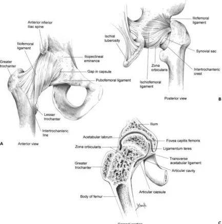
(ligaments) (Figure 1-2) and dynamic (muscles) (Figure 1-3) stabilizers around the hip
joint help accomplish this task, while preventing dislocation and maintaining efficiency.
Figure 1-3 - Muscles or dynamic stabilizers around the hip joint A) Intrapelvic and anterior proximal femur B) Extra pelvic and posterior proximal femur (Permission from Callaghan JJ, Rosenberg AG, Rubash HE, Clohisy J, Beaule P, DellaValle C. The Adult Hip: Hip Arthroplasty Surgery. Wolters Kluwer Health; 2015.)
The acetabulum is hemispherical in shape and accommodates a nearly spherical femoral
head. Acetabulum has an abduction (inclination) angle (Figure 1-4) of approximately 38
degrees in males and 40 degrees in females in the anterior posterior plane. This allows
for abduction of the hip, while limiting its adduction[2]. Moreover, the acetabulum has an
approximate anteversion of 16 degrees in men and 19 degrees in women to allow for
flexion and to provide posterior coverage preventing dislocation in a flexed position.
Furthermore, the ligaments, capsule, labrum and muscles surrounding the hip joint play a
substantial role on maintaining stability of the hip joint. During a single leg stance or
stance phase of the gait cycle, the hip experiences a downward force exerted by the body
level and depends on the magnitude of force due to the body weight and the pull of the
abductors, and hip center - which can alter the lever arm of each force.
Figure 1-4 - Inclination, abduction or coronal tilt angle demonstrated on the
Figure 1-5 - Demonstrated above are the forces of body weight and abductor pull (red arrows) and their direction. Joint reaction force (blue arrow) is the net force experienced by the hip joint and can be influenced by the lever arms a and b depending on the hip/femoral head center. (Permission from Sariali E, Veysi V, Stewart T. MINI-SYMPOSIUM: ESSENTIAL BIOMECHANICS OF HIP REPLACEMENT (i) Biomechanics of the human hip e consequences for total hip replacement. Curr Orthop n.d.;22:371–5. doi:10.1016/j.cuor.2008.10.005.)
1.2
Osteoarthritis
Osteoarthritis (OA) is a disorder of a joint, involving the cartilage, bone, synovium and
capsule[1]. Damage to focal areas of the articular cartilage, resulting in loss of volume of
cartilage is the hallmark of OA. OA is the most common cause of musculoskeletal pain
and disability surrounding the hip. The main risk factor for development of hip OA is
age[3]. The treatment of hip OA includes non-surgical and surgical management
nonsteroidal anti-inflammatory medications (NSAIDs) and intra-articular corticosteroids.
Failure of non-surgical management is the prime indicator for surgical intervention in the
setting of painful and debilitating OA. Total hip arthroplasty (THA) is the operation of
choice for the treatment of hip OA in most of the patients.
1.3
Total Hip Arthroplasty
Low Friction total hip arthroplasty (THA) was first performed by Sir John Charnley in
1962 for treatment of arthritic conditions of the hip[5]. The indications for THA, as
reported by Charnley, included primarily advanced osteoarthritis and rheumatoid
arthritis, and secondarily ankylosing spondylitis, femoral neck fractures and Paget’s
disease of the bone[5]. The goals of the surgery involve improvement of pain and
function while restoring normal anatomy, biomechanics and kinematics of the hip joint.
Metal femoral head on ultra-high-molecular-weight-polyethylene (UHMWPE) acetabular
liner has been the most common bearing surface in THA (Figure 1-6). In THA, the
acetabular cartilage is replaced with a hemispherical metal acetabular shell which fits into
the patient’s acetabulum. A UHWMPE polyethylene liner is locked into the acetabular
metal shell. On the femoral side, the native head is removed and a metal stem is inserted
into the intramedullary canal of the femur. A metal head is placed on to the neck portion
of the stem. The modularity in the modern components gives multiple options to match
the patient’s anatomy and restore function. From a fixation standpoint, cementless and
cemented fixation options exist for the acetabular shell and the femoral stem. Cementless
fixation is preferred for young patients with good bone quality, while cemented fixation
is used for older patients with osteopenic/osteoporotic bone.
The placement of the components in the proper orientation, restoration of leg length and
offset of the hip has implications in restoring function and determining longevity of the
hip replacement. The considerations in component placement on the acetabular side and
Figure 1-6 - Metal on polyethylene total hip replacement. (Permission from Total Hip Arthroplasty (THA). OrthopaedicsOne Clerkship. In: OrthopaedicsOne - The Orthopaedic Knowledge Network. Created Dec 13, 2010 21:12. Last modified Dec 14, 2010 09:10 ver.3. Retrieved 2017-06-01, from
http://www.orthopaedicsone.com/x/-oDYAg.)
1.3.1
Acetabulum
The acetabular component position can influence center of rotation of the articulating
femoral head. The center of rotation of the femoral head should be restored to its
pre-disease position, usually by medial positioning of acetabular shell. This effectively
increases the lever arm of the abductors, decreasing joint reactive forces, ultimately
resulting in decreased wear of the UHMWPE liner. The abduction angle is chosen to
match the patient’s anatomy of around 40 degrees to maximize the range of motion. The
more vertical placement or increased abduction angles can result in edge loading of the
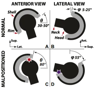
acetabular component and decreased range of motion before impingement (Figure 1-7).
Excessive anteversion can result in impingement of the prosthetic neck on the posterior
side of the acetabular rim during extension and external rotation, resulting in anterior
acetabular component leads to neck impingement on the anterior acetabular rim during
flexion and can result in posterior dislocation.
Figure 1-7 - A and B show an acetabular component with normal abduction and anteversion angle respectively. C and D show an excessively abducted and
anteverted acetabular component. (Permission from Furmanski J, Anderson M, Bal S, et al. Clinical fracture of cross-linked UHMWPE acetabular liners. Biomaterials. 2009;30(29):5572-5582.)
1.3.2
Femur
Femoral neck anteversion, which is the rotation of the neck about the femoral shaft, must
be considered and restored. This preserves the flexion and internal rotation of the hip
can result in impingement of the neck with the anterior acetabular rim and result in
posterior dislocation of the femoral head from the acetabulum. Conversely, a relative
excessive anteversion can lead to impingement of the prosthetic neck with posterior
acetabular shell during extension and external rotation of the hip resulting in an anterior
dislocation.
Femoral offset is defined as the distance between the center of the femoral head and the
long axis of the femur. Restoration of offset is important in hip biomechanics by
appropriate tensioning of the abductor muscles (Figure 1-8).
Figure 1-8 - Femoral offset is represented by the perpendicular distance "A" from the center of the femoral head to the long axis of the femur. The neck-shaft angle is represented by angle "B," which subtends the long axis of the femoral neck and the long axis of the femoral shaft. (Permission from Charles M, Bourne R. Soft-tissue balancing of the hip. J Bone Jt Surg 2004;86:1078–88. doi:10.1054/arth.2002.33263.)
The head to neck ratio, which is the ratio between the diameter of the prosthetic head and
neck, is another important factor to consider. The increase in head diameter relative to the
neck increases the jump distance before dislocation, reducing the incidence of dislocation
(Figure 1-9). However, this comes at the expense of increasing volumetric wear of
Figure 1-9 - Demonstrated above is the increase in jump distance with an increase in femoral head size. (Permission from Veitch SW, Jones SA. (v) Prevention of
dislocation in hip arthroplasty. Orthopaedics and Trauma 2009 200902;23(1):35-39.)
While THA has been extremely successful clinically in improving quality of life and
function[6–9], the revision THA burden has continued to rise[10–13]. The clinical failure
of THA, and thus the increased need for revision THA, has been a result of surgery in
younger patients, active population, and issues with implant longevity[13]. Despite the
established cost effectiveness of performing THA[14], the increasing number of revisions
is placing a tremendous clinical and economic burden on the healthcare
system[10,13,15]. The most common causes for revision surgery has been reported as
aseptic loosening, instability/dislocation and infection[10,16,17]. Registry data from
Europe has suggested that aseptic loosening, bearing surface wear and osteolysis
comprise the majority of the reasons for revision[18].
Wear of conventional UHMWPE and the reactivity of the generated wear particles in
vivo has been the major player in osteolysis and aseptic loosening in THA[19,20]. The
polyethylene debris generated because of wear leads to macrophage induced osteolysis
via a complex inflammatory cascade. Osteolysis results in aseptic loosening of the hip
In an effort to reduce wear and osteolysis, highly cross-linked UHMWPE (HXLPE) was
developed in 1990s[21]. Since the inception of HXLPE, the wear rates and incidence of
osteolysis have significantly decreased[22–25]. Furthermore, the revision THA rates with
HXLPE are lower than the conventional UHMWPE[23,26,27].
Development of HXLPE and the lower wear rates comes with a compromise of
mechanical properties of polyethylene[21,28]. Formation of cross-linking requires
irradiation of UHMWPE, thereby reducing its mechanical strength to failure[28]. In
addition, concerns have developed regarding further degradation of mechanical properties
with time in vivo, as a result of oxidation[29–32]. Numerous case reports have been
published reporting fractures of the polyethylene at the rim of the polyethylene
liners[16,33–35] (Figure 1-10). Increasing head sizes, to minimize dislocation, and
subsequent use of thinner HXLPE acetabular liners has been named a contributing factor.
Furthermore, increased susceptibility to in vivo degradation of HXLPE liners, and further
compromise of mechanical properties raises significant concerns of implant longevity in
Figure 1-10 - Demonstrated above are the fractures at the rim of HXLPE liners. (Permission from Furmanski J, Anderson M, Bal S, et al. Clinical fracture of cross-linked UHMWPE acetabular liners. Biomaterials. 2009;30(29):5572-5582.)
1.4
Research Objectives and Impact
With the overall aging population, maximizing implant longevity in THA is especially
important. OA accounts for 93% of the THA procedures[36], with the main risk factor for
development of OA being age[37]. Globally, the number of persons aged 60 and above is
expected to more than double by 2050 and more than triple by 2100, increasing from 901
million in 2015 to 2.1 billion in 2050 and 3.2 billion in 2100[38].
This will result in an increasing incidence of OA and a higher need for THA[39–41].
Over 1 million THA are performed worldwide and this number is expected to double in
the next two decades[36]. In the United States, the number of THA is projected at
increase in the number of THA performed as compared to 5 years ago[42]. Additional
implant retrieval studies, to understand the impact of in vivo degradation of HXLPE, are
necessary to improve implant longevity, as the number of THAs performed continues to
rise.
Figure 1-11 - Different regions of the polyethylene liner are labeled.
The objective of this masters is to better our understanding of the fractures at the rim
(Figure 1-11) of the HXLPE liners. Implant retrieval analysis have shown clear evidence
However, no studies have investigated the mechanical properties at the rim of the
HXLPE after time in vivo. Some retrieval studies have reported on preservation of
mechanical properties at the articular surface and backside of the HXLPE liners with
increasing in vivo time. In this masters, we will develop a way of testing mechanical
properties at the rim of the HXLPE liners. Furthermore, we will test the mechanical
properties of retrieved HXLPE acetabular liners with varying in vivo times to investigate
the impact of in vivo time on mechanical properties. This will help with the
understanding of mechanical failures of HXLPE acetabular liners at the rim.
1.5
References
[1] Callaghan JJ, Rosenberg AG, Rubash HE, Clohisy J, Beaule P, DellaValle C. The Adult Hip: Hip Arthroplasty Surgery. Wolters Kluwer Health; 2015.
[2] Sariali E. (i) Biomechanics of the human hip – consequences for total hip replacement. Curr Orthop 2008;22.
[3] Zhang Y, Sc D, Jordan JM, Edu Y. Epidemiology of Osteoarthritis n.d. doi:10.1016/j.cger.2010.03.001.
[4] Altman RD, Hochberg MC, Schnitzer TJ, Marc Hochberg CC, Lilly E, Plough Roland Moskowitz SW, et al. RECOMMENDATIONS FOR THE MEDICAL MANAGEMENT OF OSTEOARTHRITIS OF THE HIP AND KNEE 2000 Update AMERICAN COLLEGE OF RHEUMATOLOGY SUBCOMMITTEE ON OSTEOARTHRITIS GUIDELINES. ARTHRITIS Rheum 2000;43:1905–15.
[5] Charnley J. THE LONG-TERM RESULTS OF LOW-FRICTION ARTHROPLASTY OF THE HIP PERFORMED AS A PRIMARY
INTERVENTION. J Bone & Jt Surgery, Br Vol 1972;54–B:61 LP-76.
[6] Shan L, Shan B, Graham D, Saxena A, Learmonth ID, Young C, et al. Total hip replacement: a systematic review and meta-analysis on mid-term quality of life. Osteoarthritis Cartilage 2014;22:389–406.
doi:http://dx.doi.org/10.1016/j.joca.2013.12.006.
[7] Laupacis a, Bourne R, Rorabeck C, Feeny D, Wong C, Tugwell P, et al. The effect of elective total hip replacement on health-related quality of life. J Bone Joint Surg Am 1993;75:1619–26.
[8] Ng CY, Ballantyne J a, Brenkel IJ. Quality of life and functional outcome after primary total hip replacement. A five-year follow-up. J Bone Joint Surg Br 2007;89:868–73. doi:10.1302/0301-620X.89B7.18482.
[10] Bozic KJ, Kurtz SM, Lau E, Ong K, Vail TP, Berry DJ. The epidemiology of revision total hip arthroplasty in the United States. J Bone Jt Surg Am
2009;91:128–33. doi:10.2106/JBJS.H.00155.
[11] Kurtz S, Mowat F, Ong K, Chan N, Lau E, Halpern M. Prevalence of primary and revision total hip and knee arthroplasty in the United States from 1990 through 2002. J Bone Joint Surg Am 2005;87:1487–97. doi:10.2106/JBJS.D.02441.
[12] Kurtz S, Ong K, Lau E, Mowat F, Halpern M. Projections of primary and revision hip and knee arthroplasty in the United States from 2005 to 2030. J Bone Jt Surg Am 2007;89:780–5. doi:10.2106/JBJS.F.00222.
[13] Ong KL, Mowat FS, Chan N, Lau E, Halpern MT, Kurtz SM. Economic burden of revision hip and knee arthroplasty in Medicare enrollees. Clin Orthop Relat Res 2006;446:22–8. doi:10.1097/01.blo.0000214439.95268.59.
[14] Chang RW, Pellissier JM, Hazen GB, Harris WH SC, Replacement NCDP on TH, Friedman B EA, et al. A Cost-effectiveness Analysis of Total Hip
Arthroplasty for Osteoarthritis of the Hip. JAMA J Am Med Assoc 1996;275:858. doi:10.1001/jama.1996.03530350040032.
[15] Bozic KJ, Kamath AF, Ong K, Lau E, Kurtz S, Chan V, et al. Comparative Epidemiology of Revision Arthroplasty: Failed THA Poses Greater Clinical and Economic Burdens Than Failed TKA. Clin Orthop Relat Res 2015;473:2131–8. doi:10.1007/s11999-014-4078-8.
[16] Tower SS, Currier JH, Currier BH, Lyford KA, Van Citters DW, Mayor MB. Rim cracking of the cross-linked longevity polyethylene acetabular liner after total hip arthroplasty. J Bone Jt Surg Am 2007;89:2212–7. doi:10.2106/JBJS.F.00758.
[17] Berry DJ, Harmsen WS, Cabanela ME, Morrey BF. Twenty-five-year survivorship of two thousand consecutive primary Charnley total hip replacements: factors affecting survivorship of acetabular and femoral components. J Bone Joint Surg Am 2002;84–A:171–7. doi:10.1016/0883-5403(94)90076-0.
[18] Malchau H, Herberts P, Garellick G, Söderman P, Eisler T. Update of Results and Risk-Ratio Analysis for Revision and Re-revision from the Swedish National Hip Arthroplasty Register 1979-2000 n.d.
[19] Willert HG, Bertram H, Buchhorn GH. Osteolysis in alloarthroplasty of the hip. The role of ultra-high molecular weight polyethylene wear particles. Clin Orthop Relat Res 1990:95–107. doi:10.1097/00003086-199009000-00013.
[20] Cooper RA, McAllister CM, Borden LS, Bauer TW. Polyethylene debris-induced osteolysis and loosening in uncemented total hip arthroplasty. A cause of late failure. J Arthroplasty 1992;7:285–90. doi:10.1016/0883-5403(92)90050-Z.
[22] Jacobs C a, Christensen CP, Greenwald AS, McKellop H. Clinical performance of highly cross-linked polyethylenes in total hip arthroplasty. J Bone Joint Surg Am 2007;89:2779–86. doi:10.2106/JBJS.G.00043.
[23] Lachiewicz PF, Soileau ES. Highly Cross-linked Polyethylene Provides Decreased Osteolysis and Reoperation at Minimum 10-Year Follow-Up. J Arthroplast 2016;31:1959–62. doi:10.1016/j.arth.2016.02.038.
[24] Kurtz SM, Gawel HA, Patel JD. History and Systematic Review of Wear and Osteolysis Outcomes for First-generation Highly Crosslinked Polyethylene. Clin Orthop Relat Res 2011;469:2262–77. doi:10.1007/s11999-011-1872-4.
[25] Glyn-Jones S, Thomas GE, Garfjeld-Roberts P, Gundle R, Taylor A, McLardy-Smith P, et al. The John Charnley Award: Highly crosslinked polyethylene in total hip arthroplasty decreases long-term wear: a double-blind randomized trial. Clin Orthop Relat Res 2015;473:432–8. doi:10.1007/s11999-014-3735-2.
[26] Paxton EW, Inacio MC, Namba RS, Love R, Kurtz SM. Metal-on-conventional polyethylene total hip arthroplasty bearing surfaces have a higher risk of revision than metal-on-highly crosslinked polyethylene: results from a US registry. Clin Orthop Relat Res 2015;473:1011–21. doi:10.1007/s11999-014-4105-9.
[27] Paxton E, Cafri G, Havelin L, Stea S, Palliso F, Graves S, et al. Risk of revision following total hip arthroplasty: metal-on-conventional polyethylene compared with metal-on-highly cross-linked polyethylene bearing surfaces: international results from six registries. J Bone Jt Surg Am 2014;96 Suppl 1:19–24.
doi:10.2106/JBJS.N.00460.
[28] Baker D a, Bellare a, Pruitt L. The effects of degree of crosslinking on the fatigue crack initiation and propagation resistance of orthopedic-grade polyethylene. J Biomed Mater Res A 2003;66:146–54. doi:10.1002/jbm.a.10606.
[29] Kurtz SM, Hozack W, Marcolongo M, Turner J, Rimnac C, Edidin A.
Degradation of mechanical properties of UHMWPE acetabular liners following long-term implantation. J Arthroplasty 2003;18:68–78. doi:10.1016/s0883-5403(03)00292-4.
[30] Kurtz SM, Hozack WJ, Purtill JJ, Marcolongo M, Kraay MJ, Goldberg VM, et al. 2006 Otto Aufranc Award Paper: significance of in vivo degradation for
polyethylene in total hip arthroplasty. Clin Orthop Relat Res 2006;453:47–57. doi:10.1097/01.blo.0000246547.18187.0b.
[31] Wannomae KK, Bhattacharyya S, Freiberg A, Estok D, Harris WH, Muratoglu O. In vivo oxidation of retrieved cross-linked ultra-high-molecular-weight
polyethylene acetabular components with residual free radicals. J Arthroplast 2006;21:1005–11. doi:10.1016/j.arth.2005.07.019.
[32] Currier BH, Van Citters DW, Currier JH, Collier JP. In vivo oxidation in remelted highly cross-linked retrievals. J Bone Jt Surg Am 2010;92:2409–18.
[33] Furmanski J, Anderson M, Bal S, Greenwald AS, Halley D, Penenberg B, et al. Clinical fracture of cross-linked UHMWPE acetabular liners. Biomaterials 2009;30:5572–82. doi:10.1016/j.biomaterials.20.
[34] Furmanski J, Kraay MJ, Rimnac CM. Crack initiation in retrieved cross-linked highly cross-linked ultrahigh-molecular-weight polyethylene acetabular liners: an investigation of 9 cases. J Arthroplast 2011;26:796–801.
doi:10.1016/j.arth.2010.07.016.
[35] Currier BH, Currier JH, Mayor MB, Lyford KA, Collier JP, Van Citters DW. Evaluation of oxidation and fatigue damage of retrieved crossfire polyethylene acetabular cups. J Bone Jt Surg Am 2007;89:2023–9. doi:10.2106/JBJS.F.00336.
[36] Pivec R, Johnson AJ, Mears SC, Mont MA, Kurtz S, Ong K, et al. Hip
arthroplasty. Lancet 2012;380:1768–77. doi:10.1016/S0140-6736(12)60607-2.
[37] Arden N, Rheumatologist M, Nevitt MC. Osteoarthritis: Epidemiology n.d. doi:10.1016/j.berh.2005.09.007.
[38] Melorose J, Perroy R, Careas S. World population prospects. vol. 1. New York: 2015. doi:10.1017/CBO9781107415324.004.
[39] Badley EM, Crotty M. An international comparison of the estimated effect of the aging of the population on the major cause of disablement, musculoskeletal disorders. J Rheumatol 1995;22:1934–40.
[40] Birrell F, Johnell O, Silman A. Projecting the need for hip replacement over the next three decades: influence of changing demography and threshold for surgery. Ann Rheum Dis 1999;58:569–72. doi:10.1136/ard.58.9.569.
[41] Fear J, Hillman M, Chamberlain MA, Tennant A. Prevalence of hip problems in the population aged 55 years and over: access to specialist care and future demand for hip arthroplasty. Rheumatology 1997;36:74–6.
doi:10.1093/rheumatology/36.1.74.
Chapter 2
2
Clinical performance of highly cross-linked polyethylene
in total hip arthroplasty, a literature review
2.1
Introduction
Total hip arthroplasty (THA) has been termed “The operation of the century” for its
excellent improvement in pain and function of patients with end-stage arthritis[1]. An
ultra-high-molecular-weight polyethylene (UHMWPE) liner articulating against a metal
femoral head has been the predominant bearing surface in THA. Despite the clinical
success, the longevity of THA using conventional UHMWPE is limited due to
polyethylene wear, and the resultant osteolysis and aseptic loosening[2]. This led to the
development of a highly cross-linked polyethylene (HXLPE) [3]. Since the adoption of
HXLPE for THA in the early 2000s, a significant decline in polyethylene wear, osteolysis
and wear related revisions has been reported[4–8]. However, irradiation and thermal
treatment utilized in manufacturing of HXLPE leads to reduced mechanical properties of
polyethylene [3,9]. In addition, presence of free radicals in irradiated annealed HXLPE
predispose it to oxidation, further compromising its mechanical properties[10]. With case
reports of fractures at the rim of the polyethylene liners, concerns related to mechanical
properties and oxidation of first-generation (irradiated melted and annealed) HXLPE
liners led to the development of second-generation (sequentially annealed, mechanically
annealed, and vitamin-E containing) HXLPE [10–13].
The projected increase in demand of primary and revision THA warrants continued
efforts to improve implant longevity[14] – especially since the number of younger and
active patients undergoing THA is increasing[15]. Furthermore, revision THA poses a
significant clinical and economic burden [16]. In this review, we will discuss the
evolution of polyethylene, manufacturing processes, mechanical properties and clinical
2.2
Methods
We performed a comprehensive literature search using PubMed and Medline. The
keywords “hip”, “arthroplasty”, “crosslinked”, “polyethylene”. The terms/phrases “wear”, “osteolysis”, “revision”, “oxidation”, “mechanical properties. Studies from 2000
to 2016 were included. Study titles and abstracts were reviewed to ensure relevant and
high-quality literature was included. Articles prior to 2000 were included if they provided
relevant background information. Furthermore, “UHMWPE Biomaterials Handbook”, 3rd
edition, by Kurtz was utilized for background information[17].
2.3
Conventional UHMWPE
2.3.1
Introduction
Contemporary THA, as started by Charnley, used metal on polyethylene as the bearing
surface. Charnley initially (1958-1962) picked polytetrafluoroethylene (PTFE) as the
bearing surface on the acetabular side due to its general chemical inertness and its low
coefficient of friction[18]. PTFE acetabular cups failed early in 1-2 years secondary to
low resistance to creep deformation of the PTFE resins and relatively poor abrasive
properties[18,19]. Poor wear characteristics and clinical failure of PTFE led to use of
alternative materials[19,20]. In 1962, UHMWPE was introduced to Charnley’s technician
Craven, who tested the material in a self-designed wear tester with encouraging
results[19,21]. The first hip made of UHMWPE was implanted in November 196219. As
Charnley reported his outcomes in 1975, measurable wear rates up to 0.18 mm/year were
reported in the first five years, and 0.10 mm/year for the next 5-10 years of
implantation[18].
2.3.2
Evolution of polyethylene
In an effort to reduce wear, attempts were made to replace UHMWPE. Between 1970 and
1986, two new materials, polyacetal and Poly Two, were the most clinically relevant
materials used to make acetabular cups[19,20]. However, due to early failure, the use of
these was abandoned shortly after introduction[19,20]. Hylamer, a polyethylene with
Depuy Orthopaedics[19]. Only 2 years after implantation, the wear rates in vivo were
found to be unacceptable in THA, and it was discontinued[21].
Meanwhile, it was not until the 1990s that osteolysis was identified as a direct result of
macrophage induced process from exposure to UHMWPE wear particles[22]. This led to
increased efforts to understand the process of UHMWPE manufacturing[23]. Through
these efforts, the deleterious effects of oxidation of UHMWPE from gamma sterilization
in air were discovered[24], and led to the introduction of sterilization using ethylene
oxide, gas plasma and gamma irradiation in an inert environment . In vitro studies
demonstrated increased wear rates in conventional UHMWPE that were sterilized using
ethylene oxide and gas plasma, as the cross-linking induced by sterilization in gamma
irradiation in air was lost[25,26]. This led to the development of HXLPE in the late
1990s.
2.4
HXLPE
2.4.1
Introduction
The inception of HXLPE in THA in the late 1990s has led to a considerable decline in
wear rates. Revision rates of THA when comparing conventional UHMWPE and HXLPE
has shown a clear advantage since the routine use of the latter. Sterilization of
conventional UHMWPE required 2.5-4.0 megarads (MRad) of gamma irradiation.
However, cross-linking is induced in HXLPE using an irradiation dose between 5-10
MRad, which compromises its mechanical properties (discussed later). Moreover,
formation of reactive species or free radicals increased the susceptibility of the HXLPE to
oxidation after exposure to the environment or in vivo bodily fluids, with further
deterioration of mechanical properties. Thermal processing was developed as a step in
HXLPE production to reduce or remove the free radicals by annealing or remelting the
irradiated HXLPE, respectively. Thermal processing led to further degradation of
mechanical properties by inducing alterations in the microstructure of polyethylene. This
led to the development of antioxidant stabilized, mechanically annealed, and sequentially
annealed, second generation HXLPE – which will be discussed in further detail later.
published, this has largely been postulated to be a multifactorial problem. No reports of
mechanical failure of second generation HXLPE have been published since its inception
in 2005.
2.4.2
Manufacturing
2.4.2.1
Overview
To fully understand the chemical, mechanical and, thus clinical behavior of the
polyethylene, one must understand the manufacturing process of UHMWPE. UHMWPE
is a linear, semi crystalline polymer (45-65% crystallinity) with a molecular weight in the
range of 4-6 million g/mol, and exist in crystalline and amorphous states[17](Figure 2-1).
Its chemical inertness, lubricity, impact resistance, and abrasion resistance make it an
excellent choice for use in total hip arthroplasty. Manufacturing of a finished UHMWPE
implant requires three steps, which are generally carried out by three different highly
specialized processes. First, UHMWPE powder or resin is polymerized from ethylene gas
by polymer resin producers. Second, the powder is consolidated into a sheet, rod, or
near-shape of the implant. Third, implant is machined into its final near-shape [27]. The overview
of manufacturing process of conventional and various HXLPE is shown in Figure 2-2.
Figure 2-1 - A. Molecular structure of the monomer ethylene; B. The structure of UHMWPE, showing the crystalline and amorphous states, and crystalline lamellae. (Permission from Kurtz SM, 1 - A Primer on UHMWPE, In UHMWPE
Figure 2-2 - Schematic depiction of manufacturing of UHMWPE in THA. (Modified with permission from Oral E, Muratoglu OK. Vitamin E diffused, highly
crosslinked UHMWPE: a review. Int Orthop 2011; 35: 215–23.)
2.4.2.2
Resins
HXLPE implants used currently are fabricated out of three resins – GUR 1020, GUR
1050 and 1900H – which differ based upon their molecular weight and producer[27]. The
condensed UHMWPE contains amorphous and crystalline regions, and the percentage
and distribution of each can influence its properties and this can be affected by its resin
type[28]. 1900H resin is no longer produced but stocks of this resin has been stored by
some manufacturers and this may continue to be used[27,28]. Although variations in
mechanical properties exist in terms of resin type used to manufacture an implant, no
consensus is present as to which resin type is superior in terms of clinical
performance[27].
2.4.2.3
Conversion from resin to bar or sheet
The resin is first converted into a molded-sheet, ram extruded bar or a preliminary
vary based on the temperature, pressure and cooling rate used in this process[28]. Based
on a survey conducted of commercial suppliers, differences in conversion method were
found to influence impact strength and the tensile mechanical properties when comparing
implants made out of GUR 1050 and 1020 resins[27].
2.4.2.4
First-generation HXLPE
In the production of HXLPE implants, the converted preliminary product undergoes
irradiation treatment with either gamma radiation or electron beam with various doses
ranging from 5-10 MRad depending on the manufacturers. The details of this process
much like other manufacturing processes are largely proprietary. The amount of
irradiation dose increases the amount of cross-linking and improves wear resistance, but
the amount of cross-linking plateaus around 10 MRad to maintain tensile strength and
fatigue properties[3]. Both gamma and electron beam irradiation have been accepted
techniques and do not show relevant differences in wear rates[29].
The gamma irradiation process is slow but easily accomplished, the dose is expected to
be uniform throughout the thickness of the polyethylene. Conversely, electron beam
requires much shorter duration, but the depth of penetration is limited and sectioning of
the material may be necessary. A much higher dose is typically necessary for electron
beam irradiation and results in considerable heating of polyethylene. Therefore, the steps
of cross-linking by electron beam irradiation and thermal processing (annealing or
remelting) can be performed simultaneously[30].
Thermal processing of the irradiated material is then carried out to extinguish free
radicals by mobilizing them through the cross-linked regions of the polyethylene[3].
Heating of the irradiated polyethylene below the melting temperature (~137°C) is known
as annealing whereas above that is known as remelting. The choice of thermal processing
affects the crystallinity, mechanical properties, radical content, resistance to in vivo
oxidation[29].
Machining of the cups is then performed followed by terminal sterilization. Terminal
using gamma irradiation in an inert environment. Chemical sterilization produces no
detectable free radicals whereas gamma irradiation can reintroduce free radicals and
increase susceptibility to oxidative damage[31].
2.4.2.5
Second-generation HXLPE
Second-generation HXLPEs include mechanically annealed, sequentially annealed and
antioxidant-stabilized liners, and were developed to improve the oxidation resistance and
mechanical properties of the first generation HXLPE. Stryker Inc. introduced
sequentially irradiated and annealed HXLPE called X3, which was cleared in 2005 for
use by the FDA. GUR 1020 compression molded sheet was chosen for its production.
Irradiation and annealing is performed in three repeating steps, in which 3 MRad of
radiation is used for irradiation followed by annealing for 8h at 130 C, resulting in
cumulative dose of 9 MRad. The three steps were shown to provide a desired balance
between cross-linking and material properties. Following the sequential irradiation and
annealing, the components are machined, and packaged for gas plasma sterilization[32].
ArCom XL Polyethylene is the only mechanically annealed second generation HXLPE
which was introduced in 2005 for application in THA by Biomet Inc. Mechanical
deformation is used to enhance mechanical properties during the manufacturing process.
Isostatically molded compression molded GUR 1050 rods are used as the starting stock
material. Cross-linking is performed at room temperature using 5 MRad of gamma
irradiation. Mechanical deformation is performed by heating just below the melting point
to 130 C, and followed by ram extrusion with diametral compression ratio of 1.5. The rod
is heated again to annealing temperature to relieve residual stresses. Final machining into
components is carried out, followed by gas plasma sterilization[33].
Vitamin-E stabilized HXLPE was introduced to provide oxidation resistance without
compromising the fatigue strength, as a result of oxidation in annealed liners and
decreased crystallinity in melted liners[34]. Vitamin E can be introduced in HXLPE in
Vitamin E-blended HXLPE is produced by mixing the liquid antioxidant with the
UHMWPE resin and consolidating the mixture by compression molding, followed by
irradiation of the consolidated blend. However, vitamin E acts as a radical scavenger and
decreases the cross-linking efficiency of UHMWPE. This limits the vitamin E
concentration that can be used, and increases the radiation dose required for effective
cross-linking. Vivacit-E, introduced by Zimmer in 2012, is produced by blending GUR
1020 resin with vitamin E followed by compression molding. Warm e-beam irradiation is
then performed for cross-linking with an undisclosed amount of dose. No thermal
processing is performed. The molded sheets are then machined into components,
followed by packaging and ethylene oxide sterilization[35].
The second approach of introducing vitamin E involves diffusion into an already
irradiated and cross-linked UHMWPE, circumventing the issues of vitamin E dose that
can be used and the cross-linking efficiency. E1 polyethylene, introduced by Biomet in
2007, is produced by this method. Crosslinking of GUR 1050/1020 resin is carried out
with approximately 10 MRad of gamma irradiation. This is followed by diffusion of
vitamin E by soaking the irradiated HXLPE in a vitamin E at an elevated temperature
below the melting point with subsequent homogenization at an elevated temperature
below the melting point – both performed in an inert environment. A high temperature
during the diffusion and homogenization has a secondary effect on decrease of free
radical concentration in the material[36]. Sterilization is performed with gamma
irradiation in Argon with a 3 MRad dose[36].
2.4.3
Clinical Performance
2.4.3.1
Wear Rates
Numerous reports have shown significant decrease in wear rates with metal on first
generation HXLPE as the bearing surface compared to UHMWPE [6,37–47]. Kurtz et
al[39], in a systematic review of 28 studies, showed that at minimum 5-year follow-up,
HXLPE was consistently reported to have lower wear rates and an 87% lower risk of
osteolysis. A meta-analysis of 1038 total hip replacements (THR) from 12 randomized
years) showed decreased linear and volumetric wear rate in patients with HXLPE versus
conventional UHMWPE. In a double blind randomized controlled trial, Thomas et al[43]
showed that, at a minimum of 7 years postoperatively, the mean total femoral head
penetration was significantly lower in HXLPE group than UHMWPE (0.33 mm versus
0.55 mm). The mean steady-state wear rate of HXLPE was 0.005 mm/yr, compared with
0.037 mm/yr for conventional UHMWPE. There were no patients in the HXLPE group
that had a wear rate above the osteolysis threshold of 0.1 mm/yr, whereas 9% of patients
were above this threshold in the conventional group. Glyn-Jones et al[6] showed, in a
double randomized controlled trial, that the volumetric femoral head penetration from
1-10 years for the UHMWPE was 98 mm3 compared with 14 mm3 for HXLPE (p=0.01).
Bragdon et al[45], in a multicenter retrospective analysis of 2991 THRs, showed that at
minimum 7-10 year follow up HXLPE continued to demonstrate low femoral head
penetration rate/yr as opposed to historical controls of UHMWPE, which demonstrated
increased wear/yr with in vivo time.
2.4.3.2
Osteolysis
Osteolysis is a result of a complex inflammatory cascade, resulting from activation of
macrophages in presence of wear particles, with the resultant bone resorption leading to
aseptic loosening[22,48]. Although it has been established that as increased wear rates
are associated with higher incidence of osteolysis[49,50], given the complex mechanism,
the association of wear rate of HXLPE and osteolysis may not be as clear. While the wear
rates in hips with HXLPE are much lower, there is a concern that the smaller wear
particles produced from HXLPE might be biologically more active and may result in a
higher incidence of osteolysis[51,52]. Thus, the lower wear rate may not correlate with
decreased osteolysis, and ultimately reduced incidence of failure due to aseptic loosening.
Despite these theoretical and in vitro concerns, a decreased incidence of osteolysis has
been reported since the introduction of HXLPE in multiple studies [4,5,40–42,45,46].
2.4.3.3
Revision Rate
Despite the established benefits demonstrated in revision rates due to wear rates and
Austrailian Joint Replacement Registry demonstrated a reduced revision rate with
HXLPE[53]. Paxton et al[7], using the data from Kaiser Permanente’s Joint Registry
showed that, at 7 years follow up, metal-on-conventional UHMWPE had an incidence of
5.4% as opposed to 2.8% for metal-on-HXLPE. Lachiewicz et al[4] reported on a single
surgeon, and single implant experience, at minimum 10 year follow up, decreased
reoperation risk with HXLPE compared with conventional UHMWPE (1% vs 13%, p
=0.03). More recently, Hanna et al[54] showed that at minimum 13 year follow up, for
patients aged 45-65 years, with revision for polyethylene wear as the end point, an
implant survivorship of 86% for conventional UHMWPE as opposed to 100% for
HXLPE. In contrast, in a meta-analysis report of combined results of six international
registries in patients with cementless fixation, and ages 45-64 years, it was reported that
metal-on-HXLPE does not reduce the risk of revision compared to metal on conventional
UHMWPE for this subgroup of patients[8]. Inconclusive evidence on revision rates, in
combination with case reports of rim fractures and degradation of HXLPE in vivo, further
raises concerns about future performance of HXLPE as a bearing surface in THA.
2.4.3.4
Reports on Mechanical Failure
Multiple reports have been published in peer-reviewed journals showing impending or
complete fractures resulting in failures of HXLPE liners near the rim[11–13,55–58]
(Figure 2-3). Tower et al[11] examined four Longevity (Zimmer) acetabular liners
retrievals after 7-27 months in vivo and noted cracking or rim failure at the superior
aspect along the groove in the polyethylene that engages the locking ring of the shell.
They concluded that failure was due to thin polyethylene at the cup rim, relatively
vertical cup alignment, and the diminished material properties of HXLPE. Furmanski et
al[13] reported on nine Longevity retrieved liners retrievals and observed six out of the
nine liners had initiated cracks at the root of the rim notches, and postulated the loading
of the unsupported and notched rim put these implants at a higher risk of crack
propagation. Decreased resistance to fatigue crack propagation of HXLPE, implant
design factors such as notches, locking mechanisms and unsupported rim, as well as edge
Figure 2-3 - Pictures demonstrating fractures of HXLPE liners (Permission from Furmanski J, Anderson M, Bal S, et al. Clinical fracture of cross-linked UHMWPE acetabular liners. Biomaterials. 2009;30(29):5572-5582.)
2.5
Oxidation
2.5.1
Overview
The chemical and physical effects of oxidation on UHMWPE have been studied before
the advent of HXLPE. Oxidation is a chemical process, which results in cleavage or chain
scission, leading to fragmentation of the large polymer into lower molecular weight units,
and also the introduction of oxygen-containing groups into the UHMWPE[19]. The
energy required for breakage of bonds can be provided by different forms, including
osteolysis and subsequent revisions led to the understanding of the process of oxidation
induced by gamma radiation sterilization in presence of air[19]. The first step after
radiation involves the formation of macroradicals, which in the presence of oxygen, can
convert to peroxyradicals. These radicals are reactive species that stimulate further
chemical changes after irradiation, although the rate declines[59]. The cleavage of
chemical bonds due to the presence of reactive species results in degradation of
mechanical properties. This includes increased hardness or embrittlement, decreased
fatigue strength and ultimate tensile strength[60]. Although the development of
irradiation in an inert environment minimized oxidation during the sterilization, oxidation
in air permeable packaging on shelf continued to be an issue[23]. Furthermore, exposure
to oxygen of UHMWPE/HXLPE during implantation and in vivo through synovial fluid
is unavoidable.
2.5.2
Resin, consolidation method, and oxidation
In considering oxidation of HXLPE in vivo, it is important to determine the resin and the
consolidation method used to manufacture the product. Studies have shown that the
amount of oxidation during shelf aging differs between GUR and 1900 resins [61]. The
significance of these variations on in vivo oxidation and clinical performance remains
unknown.
2.5.3
Irradiation and Oxidation
Gamma irradiation in air had been the most common method for UHMWPE sterilization
until the late 1990s for THA, until the oxidative degradation of UHMWPE was
identified[23]. Oxidation during and after irradiation depends on the dose and type of
irradiation, the gaseous environment, and the temperature. These variables differ between
manufacturers. Increasing dose of irradiation produces a higher number of reactive
radicals, which can result in a higher degree of crosslinking in the amorphous phase, but
free radicals in the crystalline phase become trapped[62]. The free radicals in the
crystalline regions are long-lived and can cause oxidation whenever they migrate to the
interface between amorphous and crystalline phases[63]. Without an appropriate thermal
oxidation process initiated can continue during shelf storage and implantation, with the
rate of this process depending on the surrounding temperature and the amount of
available oxygen.
2.5.4
Thermal processing and oxidation
To prevent oxidative degradation of irradiated UHMWPE, melting after irradiation was
introduced to decrease free radicals to undetectable levels[64]. However, in vivo retrieval
studies have recently shown presence of some oxidation in irradiated and melted
HXLPE[65]. It was also noted that irradiated and melted retrievals oxidized on shelf ex
vivo even after short periods of implantation[66], suggesting that initiation of in vivo
oxidation via an unknown mechanism. Cyclical loading and aging in vitro has now been
shown to promote oxidation in irradiated and melted polyethylene despite initially
undetectable free radicals[67]. Furthermore, in vitro studies have shown that even small
amount of oxidation can result in deterioration of mechanical properties in irradiated
remelted HXLPE liners[68].
Another method developed to reduce oxidation of the irradiated polyethylene was
annealing. Annealing heats the irradiated material just under the melting point, which
reduces the number of free radicals, but does not eliminate them completely[69]. A
number of retrieval studies have clearly shown increasing oxidation as in vivo time
increases, particularly affecting the rim surface[70–72]. It has been postulated that the
articular surface is protected by the femoral head, backside is protected by the shell,
while the rim remains exposed to oxygen in bodily fluids. Sequential annealing was
introduced in 2005 to reduce the likelihood of oxidation by performing irradiation and
annealing process three times sequentially, in an effort to eliminate the free radicals more
effectively[14]. Despite this, sequentially annealed HXLPE liners show evidence of in
vivo oxidation, although to a lesser extent in comparison to single annealed HXLPE
liners[73].
2.5.5
Sterilization, packaging and oxidation
As mentioned previously, gamma irradiation in air was identified as a culprit in oxidative
gamma sterilized in air-permeable packaging continued to undergo oxidative degradation
due to the presence of macro radicals that persisted for years after irradiation[74,75].
Concerns related to this degradation on shelf led to the development of chemical
sterilization methods including gas plasma and ethylene oxide (EtO), and gamma
irradiation sterilization in an inert environment[23]. The chemical sterilization methods
are effective sterilization method and yield no free radicals that can subsequently oxidize.
McKellop et al[25] reported on the wear performance of UHMWPE sterilized with
chemical and irradiation techniques, and found that the wear rates were significantly
lower in UHMWPE sterilized by gamma irradiation as opposed to gas plasma and EtO.
The protective effect of the irradiation on wear rates from cross-linking had a greater
effect than the deleterious effect of oxidation mechanical properties. However, after the
inception of HXLPE, the use of the chemical sterilization method continued since
cross-linking was obtained using a much higher dose (7.5-10 MRad) prior to sterilization. A
variety of packaging methods and sterilization techniques are employed by various
manufacturers during production of HXLPE [33]. The packaging methods are proprietary
for each manufacturer but barrier packaging is employed for gamma sterilization in inert
environment, to prevent the problem of oxidation on shelf, while gas-permeable
packaging techniques are used for EtO and gas plasma sterilized components[31]. There
is a paucity of literature concerning oxidation on shelf prior to implantation.
2.5.6
In Vivo time and oxidation
In vivo oxidation of HXLPE depends on the total irradiation dose from cross-linking and
sterilization, and the resultant free radical content after thermal sterilization[61].
Therefore, first generation irradiated and melted HXLPE should be relatively oxidative
stable as compared to the irradiated and annealed HXLPE liners.
However, recent retrieval analysis of irradiated and melted HXLPE liners question the
notion of oxidative stability in vivo[65,66] – although more long term retrievals are
necessary. Oxidation measurement as recorded by Fourier transform infrared
spectroscopy (FTIR) showed measurable oxidation in 22% of retrieved melted HXLPE
liners and inserts after an average of two years in vivo[65]. In contrast to melted liners,
possess the potential to oxidize upon exposure to oxygen. Retrieval analysis of annealed
and gamma inert sterilized HXLPE components demonstrated that oxidation occurs
preferentially at the rim of the liner, perhaps due to the greater exposure to oxygen
containing body fluids at the rim[70,76] (Figure 2-4). Clinical significance of in vivo
oxidation of the liners remains open to debate, as no THA failures have been reported
secondary to oxidation alone. The degree of oxidation has been related to degradation in
mechanical properties (Figure 2-5). A threshold of oxidation index (OI) level of 1.0 to 1.5
has been shown to correlate with sufficient lost in mechanical strength such that it falls
60% below ASTM minimum standards for implantable polyethylene[77]. Other literature
suggests an OI > 3 as a threshold where the ability to withstand mechanical loading in
vivo has been compromised[61]. More recently, Oral et al[68] showed that even small
OI of 0.1, can have detrimental effects on the mechanical properties and wear rate of
irradiated melted HXLPE.
Figure 2-5 (a) The elongation at break and (b) ultimate tensile strength decrease as oxidation increases in irradiated melted HXLPE with in vitro thermo-oxidative aging. (Permission from Oral E, Neils AL, Doshi BN, et al. Effects of simulated oxidation on the in vitro wear and mechanical properties of irradiated and melted highly crosslinked UHMWPE. J Biomed Mater Res B Appl Biomater 2016; 104: 316–322.)
As mentioned previously, second generation HXLPE liners were developed to prevent
mechanical degradation due to in vivo oxidation and to retain mechanical properties lost
due to melting. Retrieval analysis on sequentially annealed (X3), second generation,
HXLPE liners have decreased but detectable oxidation levels in comparison to annealed
(Crossfire) liners[73]. Meanwhile, Vitamin E-diffused (E1, Biomet) components have
shown relative resistance to oxidation in comparison to sequentially annealed and
2.6
Mechanical Properties
2.6.1
Overview
The concern related to polyethylene wear and osteolysis has largely disappeared since
HXLPE was introduced. With reports of mechanical failure at the rim, the study of
mechanical properties particularly, fatigue and fracture properties is becoming more
important[11–13,55,56].
The microstructural variations that result from the molecular weight of UHMWPE,
percent crystallinity, lamellar thickness, cross-linking can influence the mechanical
properties[79]. Various manufacturing processes, including the consolidation method,
irradiation, thermal stabilization, and sterilization can affect the microstructure and alter
the mechanical properties. In addition, as discussed previously oxidation during
implantation and biomechanical cyclic stresses may play a role in degradation of
mechanical properties in vivo.
2.6.2
Mechanical Testing
The FDA requires testing of ultimate tensile strength, yield strength, young’s modulus, Poisson’s ratio and percent elongation as part of stage 1 testing of a new UHMWPE
material submission. If stage 1 testing reveals properties identical to another material on
the market, no further testing maybe necessary. If differences exist, stage 2 testing
includes fatigue, crack propagation and J-integral testing. In addition to the testing on the
resin, the manufacturers must test the finished components for clinical failure modes
under the in vivo loading conditions to ensure the material will perform adequately[80].
Tensile testing is the most common mechanical test conducted on UHMWPE. A dog
bone or dumbbell shaped specimen is machined or punched out and loads are applied at a
constant speed until specimen failure, and the load and axial displacement are observed.
Stress and strain are calculated, followed by determination Young’s modulus, yield
stress, ultimate tensile stress and elongation at failure from the stress/strain curve. Small
punch testing is a method that makes use of the biaxial deformation of small disks by
indentation by a hemispherical punch, thereby creating tension in the material. This test
components as opposed to the generic UHMWPE materials. Gilbert et al[82] pioneered
hardness testing of polyethylene using micro indentation to study the surface mechanical
properties of UHMWPE, as it relates to wear resistance and oxidation.
2.6.3
Resin and Consolidation
As demonstrated in Table 2-1, the resin type and consolidation method influence
mechanical properties. Although statistically different, these differences do not appear to
be clinically substantial at this stage, but must be considered when testing retrievals.
Table 2-1 Demonstrated are the variations in mechanical properties of yield strength, ultimate tensile strength, elongation at break (%) and Izod impact of UHMWPE based on resins, consolidation method, and irradiation dose.( Modified with permission from 1. Greer KW, King RS, Chan FW. The Effects of Raw
2.6.4
Cross-linking
While high level of cross-linking improves adhesive and abrasive wear, it diminishes the
mechanical properties such as ultimate strength, ductility, fracture toughness and fatigue
crack propagation[83,84]. Irradiation with doses of 5-10 Mrad results in cross-linking of
long chains in the amorphous regions, limiting molecular chain mobility in the
amorphous region, decreasing creep and ultimately increasing resistance to wear debris
release from contact surface[85]. However, this results in decreased plastic deformation,
manifesting as a reduced resistance to fatigue crack propagation, rendering components
more susceptibility to fatigue fracture[12,58]. As irradiation dose increases fatigue crack