CLINICAL ANDVACCINEIMMUNOLOGY, Sept. 2008, p. 1436–1449 Vol. 15, No. 9 1556-6811/08/$08.00⫹0 doi:10.1128/CVI.00123-08
Copyright © 2008, American Society for Microbiology. All Rights Reserved.
Gender-Dependent HLA-DR-Restricted Epitopes Identified from
Herpes Simplex Virus Type 1 Glycoprotein D
䌤
Xiuli Zhang,
1Florence A. Castelli,
2Xiaoming Zhu,
1Michele Wu,
1Bernard Maille
`re,
2and Lbachir BenMohamed
1,3*
Laboratory of Cellular and Molecular Immunology, The Gavin S. Herbert Eye Institute, University of California Irvine School of Medicine, Irvine, California 92697-43751; CEA, iBiTecS, Service d’Inge´nierie Mole´culaire des Prote´ines (SIMOPRO), Gif Sur Yvette F-91191,
France2; and Center for Immunology, University of California Irvine, Irvine, California 92697-14503
Received 6 April 2008/Returned for modification 29 May 2008/Accepted 18 July 2008
In recent clinical trials, a herpes simplex virus (HSV) recombinant glycoprotein D (gD) vaccine was more efficacious in woman than in men. Here we report six HLA-DR-restricted T-cell gD epitope peptides that bind to multiple HLA-DR (DR1, DR4, DR7, DR13, DR15, and DRB5) molecules that represent a large proportion of the human population. Four of these peptides recalled naturally primed CD4ⴙT cells in up to 45% of the 46 HSV-seropositive, asymptomatic individuals studied. For the gD49-82, gD77-104, and gD121-152peptides, the
CD4ⴙT-cell responses detected in HSV-seropositive, asymptomatic women were higher and more frequent
than the responses detected in men. Immunization of susceptible DRB1*0101 transgenic mice with a mixture
of three newly identified, gender-dependent, immunodominant epitope peptides (gD49-82, gD77-104, and
gD121-152) induced a gender- and CD4ⴙT-cell-dependent immunity against ocular HSV type 1 challenge. These
results revealed a gender-dependent T-cell response to a discrete set of gD epitopes and suggest that while a T-cell epitope-based HSV vaccine that targets a large percentage of the human population may be feasible with a limited number of immunodominant promiscuous HLA-DR-restricted epitopes, gender should be taken into account during evaluations of such vaccines.
The vast majority of the world’s human population is in-fected with herpes simplex virus type 1 (HSV-1) and/or HSV-2, which cause diseases in every stage of life, ranging from fatal disseminated disease in newborns to skin lesions (cold sores), genital ulcerations, blinding eye lesions, and fatal encephalitis in adults (21, 43, 47, 85). More than 450,000 people in the United States have a history of recurrent ocular herpes disease that can lead to sight-threatening herpetic stromal keratitis caused by an immunopathological response in the cornea (2, 25, 31, 66). Recurrent genital herpes disease also has an im-munopathological course that leads to the development of genital lesions, ulcerations, and scarring (2). The rapid spread of genital herpesvirus infection, which occurs mostly during unrecognized or asymptomatic shedding, is reflected in more than one million new cases per year in the United States (44). Although costly drug therapy (e.g., acyclovir and derivatives) can limit morbidity and mortality from active disease, it cannot cure infection (38). Thus, developing an effective immunother-apeutic vaccine against herpesvirus infection must be an im-portant global health priority (52, 53, 55, 85, 86). Such a vac-cine would be a cost-effective approach and would be beneficial not only in developed nations but also in underde-veloped regions such as some sub-Saharan African regions, where 70% of high-risk, human immunodeficiency virus (HIV)-negative individuals and 85% of HIV-infected individ-uals are seropositive for herpesvirus (46, 61, 75). However,
progress toward a clinical herpes vaccine faces significant chal-lenges including the identification of protective antigens (Ags). Of the 11 HSV glycoproteins (33), glycoprotein D (gD) is a leading vaccine candidate (20, 53, 72, 73, 85). gD has been successful in eliciting T-cell-mediated protective immunity in mouse, rabbit, and guinea pig herpes models (13, 15, 53). Moreover, in the most recent clinical trial, immunization with a recombinant gD vaccine protected previously uninfected women while failing to protect men or HSV-1-seropositive women (8, 68, 69). In part, the results of this clinical trial stimulated us to look for the gender-dependent immunological response we report here. The immune mechanism and the protective gD epitopes that might be targeted during this ap-parent gender-dependent immunity remain to be elucidated. In this large trial, the HSV-specific antibody response levels induced by gD were found to be similar to those induced by natural HSV infection (8, 68, 69). However, the full breadth and specificities of the T-cell responses induced by these gD vaccines are unknown, mainly because of a lack of information about human T-cell epitopes on gD (13, 21, 68). Mapping of human T-cell epitopes on gD would (i) contribute to a better understanding of the immune correlates of protection and (ii) help in developing effective immunotherapeutic vaccine strat-egies.
The two main goals of the present study were (i) to identify HSV-1 gD-derived human CD4⫹T-cell epitopes, with a spe-cial interest in promiscuous HLA-DR-restricted epitopes, that can be recognized by the majority of humans, irrespective of ethnicity, and (ii) to determine if there are gender-dependent gD T-cell responses to these epitopes in HSV-seropositive individuals.
With the computer program TEPITOPE, we previously
pre-* Corresponding author. Mailing address: Laboratory of Cellular and Molecular Immunology, University of California Irvine Medical Center, 101 The City Drive, Bldg. 55, Room 202, Orange, CA 92868. Phone: (714) 456-7371. Fax: (714) 456-5073. E-mail: Lbenmoha@uci .edu.
䌤Published ahead of print on 30 July 2008.
1436
on August 17, 2020 by guest
http://cvi.asm.org/
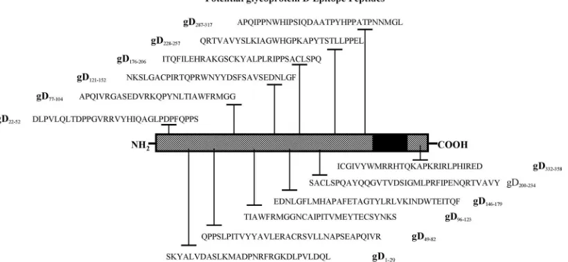
dicted 12 potential epitope peptides from HSV-1 gD that are specific to 10 HLA-DR molecules representing the majority of the world population (2). Here we demonstrated that 6 of the 12 potential epitope peptides bind in vitro with high affinity to 6 of the 10 HLA-DR molecules tested. Four gD epitope pep-tides recalled functional HSV-specific gamma interferon
(IFN-␥)-producing CD4⫹ T cells in healthy, HSV-seropositive, asymptomatic individuals and were recognizable by virus-primed CD4⫹T cells in HLA-DR*0101 and HLA-DR*0401 transgenic (Tg) mice following ocular infection with HSV. In-terestingly, three gD peptides (gD49-82, gD77-104, and
gD121-152) that recalled stronger CD4⫹ T-cell responses in
HSV-seropositive women than in men also recalled stronger CD4⫹T-cell responses in HSV-infected age-matched female compared to male HLA-DR*0101 and HLA-DR*0401 Tg mice. Finally, a cocktail of three immunodominant, gender-dependent gD peptide epitopes elicited CD4⫹ T-cell-depen-dent immunity against lethal herpesvirus infection in HLA-DR*0101 Tg mice.
MATERIALS AND METHODS
Study population. From August 2003 to October 2007, we screened 283 HSV-1- and/or HSV-2-seropositive individuals. Among these, a cohort of 87 immunocompetent individuals, with an age range of 18 to 63 (median, 31) years, who were seropositive or seronegative for HSV-1 and/or HSV-2 were enrolled in the present study. For the characteristics of this study population with respect to sex, age, and HSV serology, see Table 3. Fifty-seven individuals were white, 30 were nonwhite (African, Asian, Hispanic, or other), 43 were females, and 44 were males. All patients were negative for HIV and hepatitis B virus and had no history of immunodeficiency. Twenty-eight patients were HSV-1 seropositive and HSV-2 seronegative, 10 patients were HSV-2 seropositive and HSV-1 seroneg-ative, 9 patients were both HSV-1 and HSV-2 seropositive, and 40 were HSV seronegative. All of the patients selected for this study were healthy and asymp-tomatic (i.e., seropositive with no history of recurrent ocular, orofacial, or genital herpes disease). All subjects were enrolled at the University of California Irvine under an institutional review board-approved protocol. All subjects provided written informed consent.
HSV-1 and HSV-2 seropositivity screening.The sera collected from 61 pa-tients were tested for HSV-1 and HSV-2 status with HerpeSelect
immunoglob-ulin G1 and G2 HSV enzyme-linked immunosorbent assay (ELISA) kits (Focus Diagnostics, Cypress, CA) as previously described (30). The sensitivities and specificities of these ELISAs were 91.2% (HSV-1) to 96.1% (HSV-2) and 92.3% (HSV-1) to 97% (HSV-2). Although this assay generally gives a clear-cut result, in some instances the serotyping was also validated by Western blotting as previously described (63).
Bioinformatic analysis.The full-length gD sequence (26) was loaded into the new prediction software TEPITOPE to predict promiscuous epitopes (19). The TEPITOPE algorithm is a Windows application that is based on 25 quantitative matrix-based motifs that cover a significant part of the human HLA class II peptide binding specificity (58). The algorithm permits the prediction and par-allel display of ligands for each of the 25 HLA-DR par-alleles. The TEPITOPE prediction threshold was set at 5%, and 12 regions predicted to bind a minimum of 50% of the major histocompatibility complex (MHC) class II molecules were picked (Fig. 1). The HLA-DR-restricted peptide epitopes derived from OVA (OVA323-339) (45) and from gB (gB161-175) (ATMYYKDVTVSQVWF) were
used for control of specificity. Peptide gD253-278(LPPELSETPNATQPELAPE
DPEDSAL) from region gD257-287and peptide gB728-761(NAAMFAGLGAFF
EGMGDLGRAVGKVVMGIVGGVV), which were not picked by the
TEPITOPE program as containing potential T-cell epitopes, were synthesized and used as negative controls.
Peptides and immunization.A total of 12 gD peptides were selected by TEPITOPE as previously described (2). Each peptide, consisting of 27 to 34 amino acids, was synthesized as previously described (1, 3–7, 14, 27, 59). gB (ATMYYKDVTVSQVWF) and OVA T helper peptides were used as control peptides. The purity of peptides was 96%, as determined by reversed-phase
high-performance liquid chromatography (Vydac C18) and mass spectroscopy
(Voyager matrix-assisted laser desorption ionization–time of flight mass spec-trometry system). Stock solutions were made at 1 mg/ml in water, except for
peptide gD146-179, which was solubilized in 5% dimethyl sulfoxide. All peptides
were aliquoted and stored at⫺20°C until assayed. All in vivo immunization
studies were conducted with the immunogen emulsified in CpG1826adjuvant,
which was immediately injected subcutaneously into mice as recently described (10, 12, 53, 62).
Virus.The McKrae strain of HSV-1 and strain 333 of HSV-2 were used in this study. The virus was triple plaque purified and was prepared as previously described (54, 56). UV-inactivated HSV-1 and HSV-2 were made by exposing the live virus to a Philips 30-W UV bulb for 10 min at a distance of 5 cm. Heat-inactivated virus was made by heating a virus solution at 100°C for 5 min. HSV inactivation was confirmed by the inability to produce plaques when tested on Vero cells as previously described (11, 52).
MHC-peptide binding assays.HLA-DR molecules were purified from homol-ogous Epstein-Barr virus-infected cell lines by affinity chromatography with mono-morphic monoclonal antibody (MAb) L243 coupled to protein A-Sepharose CL-4B
FIG. 1. Illustration of HSV-1 gD showing the relative positions of T-cell epitope peptides used in this study. HSV-1 (strain 17) gD regions carrying potential human class II-restricted T-cell epitopes predicted by the TEPITOPE computer-assisted algorithm based on known HLA-peptide-TCR interactions are shown. The amino acid sequence, in single-letter code, and the peptide positions based on the full-length sequence of gD are shown. The black box represents the transmembrane domain.
VOL. 15, 2008 HUMAN CD4⫹ CYTOTOXIC T-LYMPHOCYTE EPITOPES IN HSV-1 gD 1437
on August 17, 2020 by guest
http://cvi.asm.org/
gel (Amersham Pharmacia Biotech, Orsay, France) as described previously (70, 71). Binding to HLA-DR molecules was assessed by competitive ELISA as previously reported (71). HLA-DR molecules were diluted with an appropriate biotinylated
peptide and serial dilutions of competitor peptides. Samples (100l/well) were
incubated in 96-well polypropylene plates (Nunc, Roskilde, Denmark) at 37°C for 24 to 72 h. After pH neutralization, samples were applied to 96-well MaxiSorp ELISA
plates (Nunc) previously coated with 10g/ml MAb L243 for HLA-DR molecules.
Bound biotinylated peptides were detected by streptavidin-alkaline phosphatase (Amersham, Little Chalfont, United Kingdom) with 4-methylumbelliferyl phosphate as the substrate (Sigma Chemical Co., St. Quentin Fallavier, France). Fluorescence (excitation at 365 nm, emission at 450 nm) was measured on a Wallac Victor2 1420 multilabel counter (Perkin-Elmer, Courtaboeuf, France). Maximal binding was de-termined by incubating the biotinylated peptide with the MHC II molecule in the absence of competitor. Data were expressed as the concentration of peptide that
prevented binding of 50% of the labeled peptide (IC50). Binding specificity for each
HLA II was ensured by the choice of the biotinylated peptides as described previ-ously (71). Unlabeled forms of the biotinylated peptides were used as reference
peptides to assess the validity of each experiment. Their IC50variation did not
exceed a factor of 3. Their sequences and IC50s were the following: HA 306-318
(PKYVKQNTLKLAT) for DRB1*0101 (2 nM), DRB1*0401 (42 nM), DRB1*1101 (52 nM), and DRB5*0101 (16 nM); YKL (AAYAAAKAAALAA) for DRB1*0701 (6 nM); A3 152-166 (EAEQLRAYLDGTGVE) for DRB1*1501 (13 nM); MT 2-16 (AKTIAYDEEARRGLE) for DRB1*0301 (305 nM); B1 21-36 (TERVRLVTRH IYNREE) for DRB1*1301 (276 nM); LOL 191-210 (ESWGAVWRIDTPDKLTG PFT) for DRB3*0101 (12 nM); and E2/E168 (AGDLLAIETDKATI) for DRB4*0101.
Virus challenge.Ocular challenge was performed by dropping of 2⫻105
PFU
of HSV-1 (McKrae) in 4l tissue culture medium on each eye, and gently
rubbing the lid against the eye for 30 s.
PBMC isolation and HLA-DR stereotyping.Peripheral blood mononuclear cells (PBMC) were prepared as previously described (5, 6, 27), by density gra-dient centrifugation of leukapheresis blood products provided by healthy indi-viduals and commercially available from General Clinical Research Center, University of California Irvine Medical Center (Orange, CA). The cells were washed in Hanks balanced salt solution and resuspended in complete medium (CM) consisting of RPMI 1640 medium containing 10% fetal bovine serum
(FBS) with 15 mM HEPES, 5⫻10⫺5M-mercaptoethanol, 2 mM glutamine,
50 U of penicillin, and 50g of streptomycin (GIBCO-BRL, Grand Island, NY).
Aliquots of freshly isolated PBMC were cryopreserved in 90% FBS and 10% dimethyl sulfoxide in liquid nitrogen for future experiments. The HLA-DR status
of the blood samples was confirmed by indirect staining with 10l of
anti-HLA-DR MAbs BB7.2 and MA2.1 (1:10 dilution of culture supernatant) (ATCC) at 4°C for 30 min, followed by 30 min of incubation with fluorescein isothiocyanate-conjugated goat anti-mouse antibodies (BD Bioscience, San Jose, CA) and analyzed on a FACScalibur analyzer (BD Bioscience).
HLA-DR Tg mice and immunization.Six- to eight-week-old HLA-DR1 and HLA-DR4 Tg mice, established on the susceptible BALB/c genetic background, were used in all experiments. HLA identification of HLA-DR1 and HLA-DR4 mice was confirmed by staining with fluorescein isothiocyanate-conjugated anti-mouse HLA-DR1 and HLA-DR4 antibodies at 4°C for 30 min, followed by analysis on a FACScalibur analyzer (BD Bioscience). Groups of five sex- and age-matched mice per strain were immunized subcutaneously with a mixture of
four gD peptides (100g each) in 20g CpG adjuvant on days 0 and 14.
IFN-␥ELISPOT assays.Human PBMC were cultured in 24-well plates for 5
days in a humidified 5% CO2atmosphere with heat-inactivated HSV-1
(multi-plicity of infection [MOI]⫽5) and subsequently analyzed in an IFN-␥
enzyme-linked immunospot (ELISPOT) assay. Functional T-cell recognition IFN-␥
ELISPOT assays were performed with the human CD4⫹T-cell IFN-␥ELISPOT
MAb set (BD PharMingen, San Diego, CA). Briefly, on day 5, 96-well multi-screen immunoprecipitation plates were blocked with RPMI 1640 medium
sup-plemented with 10% FBS for 2 h at room temperature. Cells (2⫻105/well) were
added in triplicate to MAb-coated plates and incubated with 12 individual gD
peptides for an additional 18 to 24 h at 37°C in 5% CO2. Plates were then washed
with phosphate-buffered saline and supplemented with a detection peroxidase-labeled antibody, followed by a substrate according to the manufacturer’s in-structions. The developed spots were counted under a light microscope. For
HLA-DRTg mice, spleen cells were cultured in 24-well plates for 5 days in a
humidified 5% CO2atmosphere with heat-inactivated HSV-1 (MOI⫽5) and
HSV-2 (MOI ⫽ 5), respectively, and subsequently analyzed in an IFN-␥
ELISPOT assay. Functional T-cell recognition IFN-␥ELISPOT assays were
performed with the mouse IFN-␥ELISPOT MAb pair (BD PharMingen, San
Diego, CA). Briefly, on day 4, 96-well multiscreen immunoprecipitation plates
were coated by incubation overnight at 4°C with 100l (1:250) of anti-IFN-␥
capture MAb. Plates were then blocked with RPMI 1640 medium supplemented
with 10% FBS for 2 h at room temperature. Cells (5⫻104/well) were added in
triplicate to MAb-coated plates and incubated for an additional 18 to 24 h at
37°C in 5% CO2. Plates were then washed with phosphate-buffered saline and
supplemented with a detection peroxidase-labeled antibody, followed by a sub-strate according to the manufacturer’s instructions. The developed spots were counted under a light microscope.
T-cell proliferation assay.We first performed a dose-response study by
incu-bating each peptide at 0.3, 1, 3, 10, and 30g/ml with either human- or
mouse-derived T cells. Although all doses of the positive peptides induced significant
T-cell responses, 1g/ml induced the optimal response of human T cells while
30g/ml induced optimal T-cell responses in mice (data not shown).
Accord-ingly, the subsequent experiments were carried out with these optimal doses.
Human PBMC were cultured in 96-well plates at 5⫻105
/well in CM with 12
individual gD peptides at a 1g/ml concentration, with heat-inactivated HSV-1
and HSV-2 (MOI, 0.3), or with phytohemagglutinin (PHA) as a positive control.
The PBMC suspensions were incubated for 72 h at 37°C in 5% CO2, and their
proliferation was determined in a [3H]thymidine incorporation assay as
de-scribed previously (1, 3, 4, 7, 14, 59). ForHLA-DRTg mice, 14 days after the
second immunization, spleen cells were removed and placed in ice-cold,
serum-free HL-1 medium supplemented with 15 mM HEPES, 5⫻10⫺5
M
-mercap-toethanol, 2 mM glutamine, 50 U of penicillin, and 50g of streptomycin
(GIBCO-BRL, Grand Island, NY) (1, 3, 4, 7, 14, 59). As for the human T-cell responses described above, we also performed a dose-response study with mouse spleen-derived T cells together with each peptide at 0.1, 0.3, 1, 3, 10, and 30
g/ml (data not shown). Although all six doses induced T-cell responses of
similar magnitudes, the optimal response was obtained with 30g/ml.
Accord-ingly, subsequent murine experiments were carried out with the middle dose of
30g/ml. The cells were cultured in 96-well plates at 5⫻105/well in CM with 12
individual gD peptides at a 30g/ml concentration as previously described (1, 3,
4, 7, 14, 59). The cell suspensions were incubated for 72 h at 37°C in 5% CO2.
One microcurie of [3
H]thymidine (DuPont NEN, Boston, MA) was added to each well for the last 18 h of culture. The incorporated radioactivity was deter-mined by harvesting cells onto glass fiber filters and counting the radioactivity on a Matrix 96 direct ionization counter (Packard Instruments, Meriden, CT) (1, 3, 4, 7, 14, 59). Results were expressed as the mean counts of cell-associated
[3
H]thymidine per minute recovered from wells containing Ag minus the mean
counts of cell-associated [3H]thymidine per minute recovered from wells without
Ag (⌬cpm; average of triplicate measurements). The stimulation index (SI) was
calculated as the mean counts of cell-associated [3H]thymidine per minute
re-covered from wells containing Ag divided by the mean counts of cell-associated
[3
H]thymidine per minute recovered from wells without Ag (average of triplicate
measurements). For all experiments, the irrelevant control peptide gB728-761and
the T-cell mitogen concanavalin A (Sigma, St. Louis, MO) were used as negative and positive controls, respectively. Proliferation results were confirmed by re-peating each experiment twice. A T-cell proliferative response was considered
positive when the⌬cpm value was⬎1,000 and when the SI was⬎2, as previously
reported (5, 6, 16, 27, 29). Three of the peptides (gD49-82, gD77-104, and
gD121-152) recalled significantly higher responses in CD4⫹T cells from
asymp-tomatic, HSV-seropositive women than in those from men. A pool of these three
epitope peptides induced a gender-dependent protective CD4⫹
T-cell-depen-dent immunity against HSV challenge in HLA-DR*0101 and HLA-DR*0401 Tg mice.
Statistical analysis.The figures represent data from two or three independent
experiments. The data were expressed as the mean⫾the standard error of the
mean and were compared by using Student’sttest on a STATVIEW II statistical
program (Abacus Concepts, Berkeley, CA) and two-way analysis of variance (ANOVA) on a GraphPad Prism 4. Differences were considered significant when
Pwas⬍0.05. AllPvalues were two tailed unless stated otherwise.
RESULTS
In vitro binding of potential CD4ⴙT-cell epitope peptides
from selected gD to HLA-DR molecules. With the computer
program TEPITOPE, we previously found 12 predicted DR-re-stricted epitope peptides from HSV-1 gD (2). The predicted pep-tides were synthesized and tested in vitro for binding to 10 soluble HLA-DR molecules (Table 1). This panel of available HLA-DR molecules consists of the most common HLA class II haplotypes, regardless of ethnicity (32). The HLA-DRB1 locus encodes seven
1438 ZHANG ET AL. CLIN. VACCINEIMMUNOL.
on August 17, 2020 by guest
http://cvi.asm.org/
of the HLA-DR molecules (HLA-DRB1*0101, HLA-DRB1* 0301, HLA-DRB1*0401, HLA-DRB1*0701, HLA-DRB1*1101, HLA-DRB1*1301, and HLA-DRB1*1501). The HLA-DRB3, HLA-DRB4, and HLA-DRB5 molecules were encode by a sec-ond HLA-DRB locus. The relative binding capacity (nanomolar IC50) for each peptide was calculated as the concentration of competitor peptide required to inhibit 50% of the binding of an allele-specific biotinylated peptide (indicator peptide) as de-scribed in Materials and Methods. Based on an upper threshold of 100 nM, which characterizes high-affinity peptide binders (17, 32), six peptides (gD1-29, gD49-82, gD96-123, gD121-152, gD176-206, and gD200-234) bound to five or more different HLA-DR mole-cules, five peptides (gD22-52, gD77-104, gD146-179, gD228-257, and
gD332-358) bound to four different HLA-DR molecules, and one
peptide (gD287-317) did not bind to any of the HLA-DR molecules tested. This suggests that 11 of the 12 predicted peptides contain at least one universal T-cell epitope or several overlapping epitopes presented by multiple HLA-DR molecules.
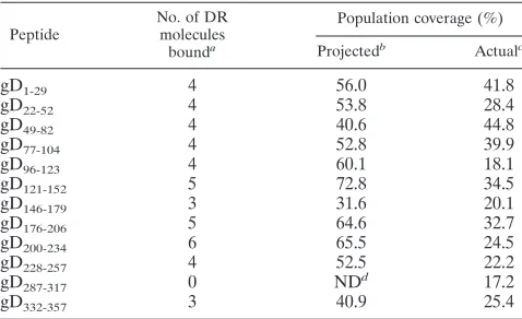
The projected population coverage of each of the 12 gD epitope peptides was calculated on the basis of the phenotypic frequencies of the HLA-DR haplotypes (Table 2), assuming that these molecules are representative of all subtypes of the same haplotype. A combination of the six promiscuous epitope peptides that bound to five or more HLA-DR molecules would be predicted to cover a large proportion of the human popu-lation, regardless of ethnicity.
Recall of gD epitope-specific CD4ⴙT cells in asymptomatic, HSV-seropositive individuals.The cohorts of 55 (22 seroneg-ative and 33 seropositive) individuals used to study T-cell pro-liferative responses (Tp-cell responses) to the 12 gD epitopes are shown in Table 3, column 2. All 33 seropositive individuals who had well-defined clinical histories with one or no episodes of recurrent disease/year (i.e., “protected asymptomatic” or “low-recurrent disease”) were healthy and hepatitis B virus and HIV seronegative. At the time of the study, none of them had had any active clinical ocular or genital herpes symptoms
for at least 1 year. Based on their ethnic backgrounds, the 55 individuals were divided into 33 Caucasians (i.e., white) and 20 non-Caucasians (i.e., nonwhites or Asians, blacks, Hispanics, and members of other ethnic minorities).
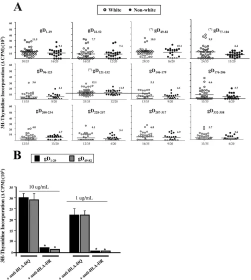
The six highest Tp-cell responses were recalled by gD1-29,
gD22-52, gD49-82, gD77-104, gD121-152, and gD176-206(Fig. 2A).
These six peptides also had medium to strong binding to mul-tiple HLA-DR molecules. This suggests that each of these peptides bears a minimum of one promiscuous T-cell determi-nant or several overlapping determidetermi-nants recognized by human T cells from multiple ethnicities. In contrast, gD96-123 and
TABLE 1. HSV-1 gD-derived human epitope binding to soluble HLA-DR molecules
Epitope
HLA-DR and HLA-DP binding capacitya(IC
50关nM兴)
HLA-DR1 B1*0101
(12.9)d
HLA-DR3 B1*0301
(11)
HLA-DR4 B1*0401
(45.2)
HLA-DR7 B1*0701
(29.0)
HLA-DR11 B1*1101
(19.4)
HLA-DR13 B1*1301
(6)
HLA-DR15 B1*1501
(8)
HLA-DRB3 B3*0101
(51.4)
HLA-DRB4 B5*0101
(45.8)
HLA-DRB5 B4*0101
(23.9)
gD1-29
c 58b 79 58 374 648 ⬎100,000 10,954 535 ⬎100,000 7
gD22-52 3 2,492 63 224 25 ⬎100,000 787 5979 397 58
gD49-82
c 3 1,249 93 173 120 ⬎100,000 18 5,000 170 66
gD77-104c 22 2,349 NDe 4 300 ND 25 ⬎100,000 ND 1
gD96-123
c 3 ND 61 37 598 4,762 167 ⬎100,000 1,672 102
gD121-152c 53 66 8 19 289 160 2 226 319 134
gD146-179 40 1,0247 632 316 175 ⬎100,000 35 2,020 743 85
gD176-206c 54 1,342 955 21 5 200 76 25,000 1,803 91
gD200-234
c 4 307 40 200 44 2,049 13 41 3,742 68
gD228-257 1,162 2,392 9,920 20 39 1,587 2 ⬎100,000 1,163 22
gD287-317 3,162 19,494 600 2,449 25,000 ⬎100,000 6,788 5,000 3,256 4,500
gD332-358 150 1,643 5,872 274 5 56 950 2,307 703 31
a
Competitive binding of synthetic gD peptides bearing potential HLA class II epitopes to solubilized HLA-DR and HLA-DP molecules was tested in vitro. Results are expressed as means of HLA binding capacities determined from three independent experiments.
b
Shown in bold are IC50s of⬍100 nM, which indicate gD peptides with high HLA binding capacities.
c
One of six MHC class II-restricted peptide sequences that bind to at least five HLA-DR and two HLA-DP4 polymorphic molecules.
d
Values in parentheses show percent population coverage (allelic frequencies in a Caucasian population). HLA-DP4 alleles (DP401 and DP402) are carried by 75%
of individuals and are the most frequent HLA II alleles worldwide. Two gD peptides (gD121-152and gD176-206) bind with high affinity to the HLA-DP401 molecule,
whereas three peptides (gD49-82, gD96-123, and gD121-152) bind to HLA-DP402.
e
ND means binding of the peptide was not determined due to lack of its solubility.
TABLE 2. Expected and actual population coverages of HLA-DR binding gD epitope peptides
Peptide
No. of DR molecules
bounda
Population coverage (%)
Projectedb
Actualc
gD1-29 4 56.0 41.8
gD22-52 4 53.8 28.4
gD49-82 4 40.6 44.8
gD77-104 4 52.8 39.9
gD96-123 4 60.1 18.1
gD121-152 5 72.8 34.5
gD146-179 3 31.6 20.1
gD176-206 5 64.6 32.7
gD200-234 6 65.5 24.5
gD228-257 4 52.5 22.2
gD287-317 0 NDd 17.2
gD332-357 3 40.9 25.4
a
The panel of HLA-DR molecules is represented by seven molecules encoded by the HLA-DRB1 locus and three molecules encoded by the HLA-DRB3, HLA-DRB4, and HLA-DRB5 loci.
b
Average projected population coverage was calculated considering the phe-notype frequencies of the HLA-DR molecules which bind the corresponding peptide epitope.
c
Actual population coverage was calculated on the basis of both Tp and IFN-␥
responses corresponding to the peptide epitope.
d
ND, not done.
VOL. 15, 2008 HUMAN CD4⫹ CYTOTOXIC T-LYMPHOCYTE EPITOPES IN HSV-1 gD 1439
on August 17, 2020 by guest
http://cvi.asm.org/
gD200-234peptides induced weaker Tp-cell responses, despite their high-affinity binding to four HLA-DR molecules. Ten of the 12 gD peptides induced similar Tp-cell responses in white and nonwhite populations. Only gD77-104 and gD176-206 ap-peared to induce higher Tp-cell responses in white than in nonwhite individuals (P ⬍ 0.05). No differences in Tp-cell responses were detected in individuals seropositive for HSV-1 compared to HSV-2 or compared to HSV-1/HSV-2 (data not shown). None of the 12 gD peptides induced positive prolifer-ative response of CD4⫹T cells from the 22 HSV-seronegative individuals, regardless of gender (Fig. 3D). As a control, CD4⫹ T cells from all 55 individuals produced positive responses to the mitogens LeuA and PHA (i.e., SIs of 32 to 45 and⌬cpm values of 54⫻103to 62⫻103). However, CD4⫹T cells alone, without Ag stimulation, produced low background responses (i.e., SIs of 3.8 to 4.3 and⌬cpm values of 3⫻103to 5⫻103). The HLA-DR dependence of the peptide-specific Tp-cell responses was confirmed in a representative HLA-DR block-ing experiment, as shown in Fig. 2B. Blockage of HLA-DR molecules resulted in a⬎95% reduction (P⬍0.005) in Tp-cell responses to either 10 or 1g/ml gD1-29 or gD49-82 peptide. However, blockage of HLA-DQ molecules did not affect Tp-cell responses induced by either gD1-29or gD49-82peptide (e.g., at 10g/ml, the gD1-29-specific Tp responses were⌬cpm values of 32.9⫻103and 32.1⫻103in the presence and absence of anti-HLA-DQ MAb, respectively).
Gender-dependent CD4ⴙT-cell responses to three gD
pep-tide epitopes in asymptomatic, HSV-seropositive individuals.
IFN-␥ secretion is a marker of T-cell activation that is fre-quently dissociated from the Tp-cell response (5, 6, 27). In addition, HSV-1 and -2 are susceptible to IFN-␥interference (2). To determine whether the T-cell response to the 12 gD peptides is gender dependent, gD epitope-specific IFN-␥ -pro-ducing CD4⫹T-cell responses were examined in a larger
co-hort of age-matched, HSV-seropositive, asymptomatic individ-uals composed 43 women and 44 men (Table 3, column 3). A preliminary analysis of IFN-␥-producing CD4⫹ T cells re-vealed that 95 or more spots/104 CD4⫹T cells gave a 97% probability of defining a positive response. Of the 960 separate ELISPOT wells used to screen 40 HSV-seronegative individ-uals, only 32 wells (3%) exceeded 95 spot-forming cells (SFC)/ 104CD4⫹T cells. Of the 1,128 separate ELISPOT assays used to screen the 47 seropositive individuals, 192 (17%) had read-ings above 95 SFC/104 CD4⫹T cells. A value of 95 SFC/104 CD4⫹T cells was therefore used as the cutoff point for sub-sequent analyses.
Three epitope peptides (gD49-82, gD77-104, and gD121-152) induced significantly higher CD4⫹Tp responses in HSV-sero-positive women than in men (P⬍0.05) (Fig. 3A). All three of the epitope peptides that induced higher Tp responses in women than in men and two additional epitope peptides
(gD200-234and gD228-257) induced significantly higher levels of
IFN-␥-producing CD4⫹ T-cell responses in women than in men (P⬍0.05) (Fig. 3B). The largest gender-dependent dif-ference appeared to occur with gD77-104(P ⬍ 0.0001). The levels of IFN-␥production in response to PHA were similar in men and women (data not shown). To ascertain the epitope specificity of gender-dependent CD4⫹T cells detected in se-ropositive individuals, we used an irrelevant HLA-DR-re-stricted peptide epitope derived from ovalbumin (OVA), i.e.,
OVA323-339(45). The geometric mean of T-cell proliferation in
response to the OVA323-339peptide was low and similarly not significant in both males and females. The IFN-␥response was also HSV epitope specific, since no IFN-␥ production was induced when HSV-specific T-cell lines were incubated with the CD4⫹Th OVA323-339peptide (Fig. 3C). Taken together, these results suggest that, compared to those from men, CD4⫹ T cells from HSV-seropositive, asymptomatic women re-sponded to the gD49-82, gD77-104, and gD121-152epitope pep-tides with higher frequency and magnitude.
Gender-dependent CD4ⴙ T-cell responses confirmed in
HLA-DRB1*0101 and HLA-DRB1*0401 Tg mice. Fourteen
days after ocular infection with HSV-1 (2 ⫻ 105 PFU/eye, strain McKrae) or intraperitoneal injection of vaccinia virus expressing HSV-1 gD (VVgD), splenocyte CD4⫹T cells from HLA-DR1 and HLA-DR4 Tg mice were recalled in vitro with the 12 individual epitope peptides and IFN-␥ELISPOT assays as described above. Studies similar to those described above with humans determined that the cutoff for a positive response in mice was 50 spots/104 cells (data not shown). The results were similar for HLA-DR1 and HLA-DR4 mice, as well as with HSV-1 and VVgD (Fig. 4). In all instances in which a positive IFN-␥ response was detected, female mice had a higher response than age-matched male mice. In addition, the same eight peptides produced a positive IFN-␥response re-gardless of the mouse strain or HSV-1 versus VVgD infection. Similar to the case for humans, gender-dependent IFN-␥ -pro-ducing CD4⫹ T-cell responses against the gD1-29, gD49-82,
gD77-104, and gD121-152peptides were detected in HLA-DR Tg
mice (Fig. 4A and B). However, the HLA-DR Tg mice re-vealed a gender-dependent T-cell response against four addi-tional peptides: gD96-123, gD146-179, gD176-206, and gD200-234. Interestingly, although a gender-dependent IFN-␥-producing CD4⫹T-cell response to gD228-257was seen in humans, this
TABLE 3. Cohort of HSV-seropositive but asymptomatic and HSV-seronegative individuals enrolled in this study
Characteristica
Value for all subjects whose sera were studied by:
T-cell proliferation
assay (n⫽55)
IFN-␥ELISPOT
assay (n⫽87)
Gender关no. (%)兴
Females 35 (63.6) 43 (49.4)
Males 20 (36.4) 44 (49.6)
Race关no. (%)兴
Whites 35 (63.6) 57 (65.5)
Nonwhites 20 (36.4) 30 (34.5)
Age (yr)
Median 31 31
Range 18–63 18–63
HSV status关no. (%)兴
HSV-1 positive 19 (34.5) 28 (32.2)
HSV-2 positive 8 (14.5) 10 (11.5)
HSV-1 and -2 positive 6 (10.9) 9 (10.3)
HSV negative 22 (40) 40 (46)
a
The HSV-seropositive, asymptomatic individuals enrolled in this study had one or no episodes of recurrent disease/year and were healthy and hepatitis and HIV negative.
1440 ZHANG ET AL. CLIN. VACCINEIMMUNOL.
on August 17, 2020 by guest
http://cvi.asm.org/
FIG. 2. CD4⫹T-cell proliferative response to HSV-1 gD-derived epitopes. (A) Fresh peripheral blood-derived CD4⫹T cells were isolated and stimulated in vitro with 12 individual gD peptides in a 6-day proliferation assay as described in Materials and Methods. The Tp-cell responses of Caucasian (white) and non-Caucasian (nonwhite) ethnic groups against each peptide, assessed by a [3H]thymidine incorporation assay, were compared. Proliferative responses were expressed as⌬cpm values (counts per minute of cells incubated with peptide⫺counts per minute of cells without peptide). The mean of each group is shown as a solid horizontal line. An analysis of T-cell responses in 22 seronegative individuals is shown. A positive T-cell proliferative response was defined on the basis of the responses of HSV-seronegative control individuals, with a⌬cpm value of
ⱖ1,000 and an SI (counts per minute of cells incubated with peptide/counts per minute of cells without peptide) ofⱖ2. The values at the bottom of each data set (i.e., 30/35) are the mean number of responders/total number of individuals tested. The value at the top of each data set (i.e., 11.5) is the mean SI of each of the two groups, i.e., white or nonwhite. ThePvalues compare the Tp-cell responses between white and nonwhite groups by one-way ANOVA. An asterisk in parentheses indicates a peptide that induced higher T-cell proliferation compared with HSV-seronegative control individuals (i.e., significantly higher SI and⌬cpm value). (B) Analysis of HLA-DR restriction and functional frequencies of HSV-1 gD peptide-specific CD4⫹T cells. Blocking of PBMC proliferation in response to the gD1-29and gD49-82peptides by specific anti-HLA-DR MAb.
1441
on August 17, 2020 by guest
http://cvi.asm.org/
1442
on August 17, 2020 by guest
http://cvi.asm.org/
epitope peptide did not induce any significant response in mice. The IFN-␥responses against gD peptides detected in HSV-1-infected HLA Tg mice were specific, since no IFN-␥ production was observed when gender-dependent gD49-82-,
gD77-104-, and gD121-152-specific T-cell lines were incubated in
vitro with a heterologous HLA-DR-restricted T-cell epitope from HSV-1 gB (i.e., gB161-175) (Fig. 4C).
The IFN-␥production was abrogated by anti-HLA-DR, but not by anti-HLA-A2.1, MAbs (i.e., in HLA-DR1, IFN-␥ ELISPOT response against gD49-82peptide; 152 spot-forming cells [SFC] without MAb reduced to 33 SFC and 148 SFC when anti-HLA-DR and anti-HLA-A, -B, and -C were added, respectively). The IFN-␥ response was gD peptide specific, since no IFN-␥ production was observed in response to a heterologous gB CD4⫹ Th peptide (Fig. 4C). Overall these results confirm that, similar to humans, a gender difference in CD4⫹T-cell responses was detected in age-matched HLA-DR1 and HLA-DR4 Tg mice against the gD1-29, gD49-82, gD77-104, and
gD121-152peptides following either HSV infection or VVgD
im-munization.
Immunization with a pool of immunodominant gD epitope
peptides induced CD4ⴙT-cell-dependent protective immunity
against lethal herpesvirus infection in HLA-DR*0101 Tg mice.
Three epitope peptides (gD49-82, gD77-104, and gD121-152) were selected as the “best” overall on the basis of three criteria: (i) high-affinity binding to multiple HLA class II molecules, in-cluding HLA-DRB1*0101 (Table 1); (ii) strong recall of CD4⫹ Tp-cell and IFN-␥responses in HSV-seropositive, asymptom-atic individuals (Fig. 2 and 3); and (iii) high recall of IFN-␥ -producing CD4⫹T cells in HLA-DRB1*0101 Tg mice (Fig. 4). A pool of these three peptides was then used to immunize susceptible HLA-DR1 Tg mice against a lethal HSV-1 chal-lenge. The previously described protective epitope gD1-29(20, 21, 53, 68, 86), which exhibited similar properties, was excluded from these experiments. A pool of the gD146-179, gD287-317, and
gD332-358subdominant peptides was used as a control.
Three groups of 20 HLA-DR*0101 Tg female mice were immunized with (i) a pool of the gD49-82, gD77-104, and
gD121-152peptides emulsified in CpG1826adjuvant; (ii) a pool
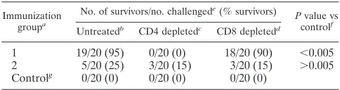
of the gD146-179, gD287-317, and gD332-358peptides emulsified in CpG1826adjuvant; or (iii) the CpG1826adjuvant alone (mock-immunized control). Two weeks after the second immuniza-tion, mice in each group were challenged ocularly with a lethal dose of 5⫻ 105 PFU of HSV-1 (McKrae strain). All of the mice that died following the challenge did so between days 8 and 14 postinfection. The HLA-DR*0101 Tg mice immunized with the immunodominant CD4⫹T-cell epitope peptide pool were protected against death, compared to those immunized with the subdominant CD4⫹T-cell epitope peptide pool (95% versus 25% survival,P⬍0.05) or adjuvant alone (0% survival) (Table 4). When the mice immunized with the immunodomi-nant CD4⫹ T-cell epitope peptide pool were depleted of CD4⫹T cells prior to virus challenge, survival was significantly
decreased (Table 4). In contrast, depletion of CD8⫹T cells had no significant effect. Depletion of either T-cell type had no significant impact on the mock immunized or subdominant immunized groups. Thus, with this immunization regimen, CD4⫹T cells were required and CD8⫹T cells were not suf-ficient for protective immunity against lethal ocular HSV-1 challenge.
To assess a gender dependence in protective immunity in-duced by gD peptides, 10 age-matched HLA-DR*0101 Tg female and male mice were immunized with the individual
gD49-82, gD77-104, and gD121-152peptides emulsified in CpG1826
adjuvant. Two weeks after the second immunization, both gen-ders were similarly challenged ocularly with a lethal dose of HSV-1 as described above. The HLA-DR*0101 Tg female mice immunized with the immunodominant CD4⫹ T-cell
gD49-82, gD77-104, and gD121-152peptides showed better
pro-tection against death compared to male mice immunized with the same epitopes (65% versus 35% survival for gD49-82, 60% versus 25% survival for gD77-104, and 55% versus 20% survival for gD121-152, respectively). This result indicates, similar to their T-cell immunogenicity, that there was also a gender-dependent protective immunity induced by the immunodomi-nant CD4⫹T-cell gD49-82, gD77-104, and gD121-152peptides.
DISCUSSION
We report here the identification of a panel of HLA-DR-restricted epitope peptides derived from HSV-1 gD, a glyco-protein that produces protective immunity against herpesvirus infection and disease in animal models and humans (20, 21, 53, 68, 86). These epitopes previously identified by screening HSV-1 gD for peptides that contain the HLA-DR supertype binding motif (2). We demonstrated that 11 of the 12 predicted peptides bound with high affinity to three or more different types of HLA-DR molecules. Three of them (i.e., gD49-82,
gD77-104, and gD121-152) recalled significantly higher responses
in CD4⫹T cells from asymptomatic, HSV-seropositive women than in those from men. A pool of these three epitope peptides induced a gender-dependent protective CD4⫹ T-cell-depen-dent immunity against HSV challenge in HLA-DR*0101 and HLA-DR*0401 Tg mice.
In humans, HSV-specific CD4⫹T cells play a crucial role in controlling both primary and recurrent infections (23, 60). In the late 1980s and early 1990s, Zarling and coworkers gener-ated several human CD4⫹T-cell clones from HSV-1-seropos-itive, asymptomatic individuals by stimulating peripheral blood lymphocytes (PBL) with a recombinant vaccinia virus express-ing HSV-1 gD (83, 84). Five of these CD4⫹T-cell clones lysed autologous HSV-1-infected lymphoblastoid cell lines, and their cytotoxicity was restricted to HLA class II molecules. Later, it was demonstrated that some of these HSV-1-specific T-cell clones were directed against both gD-1 and gD-2 by using target cells infected with wild-type HSV strains, a gC deletion
FIG. 3. Gender differences in Tp-cell responses to HSV-1 gD epitope peptides detected in healthy, asymptomatic, HSV-seropositive individ-uals. (A and D) Peptide-specific Tp-cell responses in HSV-1- and/or HSV-2-seropositive, asymptomatic, healthy individuals were examined as described in the legend to Fig. 2. ThePvalues in panel A indicate statistically significant differences in Tp-cell responses between men and women (P⬍0.05, ANOVA test). Gender dependence of IFN-␥-producing CD4⫹T cells specific to 12 gD peptides (B) and to an irrelevant CD4⫹T-cell epitope peptide from OVA (OVA323-339) (C) was detected by ELISPOT in HSV-seropositive, asymptomatic individuals. PBMC derived from HSV-1/HSV-2-seropositive, asymptomatic men and women were stimulated with 12 gD peptides for 5 days, and IFN-␥ELISPOT assays were performed as described in Materials and Methods.
1443
on August 17, 2020 by guest
http://cvi.asm.org/
1444
on August 17, 2020 by guest
http://cvi.asm.org/
mutant of HSV-1, and an HSV-1⫻HSV-2 recombinant virus (81). In another study, purified HSV-1 gD, expressed in mam-malian cells, stimulated proliferation of, and interleukin-2 (IL-2) production by, PBL of HSV-seropositive individuals, indicating the presence of memory T cells to gD in HSV-seropositive individuals (82). In addition, T-cell clones gener-ated by stimulation of PBL with HSV-1 were found to prolif-erate in response to stimulation with gD-1 in the absence of exogenous IL-2 and to lyse HSV-1- or HSV-1/HSV-2-infected autologous target cells (83).
HSV-specific cytotoxic CD4⫹clones have been recovered ex vivo from HSV-2 lesions of five patients by Corey, Koelle, and coworkers (40, 41). Cunningham and coworkers have also re-ported that HSV gD is a major target for CD4⫹cytotoxic T lymphocytes with human epidermal keratinocytes as targets (49, 50). Although the above findings demonstrated that gD is a major target for human HSV-specific CD4⫹T cells, no spe-cific gD minimal epitopes have ever been defined. Therefore, to our knowledge, the present study reports the first six immu-nodominant HLA-DR-restricted human CD4⫹T-cell epitopes on gD. However, results reported here do not imply that these are the only human CD4⫹ T-cell epitopes present on gD. Cunningham and collaborators have recently reported finding three epitopes on HSV-2 gD by the complementary overlap-ping peptide method, with some but not all predicted by the TEPITOPE algorithm (32nd Annual International
Herpesvi-rus Workshop, 2007). Our approach to epitope identification is complementary to Cunningham’s work, and because it was tailored specifically for HLA-DR epitopes, it could represent a model system that could be applied to any HLA-DR haplotype and to any other herpesvirus structural or regulatory protein. The minimum requirements needed to utilize this approach are (i) existing sequence data for the proteins in question; (ii) access to the genome (DNA or RNA) of the pathogen for the purpose of generating recombinant vaccinia virus vectors; (iii) HSV-seropositive, asymptomatic individuals; and (iv) HLA Tg mice. Moreover, the present strategy accurately identified re-gions bearing all of the previously reported gD epitopes: gD1-23 (24, 28, 36), gD241-260(34, 48, 80), and gD290-314(24). There-fore, our approach to epitope identification, as well as testing epitope peptides for their T-cell antigenicity, immunogenicity, and protective efficacy, should be nearly universal, regardless of the protein studied.
Recently, we reported four immunodominant H2d-restricted CD4⫹T-cell epitope peptides on HSV-1 gD (2). Among these, the peptide gD49-82, a naturally processed epitope, protects H2dmice against lethal ocular HSV-1 challenge (2). We have also recently found that the gD49-82epitope contains an 8-mer,
gD53-61, that represents an immunodominant
HLA-A2.1-re-stricted human CD8⫹T-cell epitope (18). Therefore, a mini-mum of two human epitopes, the gD53-61CD8⫹T-cell epitope and the CD4⫹ T-cell epitope, are nested within the gD49-82 peptide and this epitope peptide represents an excellent can-didate for inclusion in a CD4⫹and CD8⫹T-cell epitope-based human vaccine.
Humans are not immunologically naive and often develop memory T cells that cross-react with and respond to unrelated pathogens or Ags, a phenomenon termed heterologous immu-nity (reviewed in references 64 and 76). Interestingly, although an IFN-␥-producing CD4⫹ T-cell response specific to
gD228-257 was detected in HSV-seronegative individuals, no
significant T-cell responses specific to this epitope peptide were detected in HSV-infected HLA-DR mice. This result suggests that T cells specific to gD228-257detected in humans might cross-react with and respond to a yet-to-be-identified, unrelated-pathogen-derived epitope, a recently described phe-nomenon termed heterologous immunity (reviewed in refer-ences 64 and 76). The processing and presentation to T cells of the gD228-257epitope could also be different between HLA Tg mice and humans. Therefore, some apparently gD-specific CD4⫹T cells identified in this study might have arisen due to cross-reaction with an unrelated pathogen. Such scenarios have been reported in murine CD4⫹T-cell responses to cyto-megalovirus (22, 64, 76) and may be true for HSV (22, 64, 76, 77). To help ascertain whether the detected CD4⫹T-cell re-sponses against the gD epitope peptides were primed by HSV,
FIG. 4. Gender difference in gD epitope-specific CD4⫹T-cell responses detected in HLA-DR1 B1*0101 and HLA-DR4 B1*0401 Tg mice. Age-matched HLA-DR1 B1*0101 (HLA-DR1) and HLA-DR4 B1*0401 (HLA-DR4) Tg mice were infected ocularly with HSV-1 (McKrae) (A) or immunized intraperitoneally with VVgD (B). Spleens were harvest on day 14 postinfection and stimulated with each of the 12 individual peptides for 5 days, and peptide-specific IFN-␥-producing CD4⫹T cells were detected as described in Materials and Methods. (C) CD4⫹T-cell lines specific to gender-dependent gD49-82, gD77-104, and gD121-152were established from HSV-1-infected HLA Tg mice and restimulated in vitro with either the corresponding specific gD peptide or a heterologous HLA-DR-restricted T-cell epitope from HSV-1 gB (i.e., gB161-175). The numbers of SFC producing IFN-␥are shown for each peptide as mean values⫾standard deviations from two independent experiments. ThePvalues are for comparisons of the IFN-␥-producing CD4⫹T-cell responses of age-matched females and males by one-way ANOVA.
TABLE 4. Immunization with HLA-DR-restricted gD epitope peptides in CpG1826adjuvant confers protective immunity
to a lethal HSV-1 challenge in HLA-DR*0101 Tg mice
Immunization
groupa
No. of survivors/no. challengede
(% survivors) Pvalue vs
controlf
Untreatedb CD4 depletedc CD8 depletedd
1 19/20 (95) 0/20 (0) 18/20 (90) ⬍0.005 2 5/20 (25) 3/20 (15) 3/20 (15) ⬎0.005 Controlg 0/20 (0) 0/20 (0) 0/20 (0)
a
HLA-DR*0101 Tg mice, 60 per group, were immunized subcutaneously on
day 0 and day 21 with 100g each of immunodominant gD49-82, gD121-152, and
gD200-234pooled as a group of three peptides and emulsified in CpG1826adjuvant
(group 1) or with 100g each of subdominant gD146-179, gD287-317, and gD332-358
pooled as a group of three peptides (group 2). Control mice received CpG1826
adjuvant alone.
b
Twenty immunized HLA-DR*0101 Tg mice were left untreated.
c
Twenty immunized HLA-DR*0101 Tg mice were depleted of CD4⫹T cells
with a MAb specific to mouse CD4.
d
Twenty immunized HLA-DR*0101 Tg mice were depleted of CD8⫹T cells
with a MAb specific to mouse CD8.
e
Two weeks after the last immunization, all animals received a lethal ocular
challenge with 5⫻105
PFU of HSV-1 (strain McKrae).
f
ThePvalues shown are for comparisons of immunized versus control mice by
ANOVA.
g
Twenty control mice were treated similarly to those in groups 1 and 2, with an irrelevant isotype control MAb.
VOL. 15, 2008 HUMAN CD4⫹ CYTOTOXIC T-LYMPHOCYTE EPITOPES IN HSV-1 gD 1445
on August 17, 2020 by guest
http://cvi.asm.org/
the epitope mapping was extended to pathogen-free HLA-DR1 and HLA-DR4 Tg mice that were infected with either HSV-1 or VVgD. The results suggested that CD4⫹ T cells from both HLA-DR1- and HLA-DR4-infected Tg mice also recognized five of the six epitopes that were reactive to human CD4⫹ T cells (gD1-29, gD49-82, gD77-104, gD121-152, and
gD200-234). In addition, HLA-DRB1*0101 and DRB1*0401 Tg
mice also mounted CD4⫹ T-cell responses against three epitope peptides not recognized by humans. This may reflect the high level of infection in this mouse system. The ability of CD4⫹T cells generated by gD49-82, gD77-104, and gD121-152, epitope peptides to protect against HSV challenge in HLA-DRB1*0101 and DRB1*0401 Tg mice is also a critical param-eter. These mouse studies suggest that it is very likely that the
gD1-29, gD49-82, gD77-104, gD121-152, and gD200-234epitope
pep-tides detected in humans were not cross-reactive with nonher-pesvirus Ags or pathogens but rather were induced following HSV-1 or HSV-2 infection. This does not exclude the involve-ment of other infectious agents in shaping herpesvirus CD4⫹ T-cell responses. It also remains to be determined (i) whether the gender-dependent T-cell responses to gD epitopes will translate in a serotype-specific protective immunity against HSV-1 and/or HSV-2 and (ii) whether a therapeutic immuni-zation with gD epitopes will protect latently HSV-1- and/or HSV-2-infected HLA-DR Tg male and female mice.
gD is a leading herpes vaccine candidate Ag that has been successful in eliciting protective immunity in women but failed to protect men (8, 68, 69). However, the gD T-cell epitopes that might be targeted during this apparent gender-dependent protective immunity have not been reported. In this study, there were gender disparities in the CD4⫹T-cell responses of HSV-seropositive individuals to a discrete set of gD epitopes. Indeed, HSV-seropositive women exhibited higher magnitudes of CD4⫹ T-cell responses to three gD epitopes (gD49-82,
gD77-104, and gD121-152) compared to age- and
ethnicity-matched, HSV-seropositive men. In addition, a gender- and T-cell-dependent protective immunity was induced in suscep-tible HLA-DR Tg mice by the same three immunodominant CD4⫹ T-cell peptide epitopes (gD49-82, gD77-104, and
gD121-152). Thus, results reported here are in agreement with
the gender-dependent protective immunity reported in the above-cited clinical trial and both suggest that sex factors may affect the host’s immune response to herpesvirus epitopes. Although the present study was mainly focused on CD4⫹ epitopes, the possible involvement of gD CD8⫹T-cell epitopes in shaping this gender-dependent protective immunity has not been ruled out. Altogether, these data indicate that gender should be taken into account during the evaluation of T-cell epitope-based herpes vaccines. Boosting the recombinant gD immunization with a discrete set of gD T-cell epitopes may be considered particularly for women, while boosting the immu-nization of men with a different, nonoverlapping set of gD epitopes may be warranted.
The first indication of a gender difference in herpes immu-nity was reported by Cantin and coworkers, who showed that female mice have low-grade infection and disease compared with strain- and age-matched male mice (35). Besides a possi-ble association with the IFN-␥ molecule (35), the immune mechanisms contributing to gender-dependent susceptibility to herpesvirus infection and disease have not been reported. A
recent study by Diamond and coworkers demonstrated a higher frequency of memory T cells specific to human cyto-megalovirus—another virus of the herpesvirus family—that produce higher levels of Th1 cytokines in women than in men (74). On the contrary, Klein and coworkers showed that women maintain lower levels of varicella-zoster virus-specific memory T cells compared to men (37). Although the above studies suggest that gender differences in memory T-cell re-sponses exist for herpesviruses other than HSV-1 and HSV-2, no specific gender-associated Ags or epitopes have ever been defined. Identification of the viral Ags or epitopes recognized by T cells from HSV-seropositive men and women—either vaccinated individuals or individuals with naturally acquired herpes immunity (i.e., seropositive, asymptomatic individu-als)—would facilitate the process of designing an efficient vac-cine. It is possible that gender-dependent CD4⫹ T-cell re-sponses detected in this study reside at the Ag-presenting cell (APC) level rather than at the T-cell level. Indeed, in other systems, female mice that are more susceptible to experimental allergic encephalomyelitis preferentially mount Th1 immune responses and their APCs produce IL-12 but not IL-10 (78). In contrast, the APCs derived from resistant male mice produce IL-10 but not IL-12. Activation of T cells with APCs derived from the opposite sex demonstrated that these cytokines were derived from the respective APC populations (78). Other stud-ies have demonstrated that gender differences in T-cell re-sponses are mediated, in part, by differences in sex hormone-immune interactions (37, 42, 51, 57, 74, 78). Kovats and coworkers were the first to demonstrate that sex hormones such as estrogen play a crucial role in regulating dendritic cell phenotypic and functional differentiation (42, 51, 57). Whether an increase in dendritic cell phenotypic and functional matu-ration leads to the strong gD epitope-specific CD4⫹ T-cell responses detected in seropositive women or to an increase in the function of gD-specific memory T cells remains to be de-termined. Nevertheless, regardless of the mechanisms, our findings are the first demonstrating that a discrete set of gD CD4⫹T-cell epitope peptides are highly targeted in women compared to men and were able to elicit a protective immunity in female HLA-DR Tg mice but not in age-matched male mice. Altogether, these data suggest that gender restrictions to herpes vaccines may be overcome by focusing the immune response toward selected sets of epitopes.
The vast majority of the world’s human population is in-fected with HSV-1 and/or HSV-2 and may benefit from ther-apeutic vaccination approaches designed to induce vigorous HSV-specific cellular immunity at a higher magnitude and breadth than the suboptimal natural immunity. Although the primary goals of this study were to evaluate the antigenicity of the HSV-1 CD4⫹ T-cell epitope peptides and the gender-specific responses to these epitopes, a secondary but important goal is to develop the knowledge of protective herpesvirus T-cell epitopes required for the development of a lipopeptide-based vaccine. Focusing on the immune response toward such epitopes could be of value in the case of HSV-1 and HSV-2 infections, where T cells directed against the immunodominant epitopes might have been inactivated and T cells specific for subdominant epitopes might have escaped T-cell tolerance. A question of practical importance is the translation of the cur-rent immunological findings for the development of an
1446 ZHANG ET AL. CLIN. VACCINEIMMUNOL.
on August 17, 2020 by guest
http://cvi.asm.org/
epitope-based lipopeptide vaccine for a genetically heteroge-neous human population. (1, 4, 7, 53, 59, 85, 86). Accordingly, a herpes vaccine will need to induce responses against a num-ber of promiscuous epitopes that are recognized in the context of many different HLA alleles. Several studies have demon-strated that the majority of HLA-DR molecules can be grouped into broad supertypes with overlapping peptide bind-ing specificities (65). In the case of the HLA-DR molecules, a single superfamily encompassing DRB1 alleles that are ex-pressed in the majority of humans has been defined (67). HLA-DR supertypes have been studied in detail, and several CD4⫹ T-cell epitopes that belong to these supertypes have been identified and validated (9). Broad population coverage can be established, provided that epitopes corresponding to multiple HLA supertype families are incorporated into the vaccine. From the standpoint of vaccine development, the panel of immunodominant gD peptides identified in this study has the potential to induce CD4⫹ T-cell responses in up to 92.9% of an average human population. The fact that each of the peptides was antigenic in the human cohort studied is most likely a result of focusing the screening exclusively on peptides capable of binding multiple HLA-DR alleles. Similar correla-tions have been found between promiscuous HLA-DR binding capacity and antigenicity for HIV peptides (79). Because of their apparent promiscuousness, these CD4⫹ T-cell epitope peptides should prove to be useful in vaccines for the hetero-geneous human population. To maximize efficacy, a multi-epitope-based herpes vaccine should probably include several T-cell epitopes from several different structural glycoproteins and regulatory proteins, each chosen to represent the HLA supertypes known to provide recognition in a large proportion of the global population, regardless of race and ethnicity.
In the past 2 decades, there has been increasing recognition of a worldwide pandemic of herpesvirus infection despite the widespread use of antiviral drug therapies (reviewed in refer-ence 39). Although drug therapy can limit morbidity and mor-tality from active herpes disease, it cannot cure infection. In addition, considering cost, toxicity, and viral escape issues, the usefulness of drug treatment might be limited, especially in underdeveloped regions such as parts of sub-Saharan Africa, where 70% of high-risk, HIV-negative individuals and 85% of HIV-infected individuals are seropositive for herpesvirus (46, 61, 75). Developing an effective immunoprophylactic or immu-notherapeutic vaccine against herpesvirus infection must be an important global health priority. The use of multivalent epitope vaccines is a promising approach to fighting herpesvi-rus diseases, since it allows for directing the immune response to protective epitopes that might have a greater impact on disease outcome. The HLA-DR-restricted HSV-1 gD epitopes reported here meet these criteria and hence should be consid-ered in the design of an effective immunotherapeutic and im-munoprophylactic herpes vaccine for humans.
ACKNOWLEDGMENTS
This work was supported by Public Health Service grants EY14900, EY15225, and EY16663; the Discovery Eye Foundation; the Henry L. Guenther Foundation; and a Research to Prevent Blindness Challenge grant. L. BenMohamed is an RPB Special Award Investigator.
REFERENCES
1.BenMohamed, L., Y. Belkaid, E. Loing, K. Brahimi, H. Gras-Masse, and P. Druilhe.2002. Systemic immune responses induced by mucosal
administra-tion of lipopeptides without adjuvant. Eur. J. Immunol.32:2274–2281.
2.BenMohamed, L., G. Bertrand, C. D. McNamara, H. Gras-Masse, J. Hammer, S. L. Wechsler, and A. B. Nesburn.2003. Identification of novel
immunodom-inant CD4⫹Th1-type T-cell peptide epitopes from herpes simplex virus
glyco-protein D that confer protective immunity. J. Virol.77:9463–9473.
3.BenMohamed, L., H. Gras-Masse, A. Tartar, P. Daubersies, K. Brahimi, M. Bossus, A. Thomas, and P. Druilhe.1997. Lipopeptide immunization with-out adjuvant induces potent and long-lasting B, T helper, and cytotoxic T lymphocyte responses against a malaria liver stage antigen in mice and
chimpanzees. Eur. J. Immunol.27:1242–1253.
4.BenMohamed, L., R. Krishnan, C. Auge, J. F. Primus, and D. J. Diamond.
2002. Intranasal administration of a synthetic lipopeptide without adjuvant
induces systemic immune responses. Immunology106:113–121.
5.BenMohamed, L., R. Krishnan, J. Longmate, C. Auge, L. Low, J. Primus, and D. J. Diamond.2000. Induction of CTL response by a minimal epitope vaccine in HLA A*0201/DR1 transgenic mice: dependence on HLA class II
restricted THresponse. Hum. Immunol.61:764–779.
6.BenMohamed, L., A. Thomas, M. Bossus, K. Brahimi, J. Wubben, H. Gras-Masse, and P. Druilhe.2000. High immunogenicity in chimpanzees of pep-tides and lipopeppep-tides derived from four new Plasmodium falciparum
pre-erythrocytic molecules. Vaccine18:2843–2855.
7.BenMohamed, L., S. L. Wechsler, and A. B. Nesburn.2002. Lipopeptide
vaccines—yesterday, today, and tomorrow. Lancet Infect. Dis.2:425–431.
8.Bernstein, D.2005. Glycoprotein D adjuvant herpes simplex virus vaccine.
Expert Rev. Vaccines4:615–627.
9.Bertoni, R., J. Sidney, P. Fowler, R. W. Chesnut, F. V. Chisari, and A. Sette.
1997. Human histocompatibility leukocyte antigen-binding supermotifs pre-dict broadly cross-reactive cytotoxic T lymphocyte responses in patients with
acute hepatitis. J. Clin. Investig.100:503–513.
10.Bettahi, I., G. Dasgupta, O. Renaudet, A. A. Chentoufi, X. Zhang, D. Car-penter, S. Yoon, P. Dumy, and L. BenMohamed.27 June 2008. Antitumor
activity of a self-adjuvanting glyco-lipopeptide vaccine bearing B cell, CD4⫹
and CD8⫹T cell epitopes. Cancer Immunol. Immunother. [Epub ahead of
print.] doi:10.1007/s00262-008-0537-y.
11.Bettahi, I., A. B. Nesburn, S. Yoon, X. Zhang, A. Mohebbi, V. Sue, A. Vanderberg, S. L. Wechsler, and L. BenMohamed.2007. Protective immu-nity against ocular herpes infection and disease induced by highly immuno-genic self-adjuvanting glycoprotein D lipopeptide vaccines. Investig.
Oph-thalmol. Vis. Sci.48:4643–4653.
12.Bettahi, I., X. Zhang, R. E. Afifi, and L. BenMohamed.2006. Protective immunity to genital herpes simplex virus type 1 and type 2 provided by self-adjuvanting lipopeptides that drive dendritic cell maturation and elicit a
polarized Th1 immune response. Viral Immunol.19:220–236.
13.Blaney, J. E., Jr., E. Nobusawa, M. A. Brehm, R. H. Bonneau, L. M. Mylin, T. M. Fu, Y. Kawaoka, and S. S. Tevethia.1998. Immunization with a single major histocompatibility complex class I-restricted cytotoxic T-lymphocyte recognition epitope of herpes simplex virus type 2 confers protective
immu-nity. J. Virol.72:9567–9574.
14.Bossus, M., L. Benmohamed, A. Londono, B. Barbier, A. Tartar, P. Druilhe, and H. Gras-Masse.1997. Improved detection of human antibodies to a Plasmodium antigen using a peptide modified with Aib residues. J. Pept. Sci.
3:47–53.
15.Bourne, N., F. J. Bravo, M. Francotte, D. I. Bernstein, M. G. Myers, M. Slaoui, and L. R. Stanberry. 2003. Herpes simplex virus (HSV) type 2 glycoprotein D subunit vaccines and protection against genital HSV-1 or
HSV-2 disease in guinea pigs. J. Infect. Dis.187:542–549.
16.Brahimi, K., E. Badell, J.-P. Sauzet, L. BenMohamed, P. Daubersies, C. Gue´rin-Marchand, G. Snounou, and P. Druilhe.2001. Human antibodies
againstPlasmodium falciparumliver-stage antigen 3 cross-react with
Plas-modium yoeliipreerythrocytic-stage epitopes and inhibit sporozoite invasion
in vitro and in vivo. Infect. Immun.69:3845–3852.
17.Castellino, F., and R. N. Germain.2007. Chemokine-guided CD4⫹T cell
help enhances generation of IL-6R␣highIL-7R␣high prememory CD8⫹T
cells. J. Immunol.178:778–787.
18.Chentoufi, A. A., X. Zhang, K. Lamberth, G. Dasgupta, I. Bettahi, A. Nguyen, M. Wu, X. Zhu, A. Mohebbi, S. Buus, S. L. Wechsler, A. B. Nesburn, and L. BenMohamed.2008. HLA-A*0201-Restricted CD8⫹cytotoxic T lym-phocyte epitopes identified from herpes simplex virus glycoprotein D. J.
Im-munol.180:426–437.
19.Consogno, G., S. Manici, V. Facchinetti, A. Bachi, J. Hammer, B. M. Conti-Fine, C. Rugarli, C. Traversari, and M. P. Protti.2003. Identification of
immunodominant regions among promiscuous HLA-DR-restricted CD4⫹
T-cell epitopes on the tumor antigen MAGE-3. Blood101:1038–1044.
20.Cooper, D., J. C. Mester, M. Guo, F. Nasar, V. Souza, S. Dispoto, M. Sidhu, M. Hagen, J. H. Eldridge, R. J. Natuk, and M. W. Pride.2006. Epitope
mapping of full-length glycoprotein D from HSV-2 reveals a novel CD4⫹
CTL epitope located at the transmembrane-cytoplasmic junction. Cell.
Im-munol.239:113–120.
VOL. 15, 2008 HUMAN CD4⫹ CYTOTOXIC T-LYMPHOCYTE EPITOPES IN HSV-1 gD 1447