The Bag6 Complex:
Biological Complexity through Modularity
Thesis by
Jee-Young Mock
In Partial Fulfillment of the Requirements for the degree of
Doctor of Philosophy
CALIFORNIA INSTITUTE OF TECHNOLOGY Pasadena, California
2017
ã 2017
Park, whom I consider to be my second mom. My uncles, Hoyong Park and Sangyong
Park, have raised me as if I were their own and never stopped working to put me through
school. Without their hard work and persistence, I would not have had the tremendous
opportunities in the U.S. My grandfather, Donhee Park, has given me so much love and
support. He led by example to teach me how to be bold, ambitious, and loving.
Growing up, I had great examples of strong women. My grandmother, Youngsoo Kwon,
was born at the tail end of the Japanese occupation and lived through the Korean War. She
rarely spoke about it, but carried her experiences with her as she worked tirelessly to feed
and care for her family. Her sacrifice and hard work allowed me to thrive.
The greatest inspiration for my life has been my mother, who had me when she was only
22 years old. She never went to college, but she is one of the most intelligent and
hardworking people I know. She instilled in me the value of hard work and sacrificed her
own ambitions for my education. I am proud and grateful to have a mother who sees and
nurtures my potential in every way she knows how.
This thesis is dedicated to my mother and my grandmother, two intelligent and
hardworking women, who, like many Korean women of their generation, never had the
opportunity to pursue their own dreams. They instead sacrificed their lives so that we could
PUBLISHED CONTENT AND CONTRIBUTIONS
1. Mock, J.-Y., Chartron, J.W., Zaslaver, M., Xu, Y., Ye, Y., and Clemons, W.M. Jr. (2015) Bag6 complex contains a minimal tail-anchor-targeting module and a mock BAG domain. Proc Natl Acad Sci USA 106-111, doi:
10.1073/pnas.1402745112
J.-Y. Mock participated in designing experiments, conducting experiments, and writing of the manuscript.
2. Mock, J.-Y., Xu, Y., Ye, Y., and Clemons, W.M. Jr. (2017) Structural basis for nucleocytoplasmic distribution of Bag6. Submitted.
Materials and Methods ... 108 Acknowledgements ... 115
Chapter 5
Bag6: A Modular Multitasker
Figure 4.5 RNF126 knockdown stabilizes TRC35 in cells expressing mutant Bag6 ... 103
Figure 4.6 TRC35 ubiquitylation is dependent on RNF126 ... 105
Figure 4.7 Biochemical characterization of the monopartite Bag6 NLS ... 106
Figure 4.8 TRC35 binding precludes karyopherin α binding to Bag6 ... 107
CHAPTER 5 Figure 5.1 Cartoon summary of the fungal and metazoan TA sorting complexes ... 120
4 named Golgi ER Trafficking 1-3 (Schuldiner et al., 2005). After studies in the mammalian
TA targeting pathway identified Get3 as the TRC40 homologue (Stefanovic and Hegde,
2007), Get3 and its membrane associated interaction partners, Get1 and Get2, were
re-established as central players in fungal TA targeting (Schuldiner et al., 2008; Wang et al.,
2011a), which led to the renaming of the Get pathway as Guided Entry of Tail-anchored
protein pathway.
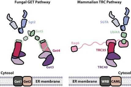
Figure 1. Overview of the TA targeting pathways in fungi and mammals. Upon translation termination and release
from the ribosome, TA is captured by Sgt2/SGTA and handed off to Get3/TRC40. This handoff step is facilitated by the Get4-5 heterotetramer in yeast and the Bag6 heterotrimer in mammals. The Get3-TA/TRC40-TA complex that results is ultimately recruited to the ER membrane by membrane receptors, fungal Get1/2 or mammalian WRB/CAML.
Unlike the co-translational SRP pathway, which is comprised of a single cytosolic factor
9 distinct binding sites; Ubl4A localizes to the putative BAG domain and TRC35 binds at
the region that includes the NLS of Bag6. The Ubl4A structure reveals that the
Bag6-BAG domain does not resemble a canonical Bag6-BAG domain, establishing Bag6 as a
misnomer. TRC35 masking NLS provides the structural basis for nucleocytoplasmic
distribution of Bag6. Chapter 3 describes the biochemical assays used to functionally
characterize the BAG domain and the isolated minimal Bag6 complex, which includes a
truncated C-terminal Bag6, truncated TRC35 and full length Ubl4A. An Hsc70 refolding
assay is used to confirm that the Bag6 “BAG” domain is not a BAG domain. We also
developed an in vitro TA transfer assay to demonstrate that the minimal Bag6 complex
identified in this study is sufficient for facilitating TA handoff, establishing it as a TA
targeting module. In Chapter 4, the regulation of nucleocytoplasmic distribution of Bag6 by
TRC35 is explored using biochemical and cell biology techniques. Chapter 5 concludes by
contextualizing the findings of this dissertation in the body of work on protein targeting and
10
C h a p t e r 2
STRUCTURAL CHARACTERIZATION OF THE
BAG6-TRC35-UBL4A COMPLEX
Parts of this chapter were first published in
Mock, J.-Y., Chartron, J.W., Zaslaver, M., Xu, Y., Ye, Y., and Clemons, W.M. Jr. (2015) Bag6 complex contains a minimal tail-anchor-targeting module and a mock BAG domain. Proc Natl Acad Sci USA. 112(1): 106-11 doi: 10.1073/pnas.1402745112
Mock, J.-Y., Xu, Y., Ye, Y., and Clemons, W.M. Jr. (2017) Structural basis for regulation of nucleocytoplasmic distribution of Bag6. Submitted.
11
Abstract
Bcl2-associated athanogene cochaperone 6 (Bag6) is a uniquely metazoan protein
involved in a diverse array of cellular processes. It is part of the heterotrimeric Bag6
complex, which also includes ubiquitin-like 4A (Ubl4A) and transmembrane domain
recognition complex 35 (TRC35). The Bag6 complex plays a central role in the
mammalian tail-anchor protein targeting pathway, mislocalized protein degradation
pathway and the endoplasmic reticulum-associated degradation pathway. Here we define
the architecture of the Bag6 complex, demonstrating that both TRC35 and Ubl4A have
distinct C-terminal binding sites on Bag6 defining a minimal Bag6 complex. The crystal
structure of the Bag6-Ubl4A dimer demonstrates that Bag6-BAG is not a canonical BAG
domain. Instead, its main function is to dimerize with the well-conserved dimerization
domain of Ubl4A. The crystal structure of Bag6 and its cytoplasmic retention factor
TRC35 reveals remarkable structural conservation of Get4/TRC35 throughout opisthokont
lineage except at the C-terminal Bag6-binding groove, which diverged to accommodate
Bag6. Together these data advance our molecular understanding of the
14 complex between the heterodimer of Bag6 and TRC35 presented here provides a
structural basis for regulation of nucleocytoplasmic distribution of Bag6 by TRC35.
Furthermore, the structures reveal that despite the changes in architecture, the overall
folds of the TA sorting complex have been remarkably conserved throughout opisthokont
21 modulated. It is likely that functionally homologous proteins with low sequence
homology exist.
Get4/TRC35 and Get3/TRC40 are conserved throughout eukaryotic evolution and seem
to occur as a pair in all opisthokonts and in most eukaryotes, suggesting the essentiality
of the two proteins in the pathway. Consistent with this notion, residues at the predicted
TRC35-TRC40 interface and the TRC35-Bag6 interface in TRC35 are highly conserved
25 These results support a model in which the primary role of the Bag6 C terminus is to
bridge TRC35 and Ubl4A. Possible Bag6 dimerization would form a heterohexamer,
creating a complex analogous to the Get4-5 heterotetramer found in yeast and providing
strong structural parallels in TA targeting (Fig. 2.12). Several unanswered questions
about the Bag6 complex remain. Does Bag6-BAG domain behave like a canonical BAG
domain? Is the structurally characterized Bag6 complex functionally equivalent to the
fungal Get4-5 complex? Can disrupting the Bag6-TRC35 interface lead to changes in the
nucleocytoplasmic distribution of Bag6? Biochemical and cell biological methods will be
26
Figures
Figure 2.1. Bag6 has distinct binding sites for TRC35 and Ubl4A at its C-terminus
(A) Scheme of the five different Bag6 fragments and of the sub-fragments of Bag6E, which
is further divided into N-terminus (EN), the NLS (ENLS), and a fragment containing only the
putative BAG domain (EBAG). (B) Yeast two-hybrid assay between Bag6 fragments and
either TRC35 or Ubl4A. The A fragment contains the UBL domain and the E fragment
contains the NLS and putative BAG domain. Yeast two-hybrid assay of TRC35 or Ubl4A
27
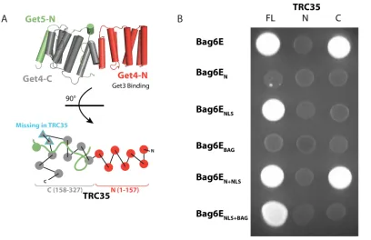
Figure 2.2 Defining the interaction between Bag6 and TRC35 by yeast two-hybrid
assay
(A, Upper) Diagram of the Get4/Get5-N complex (PDB ID code 3LKU). Get4 is shown in
gray and red, and Get5 is shown in green. (Lower) A top-view schematic representation of
the architecture. The two β-strands in Get4 that are missing in TRC35 are outlined in blue.
Ubl4A does not contain a Get5-N equivalent. (B) Bag6E fragments containing the
activating domain were combined with full-length TRC35 (FL), TRC35-N (residues 1–
157), or TRC35-C (residues 158–327) conjugated to the binding domain. TRC35-N and
28
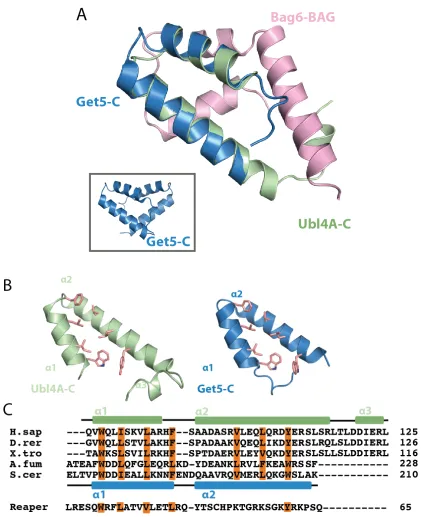
Figure 2.3. Crystal structure of Bag6-BAG/Ubl4A-C heterodimer
(A) The overall structure of Bag6-BAG/Ubl4A-C heterodimer in ribbon representation
with Bag6-BAG in cyan and Ubl4A-C in magenta. Hydrophobic residues in Bag6 involved
in packing are highlighted as orange sticks. (B) Sequence alignment of Bag6-BAG (H.sap
Homo sapiens, X.tro Xenopus tropicalis, D.mel Drosophila melanogaster, D.rer Danio
rerio, S.kow Saccoglossus kowalevskii, and T.cas Tribolium castaneum). The secondary
structure based on the structure is highlighted above the text. The conserved hydrophobic
and aromatic residues involved in the hydrophobic packing interactions between
Bag6-BAG and Ubl4A-C are highlighted in orange. The extended Drosophila melanogaster
sequence is a predicted protein sequence based on theoretical translation, and may not
29
Figure 2.4. Ubl4A-C and Get5-C are structurally homologous
(A) A monomer of Get5-C (green) (PDBID: 3VEJ) and Ubl4A-C (pink) overlaid.
30 Get5-C are juxtaposed with conserved residues involved in dimerization highlighted as
orange sticks. (C) Sequence alignment of Ubl4A-C homologs (H.sap Homo sapiens, D.rer
Danio rerio, X.tro Xenopus tropicalis, A.fum Aspergillus fumigatus, and S.cer
Saccharomyces cerevisiae) and the Drosophila apoptosis-inducing protein Reaper. The
Ubl4A-C and Get5C secondary structure are shown above (pink) and below (green),
respectively. Conserved hydrophobic residues involved in dimerization are highlighted in
31
Figure 2.5. A comparison of Bag6-BAG to canonical BAG domains
(A) Published structures of BAG domains are shown as ribbons similar to Bag6-BAG.
Residues involved in Hsp/Hsc70 binding are highlighted as magenta sticks. (B) Sequence
alignment of human Bag1-BAG, Bag5-BAG5, and Bag6-BAG with known secondary
32
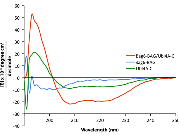
Figure 2.6. Circular dichroism spectra of Bag6-BAG, Ubl4A-C and the complex of the
two
33
Figure 2.7. The crystal structure of the Bag6-NLS/TRC35 heterodimer
(A) The structure of Saccharomyces cerevisiae Get4-Get5N complex (PDBID: 3LKU),
Get4 in rainbow and Get5 in magenta. Sequence alignment of TRC35/Get4 from helix 11
to 12 of two metazoan species, Hsap (Homo sapiens), Aque (Amphimedon queenslandica),
and six fungal species Spom (Schizosaccharomyces pombe), Ncas (Naumovozyma castelli),
Scer (Saccharomyces cerevisiae), Afum (Aspergillus fumigatus), Ncra (Neurospora
crassa), and Smus (Sphaerulina musiva). The secondary structure based on fungal Get4s is
highlighted above the text. (B) Left, the overall structure of Bag6-TRC35 heterodimer in
cylinder representation with Bag6 in light pink and TRC35 in rainbow. The nuclear
localization sequence is highlighted in sticks on Bag6. Right, a 90˚ in plane rotated
34 Comparison of the C-terminal faces of TRC35 and Get4 that bind Bag6 and Get5,
respectively. The arrows highlight the significant structural difference in the residues
between α11 and α12. (D) Zoomed in view of the regions, defined as interface I and II.
35
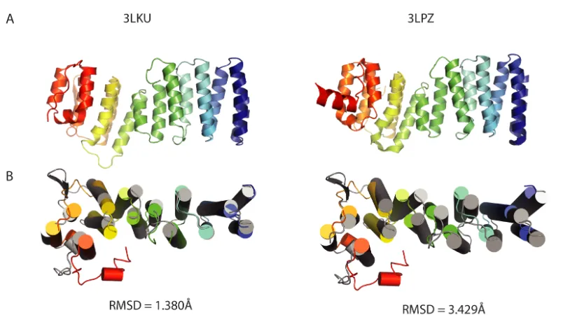
Figure 2.8.Comparison of TRC35 and Get4 structures
(A) Representative structures of fungal Get4 homologs. 3LKU is from Saccharomyces
cerevisiae (Sc) and 3LPZ is from Chaetomium thermophilum (Ct). (B) Aligned human
TRC35 (color ramped) and ScGet4 (3LKU, grey) using Pymol (Schrodinger, 2015) super
for sequence-independent structural alignment. Left, structures aligned to the six
36
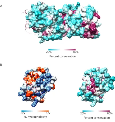
Figure 2.9 Analysis of the surface conservation and hydrophobicity of TRC35
(A) Accessible surface representation of TRC35 colored based on percent conservation as
implemented in Chimera (Pettersen et al., 2004). Conservation based on a MAFFT (Katoh
and Standley, 2013) alignment of TRC35/Get4 sequences from Homo sapiens, Xenopus
laevis, Danio rerio, Drosophila melanogaster, Nematostella vectensis, Monosiga
37 ribbons representation in pink. (B) The Bag6-binding surface of TRC35 colored based on
hydrophobicity (Kyle-Doolittle scale) and percent conservation as implemented in Chimera
39
Figure 2.10. Representative aligned sequences of eukaryotic TRC35/Get4
Species selected based on the eukaryotic phylogenetic tree by Eme et al (Eme et al.,
2009). Sequences were aligned with MAFFT (Katoh and Standley, 2013). α-helices,
based on the TRC35 structure, are highlighted in colors that correspond to the crystal
structure on Figure 2.1B and numbered on top. Residues highlighted in red boxes were
identified as critical to fungal Get4 binding Get3 (Gristick et al., 2014). Residues
highlighted in blue boxes are critical to Get4/TRC35 regulating Get3/TRC40 (Gristick et
al., 2014). The residues that comprise the fungal β-hairpin are highlighted in a black box.
The arrow indicates the end of the crystallization construct. Purple boxes highlight
40
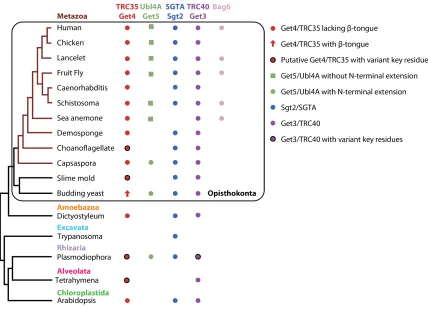
Figure 2.11 Survey of factors involved in the TRC pathway in eukaryotes
A condensed phylogenetic tree of representative eukaryotes was built based on the
maximum likelihood phylogenetic tree of eukaryotes by Eme et al (Eme et al., 2014). The
genome of each organism was searched for the presence of Get4/TRC35, Ubl4A/Get5,
Sgt2/SGTA, Get3/TRC40, and Bag6 using MEME suite motif discovery tool (MEME)
(Bailey et al., 2009) and motif scanning tool (MAST) (Bailey and Elkan, 1994) in addition
to NCBI protein BLAST (Altschul et al., 1990). Proteins are color coded and sequence
elements are highlighted. Black circles indicate homologs that are missing residues
43
49 (Collaborative Computational Project, 1994; Winn et al., 2011). Phases were determined
by molecular replacement single-wavelength anomalous dispersion (MRSAD) using
SHARP (Bricogne et al., 2003) and PHASER-MR (McCoy et al., 2007) on PHENIX
(Adams et al., 2010). The initial structure was built by PHASER as implemented by
PHENIX (Adams et al., 2010). The model was further built and refined against the native
dataset over several rounds using COOT (Emsley et al., 2010) and PHENIX (Adams et
al., 2010). Statistics are provided in Table 2.
Phylogenetic Tree of GET/TRC Components
The phylogenetic tree was modified from the maximum likelihood phylogenetic tree of
eukaryotes by Eme et al (Eme et al., 2014). The MEME suite (Bailey et al., 2009) was
used to determine the presence or absence of the GET/TRC components in the genomes
presented in the tree. The MEME motif discovery tool was used to make motifs for
Ubl4A, Get5, Get4/TRC35, and Sgt2, SGTA, and Bag6. The motifs were then used to
search the genomes using the motif-scanning tool MAST (Bailey and Elkan, 1994).
Identified proteins were confirmed using BLAST (Altschul et al., 1990) and the
50
Acknowledgements
We thank Daniel Lin and Jens Kaiser for help with data processing. We thank Dr. Meera
Rao and Dr. Michael Rome for critical reading of the manuscript. We thank members of
the laboratory for support and useful discussions. We are grateful to Gordon and Betty
Moore for support of the Molecular Observatory at Caltech. We thank the staff at the
Advanced Light Source and Stanford Synchrotron Radiation Laboratory for assistance
51
C h a p t e r 3
BIOCHEMICAL CHARACTERIZATION OF THE BAG DOMAIN
AND THE MINIMAL TA TARGETING MODULE
Parts of this chapter were first published in
Mock, J.-Y., Chartron, J.W., Zaslaver, M., Xu, Y., Ye, Y., and Clemons, W.M. Jr. (2015)
Proc Natl Acad Sci USA. 112(1): 106-11 doi: 10.1073/pnas.1402745112
Mock, J.-Y., Xu, U., Ye, Y., and Clemons, W.M. Jr. (2017) Structural basis for regulation of nucleocytoplasmic distribution of Bag6. Submitted.
52
Abstract
The metazoan protein BCL2-associated athanogene cochaperone 6 (Bag6) forms a
hetero-trimeric complex with ubiquitin-like 4A (Ubl4A) and transmembrane domain recognition
complex 35 (TRC35). This Bag6 complex is involved in tail-anchored protein targeting and
various protein quality control pathways in the cytosol as well as regulating acetylation and
histone methylation in the nucleus. Crystal structure of the Bag6 BAG domain revealed
that it does not fold into a canonical BAG domain fold. In this study, we biochemically
demonstrate that Bag6-BAG domain does not behave like a canonical BAG domain,
establishing it as a “mock” BAG domain. Furthermore, we show that C-terminal 125
residues of Bag6, which can form a stable complex with TRC35 and Ubl4A, are sufficient
54 BAG domain does not behave like a canonical BAG domain, which binds and inhibits
Hsc70 in vitro. Furthermore, the minimal Bag6-TRC35-Ubl4A complex identified and
structurally characterized in chapter 2 facilitates TA protein transfer from SGTA to
TRC40 in vitro. These findings establish the minimal Bag6 complex identified in this
60 this larger complex plays a regulatory role in TA targeting. The crystal structure of yeast
Get4 bound to Get3 highlighted a regulatory interface separate from the binding interface
(Gristick et al., 2014). When mutated, residues on either side of this regulatory interface
(the charge swaps K69D on Get3 and D74K on Get4) each resulted in a loss of ATPase
inhibition, reduction of TA insertion into microsomes, and a loss of fitness in vivo, despite
maintaining a stable complex in vitro (Gristick et al., 2014). Combining the charge swap
mutants restored the Get4 regulatory activity (Gristick et al., 2014). For TRC40, the
corresponding regulatory mutation, K86D, resulted in a reduction of the Bag6 complex
facilitated hand-off (Fig. 3.5C, compare lanes 4 & 8 and Fig. 3.7D). Similarly, the
corresponding regulatory mutation in TRC35, D84K, resulted in a reduction of facilitated
hand-off (compare lanes 4 & 5 and Fig. 3.7D). Excitingly, as seen for the yeast system
(Gristick et al., 2014), the combination of these two charge swap mutants resulted in a
rescue of the facilitated hand-off (lane 9 and Fig. 3.7D). These results highlight that this
minimal Bag6 complex acts as an independent TA targeting module and performs a similar
63 simultaneously binding ubiquitylation machinery, the proteasome, TA-targeting factors,
and proteins to be triaged. Recent biochemical characterization of the triaging process
revealed that TA substrate that is not handed off to TRC40 within ~3 cycles is rerouted to
the degradation pathway by Bag6 (Shao et al., 2017). The molecular details of how its
decision-making process relates to its other functions in apoptosis, gene regulation, and
64
65
Figure 3.1. Bag6-BAG is not a canonical BAG domain
(A) Hsc70-mediated refolding of β-galactosidase in the presence of Bag1-BAG (purple),
Bag6-BAG (orange solid), Bag6EBAG/Ubl4A (blue solid), SGTA & Bag6-BAG (orange
dashed), SGTA & Bag6EBAG/Ubl4A (blue dashed) or BSA (black) as a negative control.
(B) Affinity tagged Bag6-BAG, Bag6EBAG/Ubl4A, Bag6-BAG/Ubl4A-C or Bag1-BAG
was loaded onto cobalt resin beads and incubated with 293T whole cell lysate. Eluted
samples were immunoblotted with Hsc70 antibody (top panel) and Ponceau stained
(bottom panel). (C) FLAG-Bag6 or FLAG-Bag6ΔC81 was overexpressed in 293T cells
and anti-FLAG resin was used to capture them with bound factors. Bag6 antibody on the
66
Figure 3.2. Individual results for various in vitro refolding assays
β-Galactosidase refolding assays in the presence of Hsc70, Hdj1 and/or other factors
(labeled). Colors are based on Figure 3.1 except for assays containing Bag6-C81 that are in
67
Figure 3.3. Bag6-BAG does not bind Hsc70 nucleotide binding domain (NBD)
In vitro capture by 6xHis-Hsc70-NBD of Bag6-BAG, Bag6-C81, Bag6EBAG/Ubl4A, or
Bag1-BAG. Protein was pulled down (PD) with Ni-NTA after incubation with
6xHis-Hsc70-NBD. Four percent of total loaded protein is shown as a loading control (LC), and
68
Figure 3.4. Purification of recombinant proteins used in TA transfer assay
(A) Two-hybrid using full-length TRC35 and TRC35(23-305) with Bag6E fragment. Both
TRC35 constructs display strong two-hybrid interactions. (B) Representative Coomassie
stained 12% SDS-PAGE gel of purified hSGTA/MBP⦁Sbh1, Bag6min Complex, and
70
Figure 3.5. The Bag6min complex facilitates TA transfer from SGTA to TRC40
(A) The in vitro TA handoff reaction scheme. Recombinantly purified hSGTA-MBP⦁Sbh1
complex was incubated with GST⦁TRC40 and indicated recombinant proteins. After
incubation on ice for 10 minutes, GST⦁TRC40 and bound substrate were precipitated with
anti-GST resin followed by three wash steps and Western blotting. (B) Mutants affecting
SGTA binding to the Bag6min complex reduce TA transfer to TRC40. GST⦁TRC40 was
captured on anti-GST resin after incubation in the presence of ATP with SGTA/MBP-Sbh1
or SGTA(C38A)/MBP⦁Sbh1 alone or with the Bag6min or Bag6min(Ubl4A(L43A))
complex. Eluted samples were immunoblotted with anti-GST (red) and anti-MBP antibody
(green) then quantified by Odyssey Infrared Imaging System analysis software. Relative
values of captured Sbh1 underneath each lane with the experiment containing all wild-type
components as the reference. Sbh1 fluorescence values were normalized for each trial
based GST⦁TRC40 captured in each lane. Values are averages of six independent
experiments. Standard deviations are included in figure 3.7. The 5% rxn lane corresponds,
in all cases, to loading 5µL of the wild-type reaction prior to capture. (C) Regulatory
mutants GST⦁TRC40(K86D) and TRC35(D84K)Bag6min complex were incubated with
indicated recombinant proteins and ATP then captured on anti-GST resin and analyzed as
71
Figure 3.6. The Bag6min complex facilitates TA transfer from SGTA to TRC40 in an
ATP dependent manner
Nucleotide-dependent TA handoff facilitated by Bag6min complex. GST⦁TRC40 was
captured on anti-GST resin after incubation with SGTA-MBP⦁Sbh1 and Bag6min complex
with or without ATP. Eluted samples were immunoblotted with GST (red) and
anti-MBP antibody (green) then quantified by Odyssey Infrared Imaging System analysis
software and the Sbh1 values were normalized based on total GST⦁TRC40 captured.
MBP⦁Sbh1 signal from WT experiment was designated 1, and the rest represented as a
73
Figure 3.7. TA handoff from SGTA to TRC40
(A) Average values of nucleotide-dependent TA handoff from SGTA to TRC40 facilitated
by Bag6min complex. Error bars are from four independent experiments. (B) Average
values of TA handoff from SGTA to TRC40 facilitated by Bag6min complex in the presence
of ATP washed with buffers with varying salt concentrations. Error bars are from three
independent experiments. Fluorescence values are represented as a percentage of WT
handoff as measured by MBP fluorescence. (C) Average values of TA handoff by binding
mutants hSGTA(C38A)/MBP⦁Sbh1 and Bag6min(Ubl4A(L43A)) complex as compared to
WT. Error bars are from six independent experiments. (D) Average values of TA handoff
of regulatory mutants GST⦁TRC40(K86D) and Bag6min(TRC35(D84K)) complex as
78 is reasonable and differences could be attributed to a variety of factors such as
79
Acknowledgements
We thank Yoko Shibata and Richard Morimoto (Northwestern) for plasmids. We thank
members of the laboratory for support and useful discussions. W.M.C. is supported by
80
C h a p t e r 4
BIOCHEMICAL AND CELL BIOLOGICAL INVESTIGATION OF
THE MECHANISM FOR NUCLEO-CYTOPLASMIC
DISTRIBUTION OF BAG6 BY TRC35
A version of this chapter was first published as
Mock, J.-Y., Xu, U., Ye, Y., and Clemons, W.M. Jr. (2017) Structural basis for regulation of nucleocytoplasmic distribution of Bag6. Submitted.
81
Abstract
The metazoan protein BCL-2 associated athanogene cochaperone 6 (Bag6) acts as a
central hub for several essential cellular processes, including immunoregulation, gene
regulation, autophagy, apoptosis, and proteostasis. These roles are in both the nucleus and
the cytosol, but the mechanism by which Bag6 trafficking is regulated remains elusive.
Here we present biochemical and cell biological characterization of the cytoplasmic
retention factor of Bag6, transmembrane domain recognition complex 35 (TRC35).
Disrupting the interface between Bag6 and TRC35 results in nuclear localization of
Bag6. TRC35 binds Bag6 with higher affinity than karyopherins. Free TRC35 that cannot
bind Bag6 at its native binding site is ubiquitylated and degraded. Combined, these
results suggest a mechanism for regulation of the nucleo-cytoplasmic distribution of
84 qualitative mass spectrometry studies, suggest that nucleo-cytoplasmic distribution of
92 and bound to glutathione affinity resin beads via the GST-tag. After washing, varying
amounts of MBP•KPNA2 were added to the bound beads. The ability of MBP•KPNA2 to
displace Bag6C131-6xHis•Ubl4A from GST•TRC35 was determined by the amount of
Bag6C131-6xHis•Ubl4A that was eluted from the resin after incubation. In this case, even
at the highest concentration tested (2x molar excess), there was no significant displacement
of Bag6 from TRC35 by KPNA2 (Fig. 4.8 lanes 4-7). Performing the opposite experiment,
starting with MBP•KPNA2-Bag6C131-6xHis•Ubl4A on amylose beads, adding excess
GST•TRC35 resulted in the dissociation of the MBP•KPNA2-Bag6C131-6xHis•Ubl4A
complex (Fig. 4.8 lanes 11-14). These results highlight the stability of the TRC35-Bag6
complex and argue against the ability of KPNA regulation as a means for modulating the
95 different cell types regulate Bag6 localization, whether the TRC35-dependent regulation
can be mediated by specific stress, and the biological implications of differential
96
97
Figure 4.1. Validation of the Bag6-TRC35 interface
(A) Yeast 2-hybrid assay to validate the interface identified in the crystal structure.
Wild-type or mutant full length TRC35 conjugated to the DNA binding domain was expressed
with wild-type Bag6(951-1126) conjugated to the transcription activating domain.
Transformation was confirmed by ability to grow on SC-Ura-Leu media. Interaction was
determined by ability to grow on SC-Ura-Leu-Ade media. (B) Wild-type full-length
TRC35 conjugated to the DNA binding domain was expressed with wild-type or mutant
Bag6(951-1126) conjugated to the transcription activating domain. (C) Expression of
TRC35 and Bag6 in yeast cells used from yeast 2-hybrid was examined by Western blot.
Antibodies against Gal4 DNA binding domain or trans-activating domain were used to
detect expression of Gal4BD-TRC35 and Gal4AD-Bag6. (D) Combination of mutations at
interface I (W1004A and W1012A) and interface II (Y1036A) is sufficient for disrupting
98
Figure 4.2. Validation of Bag6-TRC35 interface in mammalian cells
Wild-type or mutant Bag6•GFP was co-expressed in Bag6-/- 293T cells with TRC35•FLAG
and immunoprecipitated using anti-GFP antibody. Amount of TRC35 retrieved by Bag6
was assessed by blotting with anti-FLAG antibody. The position of the higher molecular
99
Figure 4.3. Bag6 mutations at the TRC35 binding site results in nuclear localization of
Bag6
(A-F) Cos7 cells were transfected either with Bag6•GFP (wt or mutant) expressing
plasmid alone or co-transfected with TRC35•FLAG (wt) expressing plasmid. Cells were
100 DAPI where indicated (blue). In the Bag6 column, the percent of Bag6 calculated to be
101
Figure 4.4 Ubiquitylation of TRC35 upon mutant Bag6 expression
(A) Immunoprecipitation (IP) of TRC35 in Bag6-/- 293T cells co-transfected with plasmids
encoding TRC35•FLAG (wt), Bag6•GFP (wt or mutants), and HA•ubiquitin. Anti-GFP
102 denaturing conditions. TRC35 ubiquitination was assessed by immunoblotting with
anti-HA antibody. The exposure times were adjusted to improve visibility of the reactive bands.
(B) IP was carried out as in (A) in Bag6-/- 293T cells co-transfected with plasmids encoding
TRC35•FLAG (wt) and Bag6•GFP (wt or mutants). TRC35 ubiquitination was assessed by
immunoblotting with anti-FLAG antibody. The exposure times were adjusted to improve
visibility of the reactive bands. (C) The cell extract used for immunoprecipitation was
immunoblotted for Bag6, TRC35, and ubiquitin with Bag6 antibody, FLAG antibody and
103
Figure 4.5 RNF126 knockdown stabilizes TRC35 in cells expressing mutant Bag6
(A) wt293T cells expressing TRC35•FLAG, Bag6•GFP and HA•ubiquitin were treated
with siRNA against RNF126. Cells were lysed and TRC35 bound to Bag6 were
immunoprecipitated first with GFP antibody then with FLAG antibody in denaturing
104 immunoblotting with anti-TRC35 antibody and anti-HA antibody, respectively. (B) The
relative amounts of ubiquitylated TRC35 and unmodified TRC35 from figure 4.5 are
calculated as a ratio. (C) 293T cells expressing wtTRC35•FLAG, Bag6•GFP (wt or
W1004A/Y1036A) and HA•ubiquitin were treated 10 µM MG132. Bag6 and TRC35 were
105
Figure 4.6 TRC35 ubiquitylation is dependent on RNF126.
wt293T cells co-transfected with plasmids encoding TRC35•FLAG (wt), Bag6•GFP (wt or
mutants), and HA•ubiquitin. The cells were simultaneously treated with MG132 and
siRNA against RNF126. The cell extracts were subject to two rounds of denaturing
immunoprecipitation with anti-GFP antibody then with anti-FLAG antibody. TRC35
106
Figure 4.7 Biochemical identification of the monopartite Bag6 NLS
(A) The putative bipartite nuclear localization sequence of Bag6. The serine and leucine
mutations introduced in this study are highlighted. (B) Recombinantly purified
Bag6C131-6xHis•Ubl4A was incubated with excess GST•TRC35 or MBP•KPNA2 for 20 minutes at
room temperature. Ni-NTA beads were used to capture purified Bag6C131-6xHis•Ubl4A
107
Figure 4.8 TRC35 binding precludes karyopherin α binding to Bag6
Recombinantly purified Bag6C131-6xHis•Ubl4A was first incubated with either
GST•TRC35 or MBP•KPNA2. The resulting GST•TRC35-Bag6C131-6xHis•Ubl4A
complex was incubated with glutathione resin beads, and increasing amounts of
MBP•KPNA2 was added. The ability of MBP•KPNA2 to displace
Bag6C131-6xHis•Ubl4A from GST•TRC35 was examined by eluting the GST•TRC35 and bound
Bag6C131-6xHis•Ubl4A from the glutathione resin. The opposite experiment was also
115 7.2), 100 mM K•glutamate, 33 mM glutathione, 5 mM β-mercaptoethanol). The reverse
experiment starting with hexahistidine-tagged Ubl4A-Bag6C131 and MBP•KPNA2 was
carried out as above but using 30 µL 50% (vol/vol) slurry of amylose beads (New
England Biolabs) in 20 mM Mops (pH 7.2), 100 mM K•glutamate, 5 mM β
-mercaptoethanol and eluted with 25 µL of 20 mM Mops (pH 7.2), 100 mM K•glutamate,
116
Acknowledgements
We thank members of the laboratory for support and useful discussions. We thank
Yihong Ye and Yue Xu for useful discussions and technical assistance. W.M.C. is
117
C h a p t e r 6
119 insertion into purified microsomes (Suloway et al., 2012). Third, Get3 tetramerization
stimulates its ATPase activity by ~100 fold (Rome et al., 2013). However, the only
atomic resolution structure of a Get3-TA complex solved thus far suggests that dimeric
Get3 (Mateja et al., 2015) is sufficient for binding and targeting TA proteins. It should be
noted that the crystallized Get3-TA complex was artificially stabilized by (1) introducing
a mutation that rendered Get3 incompetent for ATP hydrolysis and (2) adding
high-affinity synthetic antibody fragments. As a result, the crystal structure could have trapped
and captured a specific intermediate or an artificial state of the Get3-TA complex.
Whether tetrameric Get3/TRC40 plays a physiological role in TA targeting remains to be
seen.
Ultimately, the observation that the C-terminal TA targeting module—the minimal Bag6
complex—characterized in this study is structurally and functionally equivalent to the
fungal Get4-5 complex begged the question: what different purpose, if any, does Bag6
120
Figure 5.1 Cartoon summary of the fungal and metazoan sorting complexes. Left: fungal Get4-5 TA sorting
complex and its binding partners, Sgt2 and Get3, are illustrated. Right: metazoan Bag6 TA sorting complex and its binding partners, SGTA and TRC40, are illustrated. The structurally conserved dimerization domains, whose structures have been solved in this study, are highlighted with grey and yellow dotted boxes.
One purpose Bag6 serves is to physically couple protein targeting and quality control. A
recent study demonstrated that purified N-terminal domain of Bag6 (Fig. 5.2), which
excludes the TA targeting module, is sufficient for substrate ubiquitylation (Shao et al.,
2017). Bag6 seems to decide the fate of TA substrate as it is handed off from SGTA by
utilizing either its C-terminal targeting module for productive synthesis of well-folded
proteins or its N-terminal quality control module for degradation of misfolded proteins.
Such coordination, enabled by modularity, likely minimizes the risk of aggregation for
123 predicted isoform is 903 residues long and is missing a large portion of the second
proline rich domain (Fig. 5.2). Analysis of RNA-seq data from the Illumina Human Body
Map Project revealed that Bag6 transcripts without NLS are abundant in breast and brain,
while transcripts with NLS are abundant in liver, lung, testes, prostate, kidney and lymph
nodes (Luce et al., 2016). In rats, different Bag6 isoforms are expressed in distinct
developmental stages (Kwak et al., 2008). Thus, in addition to the modulation of TRC35
expression, alternative splicing would affect Bag6 localization and function. The
functional and physiological consequence of different Bag6 isoforms, however, is unclear
and is a question for future studies.
Recent structural and biochemical breakthroughs have led to rapid leaps in our
understanding of TA targeting and Bag6, but challenging questions remain. Is the
Get3/TRC40 tetramer physiologically relevant? Does Bag6 change the stoichiometry of
TRC components in metazoans? How is TRC35 expression, and Bag6 localization,
modulated? Could some of the pleiotropic effects of knocking down Bag6 be explained
through the resulting destabilization of TRC35? A multifaceted approach that combines
structural and mechanistic characterization of Bag6 with cell biological and organismal