1
Development and Characterization of a Potent Tumor Necrosis Factor-Alpha Blocking Agent
Aida Jameei*, Deepesh Nagarajan*, Mohsen Sarikhani#, Nagasuma Chandra* and Anjali A. Karande*
*Department of Biochemistry, # Department of Microbiology and Cell Biology,
Abstract
Tumor necrosis factor-α (TNFα), one of the major pro-inflammatory cytokines, plays a key role in an effective immune response. However, the chronic presence of TNFα can lead to several inflammatory disorders like rheumatoid arthritis, psoriasis, Crohn’s disease etc. Inhibition of TNFα by pharmacological inhibitors or antibodies has proven to be effective in palliative treatment to some extent. The aim of this study was to develop an anti-TNFα antibody which may be used as a therapeutic option to inhibit TNFα-mediated cytotoxicity. We characterized several hybridoma clones secreting monoclonal antibodies (mAbs) to human-TNFα. Four mAbs rescued L929 fibroblast cells from TNFα-triggered cell death and one of these, namely C8 was found to have the highest affinity. To gain insights into the mechanism by which mAb C8 inhibits human TNFα-mediated toxicity, the epitope corresponding to the mAb was delineated. The antigenic determinant was found to comprise of the stretch of amino acids 99-120, of which, 102-104 (QRE) form the core epitope. The observation was supported by bioinformatics analyses of an antigen-antibody complex model. In addition, the binding affinity of mAb C8 to TNFα was found to be comparable with that of Infliximab which is a commercially available anti TNFα mAb.
2
1. Introduction
TNF is a pro-inflammatory cytokine having pleiotropic effects on immune cells and plays an indispensable role in inflammation, cell differentiation, cell proliferation, apoptosis and cell metabolism [1, 2].
There are two types of TNF, TNFα and TNFß, both of which are cytotoxic [3],[4]. They are sequentially and structurally related and compete with each other for binding to their receptors [5]. Soluble TNFα exerts a broad range of biological activities on interaction with two specific receptors, TNFα receptor-type 1 (TNFR1 or p55) and TNFα receptor-type 2 (TNFR2 or p75). TNFR1, expressed on almost all cell types, plays a major role in triggering the TNFα signaling pathways. TNFR2 expression on the other hand is mostly limited to the cells of the immune system, nerve cells and endothelial cells, under normal physiological conditions [6-8]. Upon binding to receptors, TNFα promotes a series of intracellular signaling cascades such as, activation of transcription factor NFκB, p38 mitogen activated protein kinase (p38MAPK), c-Jun N-terminal kinase (JNK) extracellular signal regulated kinase (ERK) [8, 9].
Although TNFα plays an essential role in an effective immune response, its’ unrestricted production may lead to several inflammatory disorders. Inhibition of TNFα by pharmacological agents has proven to be effective in palliative treatment [10, 11].
In the study described in this manuscript, we developed an anti-human TNFα mAb namely C8 which can neutralize human-TNFα activity. Further, using truncation and mutation analysis, we mapped the TNFα binding sites of the mAb, which was supported by docking studies using sequenced variable regions of heavy and light chain of the antibody.
A study of mAb C8 and Infliximab (a commercialized anti-TNFα antibody), demonstrated that both antibodies have comparable affinities as well TNFα neutralizing efficiency.
Methods
Animal and Cell lines
3
Expression and purification of His-tagged human-TNFα and mouse-TNFα
E. coli BL21 (DE3) strain cells were transformed with the pET15b vector containing the human-TNFα gene and pET28a vector containing the mouse-human-TNFα gene. Then cells were induced with 1 mM IPTG and were incubated at 30°C for 8 hr in an incubator shaker, at 200 rpm for the expression of each respective protein; the cells were harvested by centrifugation at 6000 rpm for 20 min at 4⁰C. The cell pellet obtained was resuspended with 50 mM Tris-HCl, pH 8.0 containing 150 mM NaCl (TBS), sonicated (sonication conditions: 4⁰C, ampt 30%, 30 min) and the soluble proteins were separated by centrifugation at 13,000 rpm for 30 min at 4⁰C. The supernatant (soluble protein) was
loaded on the Ni-NTA column. Unbound proteins were washed away with TBS containing 30 mM imidazole and the bound protein was eluted with 300 mM imidazole in TBS. After elution, the protein was dialyzed against PBS (50 mM phosphate buffer pH, 7.4, containing 150 mM NaCl). The concentration of proteins was estimated using Bradford method.
Generation of monoclonal antibodies against human-TNFα
Eight weeks old female BALB/c mice were immunized by subcutaneous injection with 5 µg of human-TNFα protein in phosphate-buffered saline PBS emulsified with Freund’s complete adjuvant and boosted twice with 5 µg of the same antigen in Freund’s incomplete adjuvant at 21 days interval. The animals were rested for a month. Three days prior to sacrificing, the mice were administrated intraperitoneally with 5 µg of antigen in PBS. The splenocytes were collected and fused with mouse myeloma cells SP2/0 (ratio 5:1) using PEG 4000 (Merck, Rahway, NJ). The fused cells were plated in IMDM supplemented with HAT (10 mM hypoxanthine, 40 µM aminopterin and 1.6 mM thymidine), 20% (v/v) FBS and 50 µM Mercaptoethanol. They were examined for secretion of anti-TNFα specific antibodies using ELISA after 10 days. The positive clones were subcloned to monoclonality by limiting dilution method. MAbs were purified by protein A (Sigma-Aldrich, St. Louis MO).
Enzyme linked immunosorbent assay (ELISA)
4 To determine comparative affinities of the mAbs, ELISA was performed as above, but after the incubation with the mAbs, ammonium thiocyanate (NH4SCN) was added at varying concentrations (0.5, 1.0, 2.0, 3.0, 4.0 and 6.0 M) in 0.1 M sodium phosphate buffer pH 6.0. The plates were incubated for 15 min at RT prior to washing with PBS, followed by blocking with 0.5% BSA in PBS for 1hr at RT, before addition of the secondary antibody conjugate. The rest of the procedure followed was as mentioned above.
For obtaining antigen and antibody dilution curves, four concentrations (500, 250, 125, 62.5 ng/well in 100 µl PBS) of hTNFα were coated in ELISA plate, blocked with 0.5% gelatin in PBS, 100 µl serially double diluted hybridoma culture supernatant was added ranging from undiluted to 1:1024 and incubated for 2 hr at RT. The remaining steps were carried out in the same way as mentioned above.
For Inhibition ELISA, 500 ng antigen/well was coated in ELISA plate overnight at RT. After blocking the plates with 0.5% gelatin in PBS for 1 hr at RT, varying concentrations of the antigen (5 µg serially double diluted to 0.0097 µg) was added in 50 µl, followed immediately with 50 µl of appropriately diluted (exhibiting a binding of ~85% of the Bmax) anti-TNFα antibodies and incubated for 2 hr at RT. The remaining steps were carried out in the same way as mentioned above.
Western blot analysis of mAb C8
HumanTNFα or its fragments, 1 µg each were electrophoresed on 12.5 % polyacrylamide gel under reducing conditions and transferred to nitrocellulose membrane using a semi-dry electro-transfer apparatus, at 125 mA. The membrane was incubated in blocking solution (5% skimmed milk in PBS) for 2 hr or overnight followed by incubation with the mAb for 2 hr. The membrane was washed with PBST and incubated with the secondary antibody (rabbit α-mouse Ig-HRP) for 45 min and developed using ECL kit (Millipore).
5
MTT assay
Viability of cells was checked using MTT 3-(4, 5-dimethylthiazol-2-yl)-2, 5-diphenyltetrazolium bromide) assay.For the assay, L929 fibroblast cells were plated in 48-well flat bottom tissue culture plate at 10000 cells/well in 200 µl DMEM supplemented with 10% FBS (fetal bovine serum). Cells were allowed to attach overnight and then incubated with different concentrations of hTNFα. Actinomycin-D (2µg/ml) was used as positive control. After 48 hr incubation, 20 µl of MTT solution (5 mg/ml in PBS) was added per well, incubated for 4 hr at 37 ⁰C in CO2 incubator. Formazan crystals formed as a result were dissolved with 100 µl of DMSO and transferred to a 96-well culture plate and OD was measured in a microplate reader, at 570 nm.
Cloning of mutant constructs of hTNFα gene
Deletion constructs of the hTNFα gene were cloned in pGEX5X2 vector between BamHI and XhoI restriction enzyme sites at the 5’ and 3’ end respectively. All clones carried an N-terminal GST-tag and were amplified employing the primers listed in Table 1.
Table 1: List of oligonucleotides used for generating the deletion constructs of hTNFα
Name Sequence (5’→3’) Restriction
enzyme site
F1 1-53 FP TAACCGGGATCCCGGTCAGATCATCTTCTC BamHI
F1 1-53 RP TGCTTCCGCTCGAGTCACTCTGATGGCACCAC XhoI
F2 26-131 FP TAACCGGGATCCCGCTCCAGTGGCTGAAC BamHI
F2 26-131 RP TGCTTCCGCTCGAGTCATCGGTCACCCTTCTCC XhoI
F3 96-157 FP TAACCGGGATCCCGCCATCAAGAGCCCCTG BamHI
F3 96-157 RP TGCTTCCGCTCGAGTCACAGGGCAATGATC XhoI
F2 core region 54-93 FP
TAACCGGGATCCCG GTGGTGCCATCAGAG BamHI
F2 core region 54-93 RP
TGCTTCCGCTCGAGTCACAGGGGCTCTTGATGG XhoI
F2-F1 72-131 FP TAACCGGGATCCCGCTCCTCACCCACACC BamHI
R1 72-107 FP CCGGGATCCCGCTCCTCACCCACACC BamHI
R1 72-107 RP CCGCTCGAGTCACTCTGGGGTCTCCCTCTGG XhoI
R2 99-131 FP CCGGGATCCcctgCCAGAGGGAGACCC BamHI
6
R3 85-120 FP CCGGGATCCcCTACCAGACCAGGGTC BamHI
R3 85-120 RP CCGCTCGAGTCACAGATAGATGGGCTCATAC XhoI
The PCR cycle used for the amplification procedure was as follows: initial denaturation - 94 °C, 5 min; denaturation - 94 °C, 1 min; annealing - 55 °C, 45 sec; extension - 72 °C, 1 min; final extension - 72 °C, 10 min. The denaturation, annealing and extension steps were carried out for 30 cycles. The hTNFα cloned in pET15b was used as the template and Deep Vent polymerase was used for amplification of DNA in the PCR reactions. The clones obtained were screened by digestion with restriction enzymes and confirmed by DNA sequencing.
Site-directed mutant genes were generated by quick change primer design method [12]. Sense and anti-sense primers carried the desired site-specific mutation along with a specific restriction enzyme site. The plasmid carrying the wild type hTNFα gene construct was amplified by PCR. The amplified product was subjected to DpnI digestion to digest the methylated plasmid template and E.coli DH5α cells were transformed with the plasmid.
The hTNFα F2 mutant clones, T72 to D; H73 to Y; T89 to E; S71deletion; TH72,73 DY; Q102 to P; R103 to K; E104 to D; R103E104 to KD; R131 to Q; S71 deletion + T 72 to D; S 71 deletion + H73 to Y and Q102R103E104 to PKD were obtained by quick-change primer design method. Phusion High-Fidelity DNA Polymerase was used for carrying out PCR reactions. In a PCR cycle, initial denaturation was carried out at 98°C for 2 min followed by 25 cycles of denaturation at 98 °C for 1 min, annealing for 1 min (at the temperature specified for each clone in the Table 2) and extension at 72 °C for 6 min. The final extension for end-filling was carried out at 72 °C for 10 min (For first 4 cycles, we have prepared reactions with forward or reverse primers separately and then pooled together for another 21 cycles).
All site-directed mutant DNA fragments obtained were cloned in pGEX5X2 vector between BamHI and XhoI restriction enzyme sites. The recombinant plasmids were isolated. The sequence of the clones obtained was verified by sequencing to confirm the presence of the desired mutations. All clones carried an N-terminal GST-tag and were amplified employing the primers listed in Table 2.
Table 2: List of oligonucleotides used for generating the site-directed mutants of hTNFα
Name Sequence (5’→3’)
S71deletion FP CCAAGGCTGCCCCGACTACGTGCTCC
S71deletion RP GGAGCACGTAGTCGGGGCAGCCTTGG
7 cDNA was prepared from total RNA isolated from mAb C8 hybridoma cells using oligo (dT) primers. The variable regions of the heavy (VH) and light chain (VL) were amplified by PCR using the cDNA as template and the respective universal primers listed in Table 3. The VH and VL DNA fragments (~300 bp) were generated using Deep Vent polymerase, cloned into the pGEMT vector system and screened by digestion with the restriction enzymes NcoI and NdeI. The DNA was sequenced.
T72D RP GAGGAGCACATGGTCGGAGGGGCAGCCTTG
H73Y FP GGCTGCCCCTCCACCTACGTGCTCCTCAC
H73Y RP GTGAGGAGCACGTAGGTGGAGGGGCAGCC
TH72,73DY FP GGCTGCCCCTCCGACTACGTGCTCCTCAC
TH72,73DY RP GTGAGGAGCACGTAGTCGGAGGGGCAGCC
S71 deletion T72D P CCAAGGCTGCCCCGACCATGTGCTCC
S71 deletion RP GGAGCACATGGTCGGGGCAGCCTTGG
S71 deletion H73Y P CCAAGGCTGCCCCGACCATGTGCTCC
S71 deletion H73Y P GGAGCACGTAGGTGGGGCAGCCTTGG
T89E FP CATCGCCGTCTCCTACCAGGAGAGGGTCAACCTCCTCTCTG
T89E RP CAGAGAGGAGGTTGACCCTCTCCTGGTAGGAGACGGCGATG
Q102P FP GGGTCTCCCTCGGGCAGGGGCTC
Q102P RP GAGCCCCTGCCCGAGGGAGACCC
R103K FP GGGGTCTCCTTCTGGCAGGGGCTCTTG
R103K RP CAAGAGCCCCTGCCAGAAGGAGACCCC
E104D FP CTCTGGGGTATCCCTCTGGCAGGGGCT
E104D RP AGCCCCTGCCAGAGGGATACCCCAGAG
RE103,104KD FP CAAGAGCCCCTGCCAGAAGGATACCCCAGAG
RE103,104KD RP CTCTGGGGTATCCTTCTGGCAGGGGCTCTTG
R131Q FP GAGAAGGGTGACCAATGACTCGAGCGG
8
Table 3: List of oligonucleotides used for generating mAb C8 variable regions of heavy (VH) and light (VL)
Name Sequence (5’→3’) Annealing
temperature PCR (⁰C)
VH FP TGAGGAGACGGTGACCGTGGTCCCTTGGCCCCA 54
VH RP AGGTGAAACTGCAGGAGTCAGG 54
VL FP GTTAGATCTCCAGCTTGGTCC 50
VL RP GACATTCAGCTGACCCAGTCTCCA 50
Expression and purification of hTNFα recombinant proteins
All recombinant hTNFα overlapping truncated and mutant forms of hTNFα F2 constructs in pGEX5X2 vector have an N-terminal GST-tag. E. coli BL21 (DE3) cells were transformed with each plasmid and were induced to express respective protein with 1 mM IPTG for hTNFα F1 and F3 for 4 hr at 30 ⁰C and 0.1mM IPTG for hTNFα F2 and F2-mutants for 14 hr at 20 ⁰C in an incubator shaker, at 200 rpm . The cells were harvested with centrifugation and the pellet obtained was resuspended and sonicated in lysis buffer (50 mM Tris, pH 8, 150 mM NaCl and 1 mM PMSF) containing lysozyme for 20 min using 5 sec On and Off pulses and then spun at 13000 rpm for 30 min at 4 ⁰C. The supernatant was passed through pre-equilibrated column (using wash buffer: 20 mM Tris, pH 7.5, 150 mM NaCl and 0.1% Triton X-100), Unbound proteins were washed away with wash buffer and the GST-fusion protein was eluted with 10 mM reduced glutathione in 50 mM Tris, pH 8. After elution, the protein was dialyzed against PBS. The concentration of proteins was estimated using Bradford method.
Bioinformatics Analysis
To determine the amino acid sequence of the variable regions of heavy (VH) and light (VL) chains of mAb C8, the amino acid sequence derived from the obtained DNA sequences were utilized for hTNFα-C8 docking study. hTNFα- mAb C8 modelling was performed using the SWISS-MODEL server [13] and superposed onto PDB ID 4g3y using Mustang [14]. All structural images were analysed and visualized using the open-source PyMOL software (www.pymol.org). Local protein-protein docking and Energy minimization for both WT TNF-Ab as well as mutated TNF-Ab complexes was performed using Rosetta [15].
Results
9
Binding of anti -TNFα monoclonal antibodies to TNFα
To screen the binding of the mAbs to hTNFα, indirect ELISA was carried out. Thirteen clones were found to bind specifically to hTNFα (Figure S1C). In spite of more than 70% sequence identity between human and mouse TNFα (mTNFα), only one out of the 13 mAbs, namely C12, bound also to mouse-TNFα (Figure S1D)
In order to identify high affinity antibodies, different concentrations of immobilized hTNFα antigen were incubated with different dilutions of the anti-TNFα mAbs. While all the mAbs bound to 500 ng h-TNFα, except mAbs C8 and C12, none bound significantly to lower concentrations of the immobilized antigen. In case of the mAb C8, there was no apparent difference in the binding of the mAb to even ~60 ng immobilized antigen (Figure 1A).
10
11 pre-determined dilution of the mAbs for 2 hr followed by incubation with anti-mouse Ig conjugated to HRP and the procedure followed as described above.
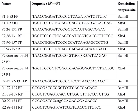
MAb C8 protects cells from TNFα-mediated toxicity
Several studies have shown that TNFα induces cell death in L929 cells via binding to TNFαR [16].To determine the activity of our purified recombinant hTNFα protein, L929 cells were subjected to different concentrations of hTNFα, and at 48 hours post- treatment the cell viability was examined using MTT assay. A concentration-dependent cell toxicity was observed and the IC50 (concentration at which there was 50% cell death) was determined to be 6.25 ng /ml (Figure 2A). To confirm that the toxicity seen in L929 cells was indeed because of TNFα, cells were challenged with heat-inactivated hTNFα protein which had no effect on the cell viability (data not shown).
12
Figure 2 A) Assessment of human-TNFα activity by MTT assay. L929 cells were incubated with different concentrations of hTNFα. After 48 hr incubation, MTT was added and further incubated for 4 hr. Formazan crystals formed were dissolved in DMSO and OD measured at 570 nm. Actinomycin D (Acd) was used as a positive control. B) Rescue from TNF-mediated toxicity with mAbs. L929 cells were incubated with 10 ng/ml hTNFα protein along with anti-TNFα purified antibodies at 100-fold molar excess. MTT assay was performed for cell viability analysis after 48 hr incubation. C, D)
Rescue of cells from TNF-mediated cell death by mAbs. L929 cells were treated with 10ng/ml hTNFα protein along with the indicated different concentrations of anti-TNFα antibodies starting from 9 µg which is 100-fold molar excess. MTT assay was performed for cell viability analysis after 48 hr incubation. C and D showed mAbs C1 and C8 dose-dependent rescue assay respectively. n = 3 independent experiments,* p<0.05, *** p<0.001.
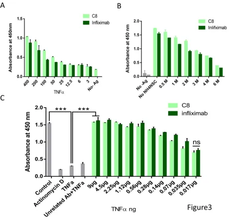
Comparison of mAb C8 and Infliximab (anti-TNFα mAb)
13 concentrations of hTNFα antigen (Figure 3A). The result revealed that not only in antigen dilution but also in antibody dilution assay, binding of C8 to TNFα is highly comparable with that of infliximab. Both mAbs were found to have high titer as determined by the dilution curves (Figure S 4). Moreover, the binding affinity of the mAbs to immobilized hTNFα was measured using the chaotrope ammonium thiocyanate ELISA to determine relative affinities of mAbs. As presented in Figure 3B the data demonstrate that the affinity of C8 and infliximab are very similar. Importantly, even the L929 cell assay showed that TNFα-mediated cytotoxicity could be rescued by mAb C8 as efficiently as Infliximab (Figure 3C).
14 incubation with the substrate, TMB H2O2. The reaction was stopped with H2SO4 and the absorbance was measured at 450 nm using an ELISA microplate reader. light green: mAb C8; dark green: Infliximab. B) Comparison of Antibody affinity using ammonium thiocyanate. 500 ng of hTNFα was coated in an ELISA plate followed by 2hr incubation with mAbs, followed by incubation with different concentrations of NH4SCN in 0.1 M sodium phosphate buffer pH 6.0 for 15 minutes and then secondary antibody conjugated to HRP. C) Rescue of cells from TNFα-mediated cytotoxicity. L929 cells were treated with 10 ng/ml hTNFα along with the indicated concentrations of mAb C8 or Infliximab. MTT assay was performed to measure cell viability after 48 hr incubation. Actinomycin- D used as a positive control.
Effect of mAb C8 on TNFα-induced signaling
TNFα mediated cell death has been shown through activation of c-Jun N-terminal kinase (JNK) [18]. TNFα triggers rapid phosphorylation of JNK and sustained JNK activation leads to cell death [19]. To confirm this, L929 cells were treated with different concentrations of TNFα and JNK phosphorylation was assessed with Western blotting. The result shows increase in JNK phosphorylation in a dose-dependent manner in TNFα treated cells, whereas heat-inactivated TNFα failed to show this (Figure 4A). To test whether mAb C8 can inhibit TNFα-mediated JNK activation, we exposed L929 cells with TNFα incubated with C8 or infliximab or mAb C12, following which pJNK level was assessed. As the immunoblot analysis suggests, though mAb C12 had no effect, mAbs C8 and Infliximab inhibited TNFα-mediated JNK phosphorylation. Interestingly, mAb C8 treated cells showed better inhibition as compared to the Infliximab treated cells (Figure 4B).
15 kit A) Cells were incubated with different concentration of TNFα as active and heat inactive forms.
B) Cells were pre-incubated with 10ng/ml TNFα along with mAbs C8/ Infliximab/ C12 or D6F10.
Localization of mAb C8 core epitope
Identification and characterization of the binding sites of neutralizing antibodies might help in the development of new vaccine, diagnostics and therapeutics [20]. Epitope mapping provides insights into the therapeutic mechanism of an individual mAb [21]. To identify the epitope corresponding to mAb C8, we made three overlapping truncated hTNFα fragments F1, F2 and F3 as GST-fusion proteins. MAb C8 bound to full length hTNFα (His-tag fusion 17 KDa) and hTNFα fragment 2 (26-131 aa) proteins but not to F1 (1-53 aa) and F3 (96-157 aa) (Figure 5A). The result was supported by indirect ELISA using different concentration of the fragments incubated with mAb C8 (Figure S 5A). The data suggest that the epitope of mAb C8 lies in TNFα fragment 2 region comprising of amino acid 26-131. Indeed F2 in solution phase bound and competed out the binding of mAb C8 to full length hTNFα as well (Data not shown).
In order to narrow down the epitope corresponding to the mAb C8 further, two truncated constructs were generated from the F2 region of hTNFα, F2 core region (54-93) excluding F1 and F3 overlap and F2-F1 (72-131). MAb bound to F2-F1 (72-131) but not to F2 Core region (54-93) (Figure 5B). The immunoblot study was confirmed by ELISA result as well (Figure S 5C).
Then, 3 overlap fragment constructs were generated from F2-F1 (72-131) region, corresponding to R1 (72-107), R2 (99-131) and R3 (85-120), as GST-fusion proteins. The antibody bound to R2 and R3 but not to R1 (Figure 5C)
16
Figure 5 A) Immunoblot analysis by ECL of the truncated protein fragments with mAb C8; fragment1 (1-53), fragment2 (26-131), fragment 3 (96-157) and full length hTNFα. 1µg of each purified protein was electrophoresed on a 12.5% polyacrylamide gel under denaturing condition. Proteins were transferred to nitrocellulose membrane, probed with mAb C8 and developed using the ECL kit (Millipore). Lanes: 1: fragment 1; 2: fragment 2; 3: fragment 3; 4: full length hTNFα. B)
17 5: 99-120 WT; 6: 99-120 QRE/PKD. (Full length hTNFα is His-fused protein; all truncated and mutated fragments are GST-fused). F) Schematic representation of hTNFα constructs F1, F2, F3, F2-F1, Core F2, R1, R2, R3 and 99-120. Truncated gene fragments were generated, cloned in pGEX5X2 vector and expressed E. coli BL21 (DE3) competent cells.
Simulation studies revealed that, Infliximab blocks TNFα activity due to binding to the E-F loop of the hTNFα structure, which hampers TNFα-receptor binding. Our study also showed that mAb C8 binds to the E-F loop in the vicinity of Infliximab epitope. This suggests that the C8 binding to TNFα being comparable with Infliximab might be due to juxtaposed epitopes (Figure 6A).
Docking
18
19 introduced in epitopic region Q102R103E104 to PKD, disturbed C8 and hTNFα interaction. The images were generated using open-source PyMOL software. E) Comparison of Rosetta energy scores for binding of wild-type (QRE) or mutated (PKD) hTNFα with mAb C8.
Discussion
TNFα is one of the most important pro-inflammatory cytokines which regulates a number of cellular processes like cell proliferation, differentiation and apoptosis [22]. Excessive production of TNFα however is detrimental to health and needs to be regulated. Specific antibodies to TNFα have proven to be useful blockers of TNFα-mediated toxicity. As it is known, anti-TNFα agents bound to TNFα may block interaction between TNFα and its receptors, thus, basically inhibit TNFα-mediated pro-inflammatory signal activation[23, 24].
Even though there are a number of anti-TNFα agents in the market, there is a demand for more TNFα specific antibodies. Like Infliximab, Adalimumab and Etanercept are currently being used in the alleviation of inflammatory immune disorders via interruption of TNFα-TNFR interaction [25]. Infliximab was the first approved anti-TNFα drug for the management of autoimmune conditions such as Crohn’s disease, rheumatoid arthritis, ankylosing spondylitis, psoriatic arthritis and various inflammatory skin problems [26, 27].
In our study, we found mAb C8 to bind with high affinity to TNFα. Since hTNFα-mediated cell death in L929 fibroblast cells is a well-accepted cell culture model [16], we utilized it to test the rescue from cytotoxicity by our panel of mAbs. MAb C8 inhibited TNFα-mediated activity significantly and comparable with that of Infliximab. Due to specificity of mAb C8 to human-TNFα only, we could not test the effect of mAb C8 in blocking the biological activity of TNFα in a mouse model. However, comparable cell rescue from TNFα-mediated toxicity and inhibition of TNFα-mediated signaling by both antibodies may suggest the capability of mAb C8 in blocking the physiological activity of TNFα also in-vivo. That mAb C8 inhibits TNFα-induced cell signaling was also demonstrated by assessing the decrease in activation of JNK.
In order to get insights into the mechanism by which C8 antibody inhibits hTNFα-mediated toxicity, we initiated studies to identify the epitope corresponding to the mAb. The core epitope of mAb C8 was delineated to amino acid sequence 99-120. Mutational analysis in this region established unequivocally that residues Gln102, Arg103 and Glu104 are the most critical for binding of mAb C8 to hTNFα. It is worth mentioning that, these residues are located in the EF loop of TNFα structure. Interestingly, the epitope of Infliximab on TNFα structure comprises of the C-D and E-F loop and include residues Glu67-His73 and Thr105-Lys112 respectively, as well as several residues in C and D strands and blocks its activity. MAb C8 and Infliximab binding to the EF loop hampers TNFα binding to its receptor, as a result inhibits its activity.
20
21
22
23 muatnts T72D; H73Y; T89E; S71de; TH72,73DY; Q102P; R103K; E104D; RE103,104KD; R131Q were determined. There are indications below x-axis which show that mutants introduced in F2 background and in F2 S71 del, H73Y background. C) Binding of mAb C8 and hTNFα truncated constructs using Indirect ELISA. The extent of binding of the antibody to immobilized full length hTNFα, F1, F2, F3, F2 Core region (54-93) and F2-F1 (72-131) were determined. D) Epitope localization of mAb C8. The extent of binding of the antibody to immobilized full length hTNFα, F1, F2, F3, 99-120 WT and 99-120 QRE/PKD mutants was determined. For sections B, C and D, 1µg per well of each protein were coated, followed by 2h incubation with 100 µl of mAb culture supernatant.
Acknowledgements
We thank the Central Animal Facility, Indian Institute of Science (IISc). We thank Dr. Manjula Das and Dr. Akanksha Dixit for valuable discussions and critical comments. We greatly acknowledge financial support from the Department of Biotechnology, DBT-IISc partnership program. We also thank IISc-student fellowship support.
References
1. Tracey, K.J. and A. Cerami, Tumor necrosis factor: a pleiotropic cytokine and therapeutic target. Annu Rev Med, 1994. 45: p. 491-503.
2. Baxter, G.T., et al., Tumor necrosis factor-alpha mediates both apoptotic cell death and cell proliferation in a human hematopoietic cell line dependent on mitotic activity and receptor subtype expression. J Biol Chem, 1999. 274(14): p. 9539-47.
3. Clark, I.A., et al., Possible importance of macrophage-derived mediators in acute malaria. Infect Immun, 1981. 32(3): p. 1058-66.
4. van der Bruggen, T., et al., Lipopolysaccharide-induced tumor necrosis factor alpha production by human monocytes involves the raf-1/MEK1-MEK2/ERK1-ERK2 pathway. Infect Immun, 1999. 67(8): p. 3824-9.
5. Kircheis, R., et al., Differences in the biological activity of TNF alpha and TNF beta correlate with their different abilities for binding to the target cells. Eur Cytokine Netw, 1992. 3(4): p. 381-90.
6. Loetscher, H., et al., Molecular cloning and expression of the human 55 kd tumor necrosis factor receptor. Cell, 1990. 61(2): p. 351-9.
7. Tartaglia, L.A. and D.V. Goeddel, Two TNF receptors. Immunol Today, 1992. 13(5): p. 151-3.
8. Aggarwal, B.B., S.C. Gupta, and J.H. Kim, Historical perspectives on tumor necrosis factor and its superfamily: 25 years later, a golden journey. Blood, 2012. 119(3): p. 651-65.
9. MacEwan, D.J., TNF receptor subtype signalling: differences and cellular consequences. Cell Signal, 2002. 14(6): p. 477-92.
10. Peyrin-Biroulet, L., Anti-TNF therapy in inflammatory bowel diseases: a huge review. Minerva Gastroenterol Dietol, 2010. 56(2): p. 233-43.
24 12. Ho, S.N., et al., Site-directed mutagenesis by overlap extension using the polymerase chain reaction.
Gene, 1989. 77(1): p. 51-9.
13. Schwede, T., et al., SWISS-MODEL: An automated protein homology-modeling server. Nucleic Acids Res, 2003. 31(13): p. 3381-5.
14. Konagurthu, A.S., et al., MUSTANG: a multiple structural alignment algorithm. Proteins, 2006. 64(3): p. 559-74.
15. Gray, J.J., et al., Protein-protein docking with simultaneous optimization of rigid-body displacement and side-chain conformations. J Mol Biol, 2003. 331(1): p. 281-99.
16. Humphreys, D.T. and M.R. Wilson, Modes of L929 cell death induced by TNF-alpha and other cytotoxic agents. Cytokine, 1999. 11(10): p. 773-82.
17. Monaco, C., et al., Anti-TNF therapy: past, present and future. Int Immunol, 2015. 27(1): p. 55-62. 18. Dietrich, N., et al., JNK2 mediates TNF-induced cell death in mouse embryonic fibroblasts via
regulation of both caspase and cathepsin protease pathways. Cell Death Differ, 2004. 11(3): p. 301-13.
19. Barbin, G., M.P. Roisin, and B. Zalc, Tumor necrosis factor alpha activates the phosphorylation of ERK, SAPK/JNK, and P38 kinase in primary cultures of neurons. Neurochem Res, 2001. 26(2): p. 107-12. 20. Gershoni, J.M., et al., Epitope mapping: the first step in developing epitope-based vaccines. BioDrugs,
2007. 21(3): p. 145-56.
21. Hasan, S.S., et al., A human antibody against Zika virus crosslinks the E protein to prevent infection.
Nat Commun, 2017. 8: p. 14722.
22. Liu, Z.G., Molecular mechanism of TNF signaling and beyond. Cell Res, 2005. 15(1): p. 24-7.
23. Wong, M., et al., TNFalpha blockade in human diseases: mechanisms and future directions. Clin Immunol, 2008. 126(2): p. 121-36.
24. Sedger, L.M. and M.F. McDermott, TNF and TNF-receptors: From mediators of cell death and inflammation to therapeutic giants - past, present and future. Cytokine Growth Factor Rev, 2014.
25(4): p. 453-72.
25. Mukai, Y., et al., Solution of the structure of the TNF-TNFR2 complex. Sci Signal, 2010. 3(148): p. ra83. 26. Buch, M.H., et al., True infliximab resistance in rheumatoid arthritis: a role for lymphotoxin alpha?
Ann Rheum Dis, 2004. 63(10): p. 1344-6.