1
Understanding
Pathogenaemia
in Man:
A Proof of Concept Study
Robert Nipah
School of Environment and Life Sciences
University of Salford, Salford, UK
A thesis submitted in partial fulfilment of the requirements of the degree
of Master of Philosophy, August
2
Contents
The Author
... 5Abstract
... 6List of Figures
... 9List of Tables
... 10Acknowledgements
... 11Abbreviations
... 12CHAPTER 1 ... 15
Background
... 16Pathogens, infection and sepsis: A relationship explored ... 16
Sepsis: a pathophysiological response to pathogenaemia ... 19
Previous in vivo Models of Sepsis ... 21
Model of investigation... 25
Introduction ... 25
Endourology ... 27
Endourology procedure - Ureteroscopy ... 28
Endourology procedure - Percutaneous nephrolithotomy (PCNL) ... 28
Antibiotic prophylaxis in urology procedures ... 30
Tools for investigation ... 32
Microbiological investigations – Blood Cultures ... 32
3
Molecular investigation – Nucleic acid based diagnostic technology ... 35
NAT assays for the detection and identification of pathogens from blood culture bottles .. 36
Direct molecular detection of pathogens in blood with PCR ... 37
SeptiFast® assay ... 39
Effect of antimicriobial administration pre testing on both microbiological and molecular approaches... 41
Cytokine driven immune reponses ... 44
Interleukin 6 ... 45
Interleukin 10 ... 47
Summary
... 48Aim of study... 50
CHAPTER 2: ... 51
Participant recruitment ... 52
Study design ... 52
Blood sampling ... 54
Data collection ... 55
Laboratory analyses ... 56
Analysis of pathogen DNA by SeptiFast® PCR assay ... 56
Pathogen DNA extraction from blood ... 57
SeptiFast® real-time PCR of pathogen DNA ... 58
SeptiFast® - Identification of pathogen species and controls... 59
Cytokine analysis ... 61
IL-6 ELISA ... 61
IL-10 ELISA ... 63
Statistical analysis ... 63
CHAPTER 3: ... 64
4
Study Population ... 66
Pathogen species identified by SeptiFast® assay and blood culture... 68
PCR positive signal patients vs. PCR negative signal patients ... 70
Relationship between pathogen isolate and clinical outcome ... 72
Analysis of circulating IL-6 and IL-10 levels ... 74
Pathogen detection and physiological response ... 76
Case specific findings - PCR positive patients ... 81
Case specific findings - PCR negative patients ... 81
Stone composition and pathogen detection... 82
CHAPTER 4 ... 83
Background of the study ... 84
Key findings of the study ... 87
SeptiFast® - Role in circulating pathogen DNA detection in infection ? ... 88
SeptiFast® - A place in clinical practice? ... 89
Circulating pathogen DNA and the immune response ... 94
The urology patient: Host response, sepsis and antibiotic prophylaxis ... 96
Strength and weakness of the study ... 98
Future work ... 99
Conclusion ... 99
5
The Author
The author graduated from the Queen Mary and Westfield College, University of
London with a First Class Honours Bachelor of Science degree in Basic Medical
Science with Clinical Immunology in 1998. He qualified as a medical doctor from St
Bartholomew and Royal London School of Medicine and Dentistry, University of
London with MB BS in 2001. He passed his MRCP post graduate examination in 2006.
He commenced his research as an out of programme placement as an intensive care
registrar at Salford Royal Foundation Trust. He has completed his specialist registrar
training and is dual accredited in general medicine and nephrology. Since March 2013,
6
Abstract
Introduction: The current understanding of the significance of circulating pathogen DNA in infection is limited. Blood cultures are the current gold standard for pathogen
detection. The administration of antibiotics can confound pathogen detection by blood
cultures. Polymerase chain reaction assays of circulating pathogen DNA has the
potential ability for rapid diagnosis of infection and may be potentially useful in a
clinical setting. However, the use of this technology has only recently been used in the
study of sepsis.
Animal models for the study of sepsis have added to our understanding. The failure to
translate results from animals to humans has been attributed to the disease
characteristics of sepsis (complexity and heterogeneity), inappropriate clinical trials
(study of ineffective drugs, inadequate clinical trial designs), and animal models that do
not fully mimic human sepsis. Therefore, the finding of an easily reproducible in vivo
human model for pathogenaemia may provide a platform for exploring the host’s
immune response to circulating pathogen material.
Infection and urosepsis are common complications in diagnostic and therapeutic
urological procedures. Urological interventional procedures for the removal of renal
stones are commonly done in a controlled operating environment and may potentially
be an in vivo model for investigating the host immune response to detected pathogen.
Antibiotic prophylaxis is routinely given across the world for these urological
procedures even though the evidence for their use is weak. If the presence of pathogen
is seen to generate an immune response in these in vivo human models, it could be
7
Aim: To investigate interventional urological stone removal as a model of pathogenaemia in man and, therefore, test the following hypotheses:
1. The detection of circulating pathogen DNA by SeptiFast® PCR is an indicator
for the infection associated with urological procedures.
2. The presence of circulating pathogen DNA correlates with the host immune and physiological response, supporting the notion that antibiotics prophylaxis is
important in urological procedures.
Method: In a prospective group of patients undergoing stone removal, blood samples were taken at five time points, peri-operatively to assess:
The presence of pathogen by blood culture and pathogen DNA by SeptiFast® PCR;
Circulating IL - 6 and IL - 10 levels to assess activation of host inflammatory
response.
Routine peri - operative observations were recorded throughout as measures of
physiological responses.
Results: Collected historical data on urological procedures by Salford Royal
Foundation Trust urology services show that the SeptiFast® assay gave full coverage for
pathogens seen in this clinical setting. While positive blood culture was rare, 50% of
patients tested positive for pathogen DNAemia and this was associated with increased
circulating IL-6 compared to patients with no circulating pathogen DNA. Linkage
between pathogen DNA positivity and patient outcome was not established.
8
inflammatory response. Endourological stone removal interventions may be a useful
model for understanding the role of pathogen DNA in triggering inflammatory
9
List of Figures
Figure 1 - Simplified clinical course of sepsis (Doi et al 2009) ... 20
Figure 2 - Percentage of kidney stones ... 27
Figure 3 - PCNL (taken from www.uroinfo.ca) ... 29
Figure 4 - Identification and genotyping of microbes by PCR/ESI/mass (Ecker et al 2010) ... 36
Figure 5 - IL - 6 and sepsis - associated mortality (Hack et al 1989) ... 46
Figure 6 - Interleukin 10 and receptor (Kotenko et al 1997) ... 47
Figure 7 - Study design ... 53
Figure 8 - Distribution of melting temperature and respective detection channels for all microorganism and internal control in Septifast assay ( Lehmann et al 2008) ... 60
Figure 9 - The principle of sandwich ELISA ( adapted from www.lenico.com) ... 62
Figure 10 - Mean IL - 6, IL10, IL-6:IL-10 ratio in PCR - positive and PCR - negative patients ... 75
Figure 11 - Variations of mean arterial blood pressures in PCR negative group ... 77
Figure 12 - Variation of mean arterial blood pressure in PCR positive group ... 77
Figure 13 - Mean MAP between PCR positive and PCR negative ... 78
Figure 14 - Variation in pulse rate in PCR negative group... 79
Figure 15 - Variations in pulse rate in PCR positive groups ... 80
10
List of Tables
Table 1 - Diagnostic criteria for sepsis according to the International Sepsis Definitions
Conference (Levy et al , 2003). ... 18
Table 2 - Possible reasons for failure in sepsis trials (Riedemann et al, 2003) ... 24
Table 3 Generally accepted risk factors for infectious complication ... 31
Table 4 - Needs and current status of methods to identify bloodstream infections (Adapted from Ecker et al 2010) ... 43
Table 5 – Timing schedule for blood sampling for RT-PCR analysis ... 54
Table 6 – Septifast® Pathogen Detection Panel ... 57
Table 7 - List of positive organism growth from mid-stream urine sample from all patients who underwent renal stone removal surgery at Salford Royal Foundation Trust between 2004 – 2011 ... 65
Table 8 - Patient demographic and operation detail ... 67
Table 9 - Detection of pathogen DNA signal RT - PCR on blood samples at different time points ... 69
Table 10 - Characteristic of PCR positive patients vs. PCR negative patients ... 70
Table 11 - Correlation between PCR positivity and clinical outcome... 72
Table 12 - Correlation between PCR negativity and clinical outcome ... 74
11
Acknowledgements
I wish to thank my principal supervisor, Professor Geoff Warhurst, and my
co-supervisor, Dr Paul Dark, for their support and advice throughout my MPhil project.
I would like to thank Ms Laura Derbyshire for assisting in data collection throughout the
project.
I would like to thank Mr Chris Betts for his input in the planning and preparation of the
project.
I would like to thank the Infection, Injury and Inflammation Research Group (3IRG),
urology services and intensive care unit at Salford Royal NHS Foundation Trust for
their support.
I would like to thank my family and friends for their encouragement, support and
12
Abbreviations
ARDS Acute Respiratory Distress Syndrome
BC Blood culture
BSI Bacterial bloodstream infection
CLP Caecal ligation and puncture
CFU Colony Forming Units
CoNS Coagulase-negative Staphylococci
COX Cyclooxygenase
CRP C Reactive Protein
DNA Deoxyribonucleic acid
EAU European Association of Urology
EPR Electronic Patient Record
ESI Electro Spray Ionisation
ESIU EAU Section for Infection in Urology
ESWL Extracorporeal Shock Wave Lithotripsy
EWS Early Warning Score
FISH Fluorescence in-situ hybridization
HMGB-1 High-mobility group box-1
ICU Intensive Care Unit
IL Interleukin
IL - 6R Interleukin 6 receptor
IQR Interquartile range
13
kDA Kilodalton
LPS Lipopolysaccharide
mls Millilitres
mg Milligrams
MODS Multiple Organ Dysfunction Syndrome
MRSA Methicillin-resistant Staphylococcus aureus
NAT Nucleic acid based diagnostic technology
NFkB Nuclear factor kB
NLR NOD-like receptor
ng Nanogram
PAI-1 promoter Plasminogen activator inhibitor-1 promoter
PAMP Pathogen-associated molecular pattern
PCWP Pulmonary capillary wedge pressure
PCNL Percutaneous nephrolithotomy
PCR Polymerase Chain reaction
pDC Plasmacytoid dendritic cells
PG Peptidoglycan
PNA-FISH Peptidic nucleic acid fluorescent in-situ hybridisation
RCT Randomized Controlled Trial
RT PCR Real time polymerase chain reaction
RNA Ribonucleic acid
rRNA Ribosomal ribonucleic acid
SCC Society of Critical Medicine
SIRS Systemic Inflammatory Response Syndrome
sIL - 6R Soluble Interleukin 6 receptor
14
SRFT Salford Royal Foundation Trust
SSC Surviving Sepsis Campaign
STARD Standards for Reporting of Diagnostic Accuracy
TNFA Tumour Necrosis Factor
TLR Toll like receptor
UNG Uracil-N-glycosylate
15
CHAPTER 1
16
Background
Sepsis is one of the oldest syndromes in medicine. Sepsis has been described in literature
since the days of the ancient Greeks. Sepsis is still one of the top 10 causes of death today.
The clinical manifestations of sepsis are highly variable, depending on the initial site of
infection, the causative organism, the pattern of acute organ dysfunction, the underlying
health status of the patient, and the interval before the initiation of treatment (Angus et al,
2013). The 'Surviving Sepsis Campaign' has been instrumental in providing the nomenclature
to help physicians understand the pathophysiology of sepsis (Dellinger et al, 2008).
The interaction of infection and immune response plays a decisive role in the pathogenesis of
sepsis. Animal and human studies in sepsis have furthered our understanding on the effect of
pathogenaemia in sepsis, such as the role of pathogen – associated molecules pattern (PAMP)
in the host response in sepsis. The significance of circulating pathogen DNA, a recognised
PAMP, in the infection and sepsis process has not been fully defined in these studies. The
advancement in nucleic acid technology (NAT) could potentially provide techniques to
explore the role of pathogen DNA in the host response in sepsis. The aim of this proof of
concept study was to explore the feasible use of a novel in vivo human model of
pathogenaemia using microbiological, immunochemistry and NAT techniques.
Pathogens, infection and sepsis: A relationship explored
Infection is the invasion and multiplication of pathogens, such as bacteria, viruses, and fungi.
If pathogen in blood is present, this represents progression of the infection to pathogenaemia. Patients’ responses to pathogenaemia differ and there is no clear answer to why some
17
sepsis (Rao et al, 1991, Mancini et al, 2010). Sepsis is defined as a suspected or proven
infection plus a systemic inflammatory response syndrome (e.g. fever, tachycardia,
tachypnoea, and leucocytosis) (Bone et al, 1992) Severe sepsis is defined as sepsis with organ
dysfunction (hypotension, hypoxemia, oliguria, metabolic acidosis, thrombocytopenia, or
obtundation). Septic shock is defined as severe sepsis with hypotension, despite adequate
fluid resuscitation.
Septic shock and multiorgan dysfunction are the most common causes of death in patients
with sepsis. The mortality rates associated with severe sepsis and septic shock are 25 to 30%
and 40 to 70%, respectively (Angus et al 2001, Vincent et al, 2006). There is an estimated
annual mortality of between 30 and 50 deaths per population of 100, 000. In Europe alone, an
estimated 135,000 patients die each year of sepsis – associated complications, with an overall
incidence of sepsis of three cases per 1000 individuals (Lever et al, 2007). In the US, the
calculated cost of sepsis is close to 20 billion pounds and is the most common cause of in
hospital mortality (Lagu, 2012).
There has been a strong international drive to provide guidelines for clinicians caring for a
patient with severe sepsis and septic shock. The on-going motivation for the international
drive is that severe sepsis and septic shock are major health care problems, affecting millions
of people around the world each year, killing one in four (and often more), and increasing in
incidence (Dellinger et al, 2013). In 2004, the surviving sepsis campaign, an international
collaboration of professional societies involved in critical care, treatment of infectious
diseases and emergency medicine, published the first internationally accepted guidelines to
improve outcomes for this group of patients (Dellinger et al, 2008). Although controversially
associated with potential conflicts of interest with the drug industry, it served as a nidus for
18
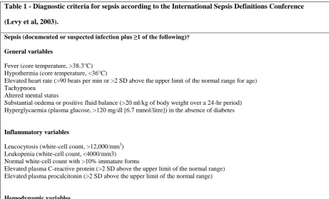
(Table 1) highlights the diagnostic criteria for sepsis, severe sepsis and septic shock derived
[image:18.595.66.528.135.414.2]from this group.
Table 1 - Diagnostic criteria for sepsis according to the International Sepsis Definitions Conference
(Levy et al, 2003).
Sepsis (documented or suspected infection plus ≥1 of the following)†
General variables
Fever (core temperature, >38.3°C) Hypothermia (core temperature, <36°C)
Elevated heart rate (>90 beats per min or >2 SD above the upper limit of the normal range for age) Tachypnoea
Altered mental status
Substantial oedema or positive fluid balance (>20 ml/kg of body weight over a 24-hr period) Hyperglycaemia (plasma glucose, >120 mg/dl [6.7 mmol/litre]) in the absence of diabetes
Inflammatory variables
Leucocytosis (white-cell count, >12,000/mm3) Leukopenia (white-cell count, <4000/mm3) Normal white-cell count with >10% immature forms
Elevated plasma C-reactive protein (>2 SD above the upper limit of the normal range) Elevated plasma procalcitonin (>2 SD above the upper limit of the normal range)
Hemodynamic variables
Arterial hypotension (systolic pressure, <90 mm Hg; mean arterial pressure, <70 mm Hg; or decrease in systolic pressure of >40 mm Hg in adults or to >2 SD below the lower limit of the normal range for age)
Elevated mixed venous oxygen saturation (>70%)
Elevated cardiac index (>3.5 litres/min/square meter of body-surface area)
Organ-dysfunction variables
Arterial hypoxemia (ratio of the partial pressure of arterial oxygen to the fraction of inspired oxygen, <300) Acute oliguria (urine output, <0.5 ml/kg/hr or 45 ml/hr for at least 2 hr)
Increase in creatinine level of >0.5 mg/dl (>44 μmol/litre)
Coagulation abnormalities (international normalized ratio, >1.5; or activated partial-thromboplastin time, >60 sec) Paralytic ileus (absence of bowel sounds)
Thrombocytopenia (platelet count, <100,000/mm3)
Hyperbilirubinemia (plasma total bilirubin, >4 mg/dl [68 μmol/litre])
Tissue-perfusion variables
Hyperlactataemia (lactate, >1 mmol/litre) Decreased capillary refill or mottling
Severe sepsis (sepsis plus organ dysfunction)
19
These definitions give a coherent language to discuss the parameters seen in sepsis which is
used globally (Levy et al, 2003). A revised and updated edition of ‘Surviving Sepsis
Campaign: International Guidelines for management of severe sepsis and shock: 2012’ was
officially released at the Society of Critical Medicine’s (SCC) 42nd
Congress (Dellinger et al,
2013), including full disclosures of potential conflicts of interest of the authors. It has been
proposed that the host response to pathogenaemia must play a vital defining role in the septic
response. To further our understanding on the pathophysiology of sepsis, an in vivo model of
human pathogenaemia could prove beneficial.
Sepsis: a pathophysiological response to pathogenaemia
Sepsis is the culmination of complex interactions between the infecting microorganism and
the host immune, inflammatory, and coagulation responses (James et al, 2006). In the last 10
years, current knowledge of the host’s ability to recognise a pathogen has increased. The
expression of common structures known as pathogen – associated molecules pattern (PAMP)
are thought to be central to the host response in sepsis (Christaki et al, 2011). These
molecules have the potential to trigger a series of events via the immune system giving us the
signs and symptoms seen in sepsis and septic shock.
Examples of PAMPs are:
Lipopolysaccharide (LPS);
Endotoxins – typically expressed by Gram negative bacteria;
Peptidoglycans;
Lipoprotein;
Flagellin;
20
The use of nucleic acid based diagnostic technology (NAT) such as polymerase chain
reaction (PCR) assays has prompted significant interest in the role of bacterial DNA as
PAMP. NAT could potentially have clinical significance as a modern day technique for
pathogen detection and in the future supplement or even supersede blood cultures in the
management of infections and sepsis.
In a recent review (Doi et al, 2009), the course of human sepsis is described as being likely to
have an initial pro-inflammatory burst responsible for hypotension and organ dysfunction,
followed by a compensatory anti-inflammatory immune response that leads to an
[image:20.595.74.526.378.659.2]immunocompromised state often called immune depression or immune dysfunction.
Figure 1 -Simplified clinical course of sepsis (Doi et al, 2009)
21
Although the progression of disease is complex and varies individually, from patient to
patient, there are a few common pivotal events seen in all septic patients. A frequent feature
in septic shock is a hyper dynamic circulation associated with diminished myocardial
function. Blood volume is continually lost into the interstitial space of tissue and intracellular
locations, and blood vessels become blocked by dysfunctional coagulation cascades
(disseminated intravascular coagulation). Infectious disease, septicaemia in particular, is the
most common clinical condition associated with disseminated intravascular coagulation
(DIC). Although, virtually all microorganisms can cause DIC, bacterial infection is most
frequently related to the development of the syndrome. Clinically onset DIC may occur in 30
to 50 percent of patients with gram negative sepsis (Levi et al, 1999). The mechanism of DIC
starts with the systemic activation of coagulation and leads to widespread intravascular
deposition of fibrin. Additionally, there is a depletion of platelets and coagulation factors. As
a result, thrombosis of small and mid-size vessels may occur, contributing to organ failure
and may lead to significant bleeding. Eventually, systolic hypotension and diffuse
vasoconstriction lead to a fatal, therapy-refractory ischemia of multiple organs and to organ
necrosis (Kruttgen et al, 2012).
It should be emphasized that the pro–inflammatory and anti–inflammatory 'stages' are not a
fixed sequence of events, nor is there an intermediate dissecting point in this diphasic
process. Inflammatory responses in sepsis are complex, dynamically evolving, pleiotropic,
synergistic and mutually reinforcing (Christaki et al, 2011). In this thesis, most of our work is
concerned with analysis of the pro-inflammatory stage/early stage of the host response to a
pathogen. In the next section, we will discuss the basis of most of the research into the
pathophysiology of sepsis through animal and human models of sepsis.
Previous in vivo Models of Sepsis
22
Injection of an exogenous toxin such as lipopolysaccharide;
Alteration of animal’s endogenous protective barrier such as intestinal leaks;
Infusion or instillation of exogenous bacteria.
Animal studies initially gave credence to the theory that death from sepsis may be due to an
overstimulated immune system. A number of animal studies have used large doses of
endotoxin or bacteria, leading to situations where circulating levels of cytokines, such as
tumour necrosis factor, were exponentially higher in animals than they are in patients with
sepsis (Hotchkiss et al, 2003).
LPS infusion/ injection model has been widely used for sepsis research. LPS administration
induces systemic inflammation that mimics many of the initial clinical features of sepsis,
including increases in pro-inflammatory cytokines such as TNFA and IL-1, but without
bacteraemia (Witcherman et al, 1980, Remick et al, 2000, Michie et al, 1988). These results
prompt work into possible therapeutic interventions. Treatment of LPS-injected animals with
neutralizing antibody against TNFA or IL-1 resulted in improved outcomes for this model
(Tracey et al, 1987, McNamara et al, 1993). The most commonly used animal models of
sepsis in the last 10 - 20 years have been models which alter the animal's endogenous
protective barrier, such as intestinal leak. Caecal ligation and puncture is very straightforward
and is the most popular technique used. CLP-induced sepsis models show a cytokine profile
similar to that in human sepsis (Remick et al, 2000,Eskandari et al, 1992), and anti–TNFA
treatment fails to alleviate sepsis in CLP models as in human sepsis (Eskandari et al, 1992).
CLP-induced sepsis increased lymphocyte apoptosis, which mimics immunosuppression at
the later phase of human sepsis (Ayala et al, 1996, Dear et al, 2006). In this respect,
CLP-induced sepsis is completely different from LPS-CLP-induced sepsis and more closely mimics
23
these findings to address sepsis in man. A major drawback with animal models is their
inability to reflect the complex clinical picture seen in humans. There are clear differences
between laboratory animals and patients. Mice and rats are housed in specific pathogen-free
areas, may often be inbred strains, have the same age and weight, and most importantly, do
not have comorbidities (such as diabetes, hypertension, and pre-existing immunosuppression
among others) seen in septic humans. Most humans with sepsis are >50 years old, and most
mice used in sepsis are <3 months old (with an average lifespan of 24 months). Furthermore,
the experimental models have a precisely known time period. In contrast, we encounter
patients of different ethnicities, ages, and weight, and most of the time, we are uncertain
when the symptoms first emerged. In addition, there are differences between rodents and
humans on the molecular level (Rittirsch et al, 2007). The predominant source of infection in
septic patients before the late 1980s was Gram negative bacteraemia. Lipopolysaccharide
(LPS), the main component of gram negative bacterial cell wall, was known to stimulate the
release of inflammatory mediators from various cell types and induce acute infection
symptoms in animals (Riedemann et al, 2003).
Administration of Gram negative bacterial LPS has been used as a model of severe infection
in man and has been shown to reliably induce a febrile systemic inflammatory response with
associated hormonal and cytokine changes (Agwunobi et al, 2000). Michie et al (Michie et al,
1988) measured plasma concentrations of circulating tumour necrosis factor alpha
(cachectin), interleukin-1 beta, and gamma interferon, together with physiologic and
hormonal responses, in 13 healthy men after intravenous administration of Escherichia coli
endotoxin (4ng per kilogram of body weight) and during a control period of saline
administration. The group showed high levels of plasma concentrations of circulating TNFA
after the infusion. Results such as these, along with animal studies, prompted the
24
sepsis but further studies have not shown this. There are a number of plausible explanations
[image:24.595.69.528.184.717.2]for the perceived failure in sepsis trials (Table 2).
Table 2 Possible reasons for failure in sepsis trials (Riedemann et al, 2003)
Development of sepsis model theory Possible reason for failure
Assumptions
1. Gram negative bacteria are cause of sepsis 2. Bacteria causing disease shed LPS
3.High levels of serum LPS achieved in septic patients
1. Incorrect assumptions based on initiating factors of disease
2. Incomplete clinical observation
Observation in animals
1. High level of TNFA achieved following LPS infusion
1. Unsuitable animal not translatable to humans
2. High serum level of TNFA not achieved in humans during sepsis
Observation of intervention
1. Anti - TNFA antibodies increase survival 1. Unable to block all TNFA
2. Results incorrect
Clinical trials in humans in sepsis
1. Anti - TNFA antibodies not protective 1. Anti - TNFA antibodies not protective
2.Study design insufficient
3.Sepsis definition insufficient
4. Drug not working (not tested, etc.)
25
However, LPS injection in humans is a valid model of endotoxemia and has been used as a
model to review stress responses and metabolic response seen in human subjects (Agwunobi
et al, 2000). For this thesis, it is proposed that urological interventional procedures involving
instrumentation could be a novel in vivo model of pathogenaemia in man and provide an
easily reproducible model assessing the host immune response to presence of pathogen.
Model of investigation
Introduction
Urology is a surgical speciality which has seen marked changes in the last few decades.
Surgical procedures have moved significantly from open to endoscopic and laparoscopic
procedures (Bootsma et al, 2008). However, it is well established in the field of urology that
there are significant levels of bacteraemia after invasive urological procedures. In one study
of 300 patients (Sullivan et al, 1972) the incidence of bacteraemia after urethral dilatation
without antibiotic prophylaxis was 24%. It is well known that in urological interventions such
as cystoscopy, ureteroscopy and percutaneous nephrolithotomy, the rate of positive blood
cultures detected up to 15% (Christanio et al, 2000, Knopf et al, 2003, Doğan et al, 2002
Turan et al, 2006, Rao et al 1991). The incidence of septic shock after endoscopic
manipulation for renal stones was about 1% but ‘less serious effects’ were more common
(Rao et al 1991). A recent study (Sohn et al, 2013) retrospectively reviewed the medical notes
of 531 patients who underwent ureteroscopy and ureteroscopic lithotripsy in their hospital
over a nine year period. A total of 20 patients (3.8%) contracted infectious complications
after various procedures in the upper urinary tract. The studies above have all been put
forward as evidence for the perceived benefit of prophylactic antibiotics before urological
procedures. It is thought the use of prophylactic antibiotics will limit post operation infection
26
One of the most common urological interventions is the removal of renal stones stuck in the
renal tract that cause persistent symptoms. Therefore, more patients have their renal stones
removed by ureteroscopy with lasering of the stone or percutaneous nephrolithotomy
(PCNL). In PCNL, one third experience some peri-operative complications, the most
common being fever secondary to a urinary tract infection (Gutierrez et al, 2013) In two other
studies looking at infectious complications in PCNL (Michel et al, 2007, Draga et al, 2009)
21 - 39.8% of patients had a post-operative fever which was transient in most cases.
However, 0.3 - 9.3% patients developed sepsis. In both studies, prophylactic antibiotics were
used. Post op fevers are not uncommon and have caused people to suggest that this could be
an indicator of an early systemic response to infection. Rao et al (Rao et al, 1991) showed
that even though post-operative fever was seen in 74% of the patients who had PCNL, only
41% actually had endotoxemia.
There is a significant burden associated with urosepsis and urinary tract infection. Urinary
tract infections are the most common cause of hospital associated infections (nosocomial
infection). Approximately 80% of nosocomial UTI have been found to be associated with
indwelling urinary catheters. Genitourinary interventions appear to be the facilitating factor
in 5 – 10% of nosocomial UTI. Patients who have been exposed to some instrumentation are
at high risk of urinary tract infections (Turan et al, 2006). In the USA, a study from early
2000s showed that urinary tract infection accounts for 1 million emergency department visits,
27 Endourology
Endourology is the branch of urologic surgery concerned with closed procedures for
visualizing or manipulating the urinary tract. It has lately grown to include all urological
minimally invasive surgical procedures. Opposed to open surgery, endourology is performed
using small cameras and instruments inserted into the urinary tract. For the purpose of this
thesis, ureteroscopy and PCNL are the endourological procedures used and the indication for
all of these procedures was for the treatment of renal calculi.
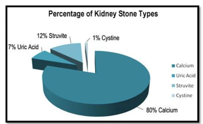
Renal calculi develop from crystals that separate from urine within the urinary tract. The
[image:27.595.125.461.479.684.2]chemical composition of renal stones, typically seen in clinical practice, is highlighted below
(Figure 2). There are well-recognised predisposing factors for stone formation which include
dehydration, lifestyle, geographical location (dry arid climate), and certain specific risk
factors.
Figure 2 - Percentage of kidney stone types
28
These factors may include anatomical / structural abnormalities (e.g. ureteropelvic junction
obstruction, urinary diversion surgery, horseshoe kidney, calyceal diverticulum), and
underlying metabolic conditions (e.g. cystinuria, oxaluria, gout), certain drugs, and
urease-producing infective organisms (Tseng et al, 2011).
Endourology procedure - Ureteroscopy
An ureteroscopy is an examination or procedure using an ureteroscope. An ureteroscope, like
a cystoscope, is an instrument for examining the inside of the urinary tract. An ureteroscope
is longer and thinner than a cystoscope and is used to see beyond the bladder into the ureters.
There are two main types of ureteroscopes (1) flexible like a thin, long straw (2) rigid and
firm. Through the ureteroscope, the obstructing stone in the ureter can be visualised and then
removed via a small basket at the end of a wire inserted through an extra channel in the
ureteroscope. In addition, a separate way to treat urolithiasis through an ureteroscope is to
extend a flexible fibre through the scope up to the stone and then, with a laser beam shone
through the fibre, break the stone into smaller pieces that can then pass out of the body in the
urine. A stent is usually placed to keep ureter patent.
Endourology procedure - Percutaneous nephrolithotomy (PCNL)
Percutaneous nephrolithotomy is a surgical procedure for removing medium-sized or larger
renal stones from the patient's urinary tract by means of a nephroscope passed into the kidney
through a track created in the patient's back via a small puncture wound (up to about 1cm).
PCNL was first performed in Sweden in 1973 as a less invasive alternative to open surgery
on the kidneys. The term "percutaneous" means that the procedure is done through the skin.
Nephrolithotomy is a term formed from two Greek words that mean "kidney" and "removing
29
With a small 1cm incision in the loin, the percutaneous nephrolithotomy (PCNL) needle is
passed into the pelvis of the kidney. The position of the needle is confirmed by fluoroscopy.
A guide wire is passed through the needle into the pelvis. The needle is then withdrawn with
the guide wire still inside the pelvis. Over the guide wire the dilators are passed and a
[image:29.595.89.503.240.526.2]working sheath is introduced (Fig 3).
Figure 3 - PCNL (taken from www.uroinfo.ca)
A nephroscope is then passed inside and small stones are taken out. A nephroscope is an
instrument with a fiberoptic light source and two additional channels for viewing the inside of
the kidney and irrigating (washing out) the area.
The surgeon may use a device with a basket on the end to grasp and remove smaller kidney
stones directly. Larger stones are broken up with an ultrasonic or electro hydraulic probe, or
30
probes and then have the stone fragments removed. The procedure can take 1 - 2 hours.
(Wynberg et al, 2012). As mentioned earlier, prophylactic antibiotics are commonly used in
this procedure and, presently, there is little evidence for their use in the urological procedures
mentioned in this proof of concept study. It can be postulated that in our novel in vivo mode,
if there is a relationship between the presence of detected pathogen and a detected host
response to the pathogen, it could be argued that antibiotic prophylaxis may be beneficial in
this setting. In the following section, there will be a brief exploration on the current use of
antibiotic prophylaxis in urological procedures.
Antibiotic prophylaxis in urology procedures
In urology, the indication for antibiotic prophylactic use is to prevent post-operative
infections. A pan-European survey was carried out by the EAU Section for Infection in
Urology (ESIU) in a large number of European countries and found that ≥ 10-12% of patients
had a healthcare-associated UTI (Bjerklund et al, 2007). The current European Association
guidelines on peri-operative antibiotic prophylaxis state that there is no evidence for any
benefits of antibiotic prophylaxis in standard non-complicated endoscopic procedures and
shockwave lithotripsy (SWL), although it is recommended in complicated procedures and
patients with identified risk factors. The EAU guidelines reference papers which are
described later in this introduction (Fourcade et al, 1990, Knopf et al, 2003, Rao et al, 1991).
Of all urological surgical procedures, there is strong evidence for a role of antibiotic
prophylaxis in transrectal prostate biopsies and transurethral resection of the prostate
(Bootsma et al, 2008). However, antibiotic prophylaxis is still widely used with marked differences in the regimens and choice of antibiotics used from one urology department to
31
There are a number of clearly established risk factors for peri-procedural infectious
[image:31.595.65.532.186.514.2]complications. These are highlighted in the table below (Table 3)
Table 3 Generally accepted risk factors for infectious complication (Grabe et al 2012)
General risk factors Special risk factors associated with an increased bacterial load
Older age Long pre-operative hospital stay or recent
hospitalisation
Deficient nutritional status History of recurrent urogenital infections
Impaired immune response Surgery involving bowel segment
Diabetes mellitus Long term drainage
Smoking Urinary obstruction
Extreme weight Urinary stones
Coexisting infection in a remote site Colonisation with microorganisms
A pan-European study on nosocomial UTI (Bjerklund et al, 2007) has identified the three
most important risk factors for infectious complications as:
An indwelling catheter;
Previous urogenital infection;
Long preoperative hospital stay.
32
There are few randomised control trials looking at antibiotic prophylaxis in ureteroscopy and
PNCL procedures. Knopf and his group (Knopf et al, 2003) studied 113 patients undergoing
uretoscopy for stone removal randomised to a single oral dose of levofloxacin versus no
antimicrobial. There was a significantly lower incidence of post-operative bacteriuria in those
who received the prophylactic antibiotic (1 patient [1.8%] vs. 7 patients [12.5%]) (p=0.026)).
Fourcade and his group (Fourcade et al, 1990) compared placebo with antibiotic prophylaxis
in both PCNL and ureteroscopy, with separate analysis performed for each intervention
group. With such small individual groups, no statistical significant difference was seen
between the groups.
Given these findings, the evidence for antibiotic prophylaxis is low to moderate in these
endourological procedures. It is proposed that our proof of concept model will identify
patients who show a peri-procedural inflammatory response associated with pathogen DNA.
This is potentially significant as it could be a first step towards developing targeted antibiotic
prophylaxis for urological procedures. This in turn may have both cost-effectiveness benefits
and reduce adverse events associated with unnecessary antibiotic use.
Tools for investigation
In this study to achieve our aims, blood cultures and polymerase chain reaction (PCR) assays
were used for pathogen and pathogen DNA detection respectively. In order to assess the host
immune response amongst the study population, serum interleukin 6 (IL-6) and interleukin 10
(IL - 10) levels were used as markers for the hosts’ immune response.
Microbiological investigations – Blood Cultures
The current gold standard of bloodstream microbial detection and identification is blood
33
liquid culture, followed by Gram stain, subculturing and use of phenotypic methods to
identify the organism and its susceptibilities. With blood culture analysis, it is very important
to differentiate between the presence of true pathogens in blood compared to detection of
contaminants. A study of 843 episodes of positive blood cultures in adult inpatients from
three hospitals in the US suggested that certain organisms should almost always be thought to
represent true bacteraemia or fungaemia when isolated from a blood culture rather than
contaminant (Weinstein et al, 1997). These organisms included Staphylococcus aureus,
Streptococcus pneumoniae, Escherichia coli and other Enterobacteriaceae, Pseudomonas
aeruginosa, and Candidaalbicans.
Certain organisms have been found to represent contamination in a significant proportion of
cases. These organisms include coagulase-negative staphylococci, Corynebacteriumspecies,
Bacillus species other than Bacillus anthracis, Propionibacterium acnes, Micrococcus
species, viridans group streptococci, enterococci, and Clostridiumperfringens (Weinstein et
al, 1997). However, it is crucial to recognize that each of these organisms can also represent
true bacteraemias with devastating consequences, particularly if untreated due to
misinterpretation as contaminants (Hall et al, 2006).
Limitation of blood cultures
Blood cultures have a central role in the detection of blood borne pathogens in patients with
evidence of a systemic inflammatory response (SIRS). SIRS defines a clinical response to a
non-specific insult of either infectious or non-infectious (e.g. ischaemia, trauma,
inflammation). The detection and identification of pathogens defines such patients as being
septic and along with the clinical presentation would prompt appropriate treatment with
antimicrobial therapy. However, there are a number of limitations associated with blood
34
1. Timing issues - A major limitation to blood culture is the time required to complete the
process, which ranges from one to five days or more. (Ecker et al, 2010). After a positive
signal is given by the automated instrument (usually within 24 to 48 h of incubation), a Gram
stain is then performed (together with a preliminary evaluation of the antimicrobial
susceptibility) directly from the blood culture bottle.
The pathogen is then identified by biochemical tests. Rapid phenotypic tests may allow the
identification of a large percentage of pathogens commonly recovered from blood cultures
(usually within 18 to 24 h); however, more time is often needed for the final identification
and for antimicrobial susceptibility evaluation of a given isolate, especially when
slow-growing pathogens such as yeasts or anaerobes are present (Mancini et al, 2010).
2. Sensitivity and false positive- Sensitivity of blood cultures for slow-growing and fastidious organisms can be poor. Blood cultures miss fastidious organisms that are difficult or
impossible to culture such as Legionella pneumophilia, Chlamydia pneumoniae, and
Mycoplasma pneumoniae (Socan et al, 1999).
The culture diagnosis of invasive fungal infections has low sensitivity and the results are not
usually available for many days in an important number of cases. From a clinical view point,
these infections are seen amongst neutropenic patients frequently and mortality from
untreated infection is high (Peters et al, 2004). Reportedly, more than 50% of blood cultures
are negative where true bacterial or fungal sepsis is believed to exist (Ecker et al, 2010). In
addition to this, as many as half of the cultures that are positive, represent contaminants
organisms inoculated from the skin into culture bottles at the time of sample collection. Such
results are false-positive blood cultures that can lead to unnecessary investigations and
35
3. Low impact on clinical management - It has been shown that there are a number of medical
disorders where blood cultures have little influence on clinical management such as non –
severe community acquired pneumonia and cellulitis (Peters et al 2004). It has been shown
that the most therapeutic interventions occur immediately after collection of blood samples
for culture and that the number of intervention decreased rapidly with time (Munson et al,
2003).
Blood cultures remain central to care of septic patients; however, molecular techniques aimed
at complementing and negating the limitations of blood cultures are likely to be pivotal in the
future, especially for ‘time-critical’ decision making and diagnosis. Polymerase chain
reaction (PCR) assays of circulating pathogen DNA is seen as a potential technology which
can be utilised for the rapid detection of infection.
Molecular investigation – Nucleic acid based diagnostic technology
Molecular methods based on nucleic acid based diagnostic technology (NAT) have been
developed for the diagnosis of infection and pathogen identification. Polymerase chain
reaction (PCR) is an example of a nucleic acid based diagnostic technology (NAT). PCR is a
biochemical technology in molecular biology, which amplifies a single (or a few copies) of a
piece of DNA across several orders of magnitude, generating thousands to millions of copies
of a particular DNA sequence. Several pathogen-specific, broad range, and multiplex
PCR-based amplification strategies have been used for positive blood cultures (Mancini et al,
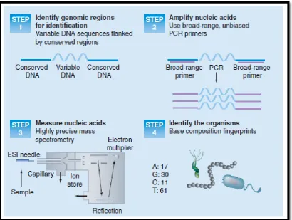
2010). An example of a more recent process used for the direct molecular detection of
36
Figure 4 - Identification and genotyping of microbes by PCR/ ESI/ mass (Ecker et al, 2010)
NAT applied to sepsis and detection of blood borne micro organisms can be divided into two
main categories which will be discussed next.
NAT assays for the detection and identification of pathogens from blood culture bottles
In the first main catergory, molecular detection and speciation after an initial growth in blood
culture medium occurs most easily with either hybridisation based or amplification based
techniques (Peters et al, 2004). After the initial growth in blood culture medium, the
hybridisation based technique provides identification of most pathogens within two hours. An
[image:36.595.73.484.137.446.2]example is fluorescence in situ hybridization (FISH) with oligonucleotide probes targeting Figure 4 – This schematic shows the stepwise processes involved in the identification
37
bacterial or fungal genes (typically rRNA genes). However, these methods can be technically
difficult and need advanced detection systems and also require skilled laboratory staff.
Pathogen-specific assays is limited due to the high variety of pathogens, potentially
responsible for blood borne infections. The main disadvantage of broad-range approaches are
that after the PCR amplification of a target sequence, further identification procedures are
necessary. The broad assay approach could be more useful for persistently negative blood
cultures in the presence of a strong clinical suspicion of bacteraemia and fungaemia, as in the
case of infective endocarditis (Mancini et al, 2010). Typically, blood cultures samples are
taken multiple times for improved detection of organsims which are located in regions with
little blood supply such as heart valves.
The risk of false-positive results due to environmental bacterial or fungal DNA contaminaton
on sampling bloods needs to be considered when these panbacterial or panfungal approaches
are used (Ng, 2006). The multiplex PCR approach targets different genes of those pathogens
most frequently isolated from blood stream infections.
After the amplification process, there is sequencing analysis of the microorganism. The above
methods of molecular identification of micro organisms on positive blood cultures are not
routinely practiced in microbiology labs as they do not show greater or parallel clinical
benefit or cost effectiveness compared with conventional methods (Peters et al, 2004).
Direct molecular detection of pathogens in blood with PCR
In 1993, two separate groups published the first use of pathogen specific PCR assay to detect
bacteria in blood. In the first study to be published (Song et al, 1993), a Salmonella typhi
PCR assays was described. The number of clinical samples was small. However, the
38
culture were positive for DNA fragment of the flagellin gene of Salmonella typhi, whereas 10
blood specimens of patients with other febrile diseases were negative. This shows that the
sensitivity of the PCR assay was 92% compared with blood cultures. In addition, Salmonella
typhi DNA were detected from blood specimens of four patients with suspected typhoid fever
on the basis of clinical features but with negative cultures. Interestingly, on the basis of the
results of the PCR, these patients were treated with ciprofloxacin for 14 days with an
excellent outcome.
There are four different molecular diagnostic approaches for detection of bacterial and fungal
DNA in whole blood samples that are currently approved for clinical use by European
regulatory authorities (Pletz et al, 2011):-
A multiplex real – time PCR that simultaneously detects a pre defined panel of the
most important sepsis pathogens by species- or genus-specific fluorescent probes (SeptiFast®;
Roche).
A eubacterial and panfungal real – time PCR that is able to detect nearly all known
bacterial and fungal pathogens by a 16S and 18S ribosomal RNA (rRNA) gene-based
universal PCR followed by sequencing of the amplification product for species identification
(SepsiTestTM; Molzym). The term eubacteria is commonly used to describe 'true bacteria'
which includes all bacteria except archaebacteria.
A multiplex PCR that detects a predefined panel of the most important sepsis
pathogens by electrophoretic separation of target-specific amplicons. An amplicons is a piece
of DNA or RNA that is the source and/or product of natural or artificial amplification or
39
A eubacterial and panfungal PCR that is able to detect nearly all known bacterial and
fungal pathogens by genome – specific targets followed by mass spectrometry for species
identification (Plex-ID; Abbott).
The majority of clinical studies which compared PCR based diagnostics with conventional
blood culture showed that more pathogens were detected by PCR techniques (Pletz et al,
2011).
SeptiFast® assay
The Roche product SeptiFast® has been available longer than other molecular-based tests and
is the assay used in our proof of concept model. SeptiFast® uses real-time PCR in a
nonquantitative mode to identify ten bacteria at the species level, several more at the genus
level, as well as five Candida species and Aspergillus fumigatus. This assay reportedly
identifies the 25 organisms that account for more than 90% of the culturable pathogens
associated with sepsis (Ecker et al, 2010). No unculturable organisms are identified nor are
most of the highly fastidious organisms that are difficult to culture.Whilst the majority of
studies have reported that SeptiFast® has a higher pathogen detection rate compared with
blood culture analysis (Dierkes et al, 2009, Lehmann et al, 2010, Louie et al, 2008, Mancini
et al, 2008, Westh et al, 2009), other studies have disputed this (Bloos et al, 2010).
Furthermore, it has been reported that SeptiFast® is more sensitive in detecting fungal
pathogens, such as Candida species and Aspergillus fumigatus when compared with
conventional blood culture (Dierkes et al, 2009, Westh et al, 2009).
A number of studies have described the occasional incidence of negative PCR results
associated with a simultaneous positive blood culture, so-called ‘false’ negative results
(Lehmann et al 2010, Louie et al 2008, Yanagihara et al, 2010). This can occur because the
40
of the assay), or because of a technical fault with the platform not detecting the presence of
the organism. Low concentration of the organisms in the blood sample can result in a ‘false’
negative result due to the limit of detection of SeptiFast® (Yanagihara et al, 2010). Excess
total DNA in the sample can lead to saturatation to the enzyme which interferes with the
amplification and signaling to produce a negative result (Louie et al, 2008).
As SeptiFast® has an analytical sensitivity of 3 to 30 CFU/ml (Lehmann et al, 2008, Pletz et
al, 2011), the diagnostic capabilities of the assay is limited to some extent, compared with the
theoretical sensitivity of one CFU per culture bottle in conventional blood culture after
inoculating approximately 10ml whole blood (Lehmann et al, 2010). This is potentially
problematic as quantitative blood culture studies have shown that the majority of clinically
significant bacteraemia in adults are characterised by circulating low numbers of bacteria
(Lehmann et al, 2010). There have been a number of studies which have looked at the clinical
application on the SeptiFast® assay in medical practice. Avolio et al (Avolio et al, 2010)
compared traditional blood cultures with SeptiFast® in patients with suspected blood stream
infections (BSI) arriving at the emergency department of a regional Italian hospital. In this
study population, not all pathogens detected by blood cultures were also found by the PCR
technique used even when those pathogens are on the PCR panel profile. Of 144 blood
samples examined, 13 cases (24.5%) blood culture identifed organisms which were not
detected by real time PCR and similar findings have been seen in other studies.
In Avolio et al’s study (Avolio et al, 2010), the SeptiFast® PCR assay gave positive results
where the blood culture was negative in 10 cases. In these10 cases there was a microbiology
confirmation of infection by pathogen isolation from other sites with diagnoses ranging from
pneumonia (two cases), meningitis (two cases), aortic prosthesis infection (one case),
41
SeptiFast® results positively impacted the therapeutic choices and clinical outcome of the
patients (Avolio et al, 2010).
Effect of antimicriobial administration pre testing on both microbiological and molecular approaches
The most recent international guidelines for sepsis (Dellinger et al, 2013), recommend
obtaining blood cultures before antimicrobial therapy is initiated if such cultures do not
significantly delay ( >45 minutes) the start of antimicrobial(s) administration. However, a
substantial number of blood cultures are taken from pretreated patients or patients developing
sepsis despite antibiotic prophylaxis, i.e. pre–operative or after solid organ transplants. A
percieved advantage of a DNA based detection system, compared to blood cultures, is that
the pathogen does not have to be viable at the time of sampling. A Danish study (Westh et al,
2009), compared SeptiFast® with blood cultures in a multicentre trial of patients with
suspected bacterial and fungal sepsis. 558 samples from 359 patients were evaluated. The rate
of positivity was 17% from blood cultures and 26% for SeptiFast®. The administration of
antibiotics did not affect the ability of DNA detection by the SeptiFast® assay and the
SeptiFast® assay had a better pick up rate for pathogen detection. This study highlights the
fact that blood culture positivity and pathogen DNA positivity are not equivalent measures of
infection.
There are situations where PCR detection occurs in those with negative cultures. It is
commonly known that our current gold standard for detecting pathogens in blood is blood
cultures. Blood cultures fail to identify more than 50% of the cases of sepsis, believed to be
caused by bacteria or fungi based on clinical and other criteria (Ecker et al, 2010). In
numerous studies, as mentioned previously, SeptiFast®consistently identified more positive specimens than blood cultures. These potential ‘false positives’ were frequently deemed
42
infection or disease severity and were often subsequently confirmed after isolation of the
pathogen from relevant clinical samples from other sources (Ecker et al, 2010). SeptiFast® -
positive / culture-negative results could conceivably come from non-viable organisms in the
blood (resulting from ongoing antibiotic treatment), cell-free DNA released from infected or
colonized remote infection sites, or antibiotic interference with culture. With this variation in
the potential role of circulating pathogen DNA, at present molecular PCR techniques would
have a role as an adjunct to blood cultures.
For the purpose of this proof of concept study, it was vital to choose a method of detecting
pathogen DNA in this model which would be robust enough to detect the organisms typical
seen in this study population. It is widely established that the SeptiFast® assay covers the
majority of bacteria and fungi seen in intensive care patients. It is also important that this
coverage is broad enough for the urology patient population in this study. At Salford Royal
Foundation Trust, where the study was undertaken, the urology service maintains a clinical
database comprising clinical infection and microbiological data on interventional renal stone
treatment dating from 2004 to 2011. The data establishes that SeptiFast® has suitable
coverage of microbial detection to make this a viable tool for investigating infection in this
study population.
Table 4summarises the comparativeadvantages and disadvantages between blood culture
43
Table 4 - Needs and current status of methods to identify bloodstream infections (Adapted from Ecker et al, 2010)
NEED CURRENT STATUS FUTURE OPPORTUNITIES
Culture Molecular methods Molecular methods
Identify all bacterial and fungal infection
Identifies only culturable organisms
Varies with method from 25 frequently cultured organisms to panbacterial; limited or no fungal
Panbacterial and panfungal identification
High sensitivity: bloodstream infections
can be caused by less than 10 CFU/ml in adults
Blood cultures are negative in >50% of clinically sepsis cases
Mixed results. Blood culture is more sensitive in some cases, but molecular methods identify organisms missed by culture; reported sensitivities as low as 3 CFU/ml
Mixed results. Blood culture is more sensitive in some cases, but molecular methods identify organisms missed by culture; reported sensitivities as low as 3 CFU/ml
Rapid identification; mortality increases hourly
in the absence of appropriate antimicrobial
therapy
Requires 1–5 days
Requires 1 day < 1 hr
Antimicrobial resistance determination Requires 1 additional day after obtaining culture isolate
A few major resistance determinants measured Significant opportunity with future understanding of molecular mechanisms Quantitative assessment
of pathogen load; load correlates with disease
severity
Quantitative culture methods too difficult for routine practice
Not quantitative Quantitative results
44 Cytokine driven immune reponses
The body has developed intricate pathways to protect and defend against pathogenic
organisms. Cytokines are one of the main protagonists in these processes. Cytokines are
small cell-signalling protein molecules that are secreted by numerous cells and used
extensively in intercellular communication. Cytokines can be proteins, peptides, or
glycoproteins; the term "cytokine" encompasses a large and diverse family of regulators
produced throughout the body by cells of diverse embryological origin (Kruttgen et al, 2012).
To understand the role of important cytokines such as IL - 6 and IL - 10, it is important to
understand the early stages of the host immune response to common pathogens such as
bacteria and fungi. The expression of common structures, known as PAMP, is thought to be
central to the host response in sepsis (see earlier). The release and increase in serum
concentration pro – inflammatory cytokines (cytokine storm) in sepsis has been shown to be
triggered by release of bacterial endotoxin (Hack, 1989, Krettgen et al, 2012).
In gram positive sepsis, a similar role is seen with lipoteichoic acid and peptidoglycans
(Schmidt et al, 2011). Both LPS and lipoteichoic acids bind to members of a family of PAMP
receptors. These receptors are known as the toll like receptors (TLR). The combination of the
receptor and its ligand alerts the innate immune response system to presence of invading
pathogen. The innate immune system comprises the cells and mechanisms that defend the
host from infection by other organisms in a non-specific manner and is the first line of the
host's defence .
TLRs trigger intracellular pathways involving the signaling molecules MyD88 or TRIF and
leading to activation of the transcription factors c – Jun N – terminal kinase and NF-kB,
thereby initiating the transcription of pro – inflammatory cytokine genes and production of
45 Interleukin 6
IL-6 is a cytokine that acts as both a pro – inflammatory and anti – inflammatory cytokine.
IL–6 is a cytokine linked to sepsis and is one of the major NF – kB target genes. IL-6 is a
member of 4 helical cytokine families, which signals via an 80 kDA cytokine receptor (IL –
6R). Once IL – 6 binds to IL – 6R, the resultant complex associates with the signalling
receptor subunit gp130. Il – 6 signals by two mechanisms (a) via the ubiquitous trans membrane gp130: ‘classic’ signalling using membrane –bound IL – 6R (gp80) and (2) via
trans-signalling using soluble IL – 6R (sIL – 6R).
Research has shown that pro – inflammatory activities of IL – 6 are mainly driven by IL – 6
trans – signalling via the sIL – 6R, whereas anti – inflammatory or regenerative functions rely
on classic IL – 6 signalling via the membrane bound receptor (Scheller et al, 2011). In a study
(Waage et al, 1989), serum samples from patients with meningococcal disease were
examined for the presence of IL - 6, TNFA and LPS. Median serum concentration of IL - 6
was 1,000 times higher in patients with septic shock (189 ng/ml) than in patients with
bacteraemia or meningitis alone. This suggests that IL - 6 has an important role in the sepsis
process. It was concluded that a complex pattern of cytokine exists in patients with fatal
sepsis in those with meningococcal infection, and that the release of IL - 6 as well as
interleukin 1 (IL - 1) is associated with a fatal outcome.
For a number of years, IL - 6 has been established as a prognostic marker for mortality in
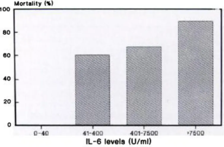
sepsis. In a study (Hack et al, 1989), a group measured levels of IL - 6 in plasma samples
from 37 patients with sepsis or septic shock obtained at the time of admission to the intensive
care unit and related these levels to hemodynamic and biochemical parameters as well as to
clinical outcome. In 32 of the 37 patients, increased levels of IL - 6 were found, occasionally
46
Figure 5 - Il – 6 and sepsis - associated mortality (Hack et al, 1989)
Importantly, IL - 6 on admission appeared to be of prognostic significance: levels were
higher in septic patients who subsequently died than in those who survived (P = .0003),
particularly when only patients with septic shock were considered (P less than .0001). All
nine septic patients with levels of less than 40 U/mL on admission survived, whereas 89% of
the nine patients with levels exceeding 7,500 U/mL died.
Research in the oncology field has raised a possible role for IL – 6 in the hemodynamic
response typically seen in the sepsis process. IL – 6 trans signalling was found to increase
endothelial permeability by phosphorylation of VE – cadherin (Kruttgen et al, 2012). This
process could lead to vascular leakage and may play a vital role in the life threatening
refractory drop in blood pressure seen in shocked patients.
47
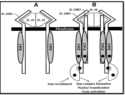
Interleukin 10
Interleukin 10 is an anti-inflammatory cytokine. Interleukin 10 levels are in-detectable in
healthy individuals.The IL – 10 protein is a homodimer; each of its subunits is 178 amino
[image:47.595.73.513.263.599.2]acids long, as shown in the figure below (Figure 6).
Figure 6 – Interleukin 10 and receptor (Kotenko et al, 1997)
IL – 10 is a pleiotropic cytokine with important immunoregulatory functions whose actions
influence activities of many cell types in the immune system (Couper et al, 2008). IL – 10 is
capable of repressing synthesis of pro-inflammatory cytokines such as TNFA, IL – 2 and
interferon gamma made by macrophages and regulatory T – cells.
48
IL-10 has the ability to suppress the antigen presentation capacity of antigen presenting cells
as well. Conversely, IL – 10 stimulates a number of inflammatory cells as well. IL – 10 has
been seen to be elevated in the state of sepsis with an association between its concentration,
severity of sepsis and death (Friedman et al, 1997, Giannodous et al, 2000).
Summary
Sepsis is the leading cause of death in critically ill patients. Sepsis has been described in
medical literature for over 2000 years but is still a leading cause of both economic burden and
patient morbidity and mortality. All episodes of sepsis are associated with infection and the
presence of pathogen in body tissue. The complex interaction between pathogen and the host
response has been extensively investigated for decades. However, the role of circulating
pathogen DNA in the early triggering of the host response to infection is not understood fully.
Blood culture is the current gold standard method for pathogen detection in blood but cannot
provide time – critical results that impact on the initial management of a patient (Dark et al,
2009). In addition, this method of pathogen detection is particularly susceptible to false
negative results following antimicrobial use. Molecular methods like PCR have the potential
ability to rapidly detect (or rule out) the presence of illness causing organisms. These
molecular methods could facilitate effective immediate management of the infection and
influence the subsequent clinical outcome. These molecular methods are not influenced by
antibiotic use. However, this expensive technology has a number of limitations.
In recent years, urology as a surgical speciality has moved away from open surgery, towards
endourological procedures for the treatment of renal stones. As with any procedure, these
interventions are not without complications. One of the most commonly seen complications
is infection and urosepsis. To combat this complication, it is common practice to give