M E T H O D
Open Access
A multi-split mapping algorithm for circular
RNA, splicing, trans-splicing and fusion
detection
Steve Hoffmann
1,2,3, Christian Otto
1,2,3, Gero Doose
1,2,3, Andrea Tanzer
4, David Langenberger
1,2,3,
Sabina Christ
5, Manfred Kunz
6, Lesca M Holdt
3,7, Daniel Teupser
3,7, Jörg Hackermüller
2,5,8and Peter F Stadler
1,2,3,4,9,10,11*Abstract
Numerous high-throughput sequencing studies have focused on detecting conventionally spliced mRNAs in RNA-seq data. However, non-standard RNAs arising through gene fusion, circularization or trans-splicing are often neglected. We introduce a novel, unbiased algorithm to detect splice junctions from single-end cDNA sequences. In contrast to other methods, our approach accommodates multi-junction structures. Our method compares favorably with competing tools for conventionally spliced mRNAs and, with a gain of up to 40% of recall, systematically outperforms them on reads with multiple splits, trans-splicing and circular products. The algorithm is integrated into our mapping toolsegemehl(www.bioinf.uni-leipzig.de/Software/segemehl/).
Background
The term splicing refers to a post-transcriptional process in which the raw transcript (pre-mRNA) is cleaved from intronic DNA fragments. In general, the splicing mech-anisms allow the recombination of protein-coding and non-coding RNA fragments and thus greatly increase the repertoire of potentially functional transcripts. While the overwhelming majority of splicing events occurs within the same pre-mRNA at consensus splice sites, some mRNAs are spliced at non-consensus sites. Many tran-scripts derived at non-consensus splice sites may have escaped detection in the past because of the assumptions built into the in silico analysis pipelines or due to the limited throughput of earlier RNA sequencing (RNA-seq) protocols.
Some species have developed mechanisms to fuse sep-arately transcribed mRNAs. These mRNAs may stem from distant loci, opposite strands or homologous chro-mosomes. A prominent physiological example is the
*Correspondence: [email protected]
1Junior Research Group Transcriptome Bioinformatics, Leipzig University, Haertelstrasse 16-18, Leipzig, Germany
2Interdisciplinary Center for Bioinformatics and Bioinformatics Group, University Leipzig, Haertelstrasse 16-18, Leipzig, Germany
Full list of author information is available at the end of the article
mod(mdg4) trans-splicing for Drosophila melanogaster [1]. Chimeric transcripts from different loci may be func-tional even in mammals [2]. Circular RNAs [3,4] are recognizable in RNA-seq data in the form of reads that contain apparent splice junctions that connect the end (start) of a split read fragment to the start (end) of a downstream (upstream) fragment. Very recently, they have been identified as an abundant class of regula-tory transcripts functioning as microRNA sponges [5,6]. In addition to physiological trans-splicing, a number of transcripts potentially derived from the fusing of genes have been observed in different types of cancer, such as melanoma [7] and breast cancer [8]. In the follow-ing, we use for brevity the term ‘fusion transcript’ to refer to RNAs that stem from a fused gene or a trans-splicing event. Although trans-trans-splicing and mRNA fusion events appear to be rare compared to the regular local and collinear splicing, fusion transcripts indicate potentially important functional entities or diagnostic marker genes. Their emerging importance mandates the use of analy-sis pipelines for RNA-seq data that ensure their efficient detection and inclusion in the subsequent data analysis workflow.
Several different algorithms for splice site detection have been devised so far. The original version ofTopHat
[9] predicts exon locations from the coverage data and attempts split read alignments across neighboring exons. This algorithm was not able to detect fusion events,
so a new algorithm, TopHat-Fusion [10], was
pub-lished and has since been integrated into TopHat2
along with some other modifications to the original
algo-rithm. SpliceMap [11] starts by splitting the reads
into fragments of 25 nucleotides and then attempts to align all fragments separately with a limited num-ber of mismatches. Subsequently, canonical splice junc-tions are searched within a genomic interval of 400 Mb. The specificity of splice junctions may be improved by
providing paired-end information. SpliceMap’s
junc-tion search is significantly distinct fromTopHat2’s. The MapSplice algorithm [12] resembles SpliceMap. It also performs a segmentation of reads into tags and han-dles each tag individually. The tags are aligned to exons and junctions inferred from tags mapping to consecutive exons.
SplitSeek[13] also uses both the 5 and 3 ends of
reads to infer spliced exons.SplitSeekdoes not make
use of canonical splice site information and is not lim-ited to a common locus. Another tool that was specifically designed for the detection of fusion transcripts,deFuse [14], makes use of paired-end information and triggers local alignments at positions of discordant paired-end reads.
Like TopHat2, SOAPsplice [15] is based on a Burrows-Wheeler transform and attempts to map the reads completely to the genome with no more than three mismatches or one gap. All unmapped reads are subse-quently subjected to a split mapping with two segments. Each segment has to fulfill a number of quality criteria. GSNAP[16] uses hash tables to retrieve position lists and subsequently merges and filters them efficiently. It is able to allow for multiple mismatches and long indels and can detect short- and long-distance splicing. The RNA-seq
mapper RUM [17] uses Bowtie [18] and BLAT [19] to
detect annotated as well asde novosplice junctions. In a first step, reads are mapped against the genome as well as the user-supplied transcriptome. All unmapped reads are forwarded toBLATand split alignments are merged. One of the latest tools for RNA-seq alignment,STAR[20], uses maximal mappable prefixes that are identified using suffix arrays. In a second step, the prefixes are ‘stitched’ together to reconstruct the isoforms. This algorithm was reported to be very fast – in fact it was shown to be more than 50 times faster than some of its competitors.
Here, we present a unified unbiased algorithm to detect splicing, trans-splicing and gene fusion events from single-end read data. The method, based on an enhanced suffix array, chaining and dynamic program-ming algorithms, is integrated into the mapping tool segemehl[21].
Results and discussion
The algorithm presented here makes use of a read match-ing method with enhanced suffix arrays (ESAs) published earlier [21]. In brief, for a read of lengthm, the algorithm evaluates the best alignments with a limited number of
mismatches, insertions and deletions for all 2(m − )
suffixes of the read and its reverse complement, where
is the minimum suffix length. An alignment qualifies
as a seed if a score-based maximum E-value criterion and a maximum occurrence threshold are met. Subse-quently, full reads will be aligned to all distinct seed locations in the reference genome using Myers’ semi-global bit-vector alignment [22]. All alignments passing a minimum accuracy threshold are reported. While the E-value, minimum accuracy and maximum occurrence parameter control the specificity and limit the number of multiple hits, the potentially large number of seeds from the beginning to the end of the read ensure a high sensitivity. For spliced or fusion transcripts, a successful semi-global alignment of the read is likely to fail. Instead, the ESA-based method will identify several seeds match-ing different locations or strands. The algorithmic strategy to identify splicing, trans-splicing or gene fusion sites is based on a greedy, score-based seed chaining followed by a Smith-Waterman-like transition alignment. This ment optimizes the total score of a number of local align-ments at different locations and strands. The algorithm does not have any effective length restrictions. Details are given in the Materials and methods section.
Simulated data
The algorithm’s performance was compared with seven alternative split read methods: TopHat2, RUM, STAR, SOAPsplice,MapSplice,SpliceMapandGSNAP. In principle, all tools were run with default parameters for split-read mapping. Where available, fusion and trans-splice sensitive alignment parameters were turned on.
With the exception of RUM, no extra annotations were
given to any of the programs (see Materials and methods and Additional file 1).
were applied to the simulated reads. These models intro-duce mismatches, insertions and deletions to the reads to emulate sequencing artifacts (see Materials and methods). The results for simulated 454 and Illumina reads are
summarized in Figure 1. segemehlperformed best in
both 454 simulations. In the data set with regular splice
junctions,segemehlconsistently recovered more than
90% of all simulated splice junctions. Its closest competi-tor, GSNAP, achieved a recall of between 81% and 92%. STARwas third, with less than 87% of recalled splice junc-tions. Probably due to length restrictions,TopHat2did not report any results after running for over 1 week and was terminated. For irregular splice events, the
differ-ence was even more striking: whilesegemehlrecovered
approximately 90% of all simulated splice junctions, the next best competitor,STAR, achieved a recall of approxi-mately 55%.
The improved performance for 454 reads did not signifi-cantly impairsegemehl’s performance for Illumina data. All tools, includingsegemehlwith a recall at the 95% level, found more than 90% of all simulated splice junc-tions.RUM, gaining an advantage by simultaneously align-ing the reads to the genome and transcriptome, performed best in this scenario. The best genome-only aligner was STARwith a recall of 98%.
When trans-splicing events were included, five of the seven alternative tools recovered less than 80% of all splice
junctions.TopHat2 had the best sensitivity and found
more than 90% of the junctions. However, its false pos-itive rate of more than 10% was quite high. In contrast,
segemehlidentified about 95% of all junctions in this data set and found only 2% false positives. For Illumina
reads, the runtime ofsegemehlwas comparable to most
of the tools tested. OnlyMapSplice,GSNAPandSTAR
were significantly faster than segemehl (Additional
file 1: Table S1).
In addition to these benchmarks with Illumina and 454 error models, we also wanted to investigatesegemehl’s behavior for reads with higher error rates, for example caused by multiple single nucleotide variations or, more importantly, less successful sequencing runs. Therefore, we carried out further benchmarks with higher error rates (up to 5% mismatches and indels) and varying coverage. As expected, the recall increased with higher coverage and
dropped with higher error rates. However,segemehl’s
specificity was consistently at a very high level for all types of splice junctions (Additional file 1: Figure S1).
The performance for artificial short circular (100 bp), long circular and long linear reads (0.5 to 5 kb) is summa-rized in Figure 2. For the short reads,segemehlachieved a recall of 85% of all circular junctions using uniquely mapping split reads. The closest competitor, TopHat2, achieved a recall of 55%.SpliceMap,RUMandSTARdid not find any circular junctions (Figure 2A). In contrast, STAR and GSNAPwere the only tools that in principle were able to handle long reads. For linear transcripts, GSNAPwas slightly ahead ofsegemehlby 6%, whereas STARwas merely able to recall 40% of all junctions. No
circular junctions were recovered by STAR or GSNAP
(Figure 2B).
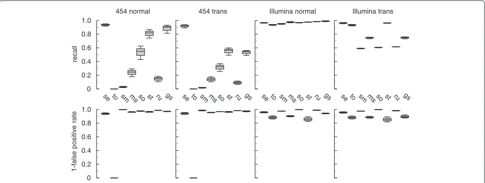
454 normal 454 trans Illumina normal Illumina trans
0 0.2 0.4 0.6 0.8 1.0
0 0.2 0.4 0.6 0.8 1.0
se to sm ms so st ru gs se to sm ms so st ru gs se to sm ms so st ru gs se to sm ms so st ru gs
recall
1-false
p
ositiv
e
[image:3.595.58.540.473.655.2]rate
Figure 1Performance of various read aligners on simulated data sets with different splice events.For simulated 454 reads (400 bp),
segemehlperformed significantly better in detecting conventional and ‘non-conventional’ (strand-reversing, long-range) splice junctions.
segemehlwas the only tool that consistently recalled more than 90% of conventional splice junctions. For ‘non-conventional’ splice events,
A
B
circular short (100bp) circular and linear long (0.5-5kbp)
reca
ll
p
reci
si
on
0 0.25 0.50 0.75 1.00
se so ms to gs se st gs
linear circular
0 0.25 0.50 0.75 1.00
se so ms to gs se st gs
10 kb
chr1: 55,250,000 55,255,000 55,260,000 55,265,000
[image:4.595.65.538.87.431.2]TTC22
Figure 2Recall and precision for short circular, long circular and long collinear transcripts.For this benchmark, we testedsegemehl’s performance with sequence reads that were generated from the RefSeq database(A). To simulate sequencing errors, we applied an Illumina error model to the short circular reads (100 bp) and a 454 error model to the long circular and collinear transcripts (0.5 to 5 kB). For short circular transcripts,segemehlachieved a recall of more than 85%, outcompeting all other tools while maintaining a high precision of 98%. Using RefSeq transcripts of length 0.5 to 5 kB,segemehlachieved a recall of more than 80% for circular and linear transcripts. Among the tools that were able to handle such long transcripts,segemehlwas the only tool that was able to detect the circularization. For long collinear transcripts,GSNAPwas slightly better thansegemehlby 6%, at the expense of a nearly twofold increase in runtime (Additional file 1: Table S1).(B)The RefSeq TTC22 transcript is an example of a simulated circularization. The arrow indicates where the transcript has been artificially circularized.SpliceMap,RUM
andSTARdid not find any circular junctions (not shown).STARandGSNAPwere the only tools able to handle long reads. gs,GSNAP; ms,
MapSplice; se,segemehl; so,SOAPsplice; st,STAR; to,TopHat2.
Real data
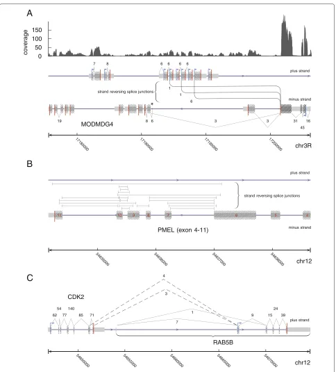
We applied segemehl to a number of real data sets.
Split-mapping of a Drosophila melanogaster RNA-seq
data set resulted in the successful recovery of the
pre-viously described trans-splicing of theMODMDG4gene
(Figure 3A) [1]. Most notably,segemehlrevealed three alternative strand-reversing junctions consistent with a splice event between a common exon on the reverse strand to three exons encoded on the forward strand.
For a human melanoma transcriptome data set, the
method identified the recently described CDK2-RAB5B
read-through transcripts on chromosome 12 (cf. [7])
(Figure 3B). In addition, segemehl identified a huge
number of strand-reversing split reads located at the
locus of the premelanosome protein (PMEL). This gene is also known as Silver Homolog (SILV). This gene located on chromosome 12 encodes a melanocyte-specific trans-membrane glycoprotein and is expressed under physiolog-ical conditions in melanosomes. It plays an essential role in the structural organization of premelansomes and has been suggested as a potential serum marker for melanoma (Figure 3C). More than 20% of the trans-splicing events detected in the sample occurred at this locus, making gen-eral errors in the RNA preparation and analysis highly unlikely. Thus, the PMEL locus might be an interesting target for further investigations.
A
B
C
19
7 8
3 6
8
6 6 6 6
3 31
45 16 6
1 1
cov
e
ra
ge
strand reversing splice junctions
plus strand
minus strand
MODMDG4
17185000 17190000 17
195 00
0
172000
00
*
chr3R 0
50 100 150
PMEL (exon 4-11)
54635000 54636000 54
637000 54638000
strand reversing splice junctions plus strand
minus strand
11 10 9 8 7 6 5 4
chr12
62 54
77 140
85 71
4
3
7 1
9 15
24
39
plus strand
CDK2
chr12 RAB5B
54650000 5465500
0
5466000
0
54665000 5467000
[image:5.595.58.542.83.618.2]0
Figure 3Examples of (re-)discovered splicing events from single-end split reads.(A)ForDrosophila melanogaster,segemehlrecovered three different previously described splice junctions linking the minus encoded exon three ofMODMDG4on chromosome 3R to exons on the opposite strand. The strand-reversing splice junctions are annotated between the plus and minus strands. The direction of the strand-reversing splice junctions, i.e. from the minus to the plus strand, was inferred from annotation and prior knowledge. This was necessary because the RNA-seq library used was not strand specific.(B)For the human melanoma transcriptome data set,segemehlidentified a very large number of
strand-reversing splice junctions in the premelanosome protein (PMEL) gene locus. The split reads that support these junctions split from the plus strand to the minus strand and vice versa. Since we lack additional information, a direction for these junctions cannot be given. Only a selection of strand-reversing PMEL junctions is shown here.(C)For the same data set,segemehlfound two alternative transcripts linkingCDK2andRAB5B
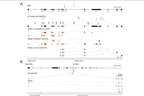
fibroblasts. As an example demonstrating the efficiency ofsegemehl, we considered the tumor suppressor gene p53 (Figure 4A). This key regulator of the cell cycle is one of the most intensely studied genes because of its importance in cancer research. The functionally distinct variants and isoforms of p53have been the focus of an intense research effort [25,26]. Despite the attention this gene has already received, a reanalysis of the raw data [24] identified three previously unrecognized canonical splice variants. We have validated all three novel isoforms in venous fibroblasts using PCR and sequencing (see Materials and methods and Additional file 1). Since
we failed to validate the p53 isoforms in HUVEC cells
(data not shown), these splice variants might be tissue-specific.
Novel transcripts were also predicted in a HUVEC 454 RNA-seq data set [27]. The example in Figure 4B shows
two isoforms whose start is located anti-sense within the intronlessTACSTD2gene. Their first intron contains on the opposite strand the entireMYSM1gene, which codes for a histone H2A deubiquitinase.
For a human prostate carcinoma cell line, we identi-fied a transcript that aligns to adjacent regions on the plus and minus strands of the genome. We validated the occurrence of this split using RT-PCR followed by cloning and Sanger sequencing. Interestingly, the split was located
in the 3 UTR of the stearoyl-CoA desaturase geneSCD
(Additional file 1 and Additional file 1: Figure S5), which has been implicated in prostate cancer [28].
For the RNA-seq data from HEK293 cells analyzed specifically for circular RNAs in [5], we were able to recover all circular RNAs that were experimentally validated by the authors of the original study (see Additional file 1).
A
B
//
p53
minus strand
all measured isoforms
β
γ α
i9
1 2 3 4 5 6 7 8 9 10 11 12 13 14
42 13 18 161 413 608 503 679 598 424 9
51 152 6 10 6
reads in annotated isoform
reads in Mercer isoforms
i
ii iv iii
reads in novel isoforms
v
vi
vii
*
*
*
chr 17 //
7,590-7,591k 7,569-7,581k
//
TACSTD2 MYSM1
minus strand
all isoforms
663
15
22 1261 1099 178
reads
// chr 1
[image:6.595.60.538.311.631.2]59,041-59,053k 59,110-59,184k
Figure 4Novel and known spliced transcript isoforms identified with long single-end 454 RNA-seq split reads.(A)Transcript isoforms of thep53gene. In addition to previously reported isoforms, (i) to (iv) [24], we identified three novel canonically spliced isoforms, (v) to (vii). Consistent with [24], theβandγisoforms were not expressed here. Each splice junction is labeled with its read support, i.e. the number of reads that map across this junction. For better comparability with [24], thep53gene, encoded on the minus strand of chromosome 17, is shown in the direction of transcription from left to right. The junctions marked with an asterisk have been experimentally validated.(B)Unannotated transcripts in the vicinity of theTACSTD2andMYSM1genes recovered from a HUVEC RNA data set [27].segemehlrevealed the exon structure of two novel transcript isoforms comprising at least four exons. One exon common to both isoforms was mapped to theTACSTD2gene. The associated introns enclose the
Finally, we tested our algorithm on the transcriptome of the nematodeCaenorhabditis elegans. The roundworm is known to have an extensive number of trans-spliced transcripts. In particular, spliced leader sequences (SLs) of about 22 nucleotides were spliced to the trimmed 5 ends of many mRNAs [29]. The spliced leader sequences were encoded as part of small non-coding RNAs, the SL RNAs, in 28 loci scattered throughout the genome (Additional file 1: Table S3). To test whether SL
trans-splicing can be detected directly from the segemehl
mapping results, we reanalyzed a small part of the publicly available sequencing data generated by [30]. All
trans-splicing junctions reported by the segemehlmapping
(see Additional file 1) were required to have a minimum split read support of three. After masking rRNAs, we obtained approximately 9,000 junctions linking loci with a distance of more than 200 kb or on different chro-mosomes. These were supported by about 139,000 split reads. More than 90% of them connect to the genomic coordinates of the 3end of one of the annotated spliced leader sequences. This simple survey accurately repro-duces results from a recent detailed analysis ofC. elegans trans-splicing [31]. In particular we found that 70% of the ce6 mRNAs annotated in the UCSC genome browser were trans-spliced. We also recovered the expected dis-tribution of spliced leader usage: SL1 is by far the most frequently used leader sequence (85.9% of all SL junctions) followed by SL2 (13%), SLf (0.4%), SLb (0.3%), SLc (0.2%), SLd (0.2%) and SLa (<0.1%) (Additional file 1: Table S5). Other trans-splice junctions found during this exercise are subject to further research.
To benchmark the speed of our split-read aligner, we aligned four different data sets withsegemehl,STAR, SOAPsplice, GSNAP, TopHat2 and RUM. With the
exception of STAR, in most scenarios segemehl was
faster or on a par with the other tools tested (Additional file 1: Table S2). Because it uses a full ESA, the memory
consumption ofsegemehldepends on the size of the
reference genome (cf. [21]). Thus, it has the highest mem-ory consumption among all tools tested. For large
mam-malian genomes,segemehlmay not be feasibly applied
on computers with less than 50 GB of memory. The size of the read library, however, does not affect the memory consumption. Note that for smaller genomes the mem-ory consumption is considerably smaller, e.g.C. elegans 1.5 GB,Drosophila melanogaster2.6 GB andArabidopsis thaliana1.8 GB.
Conclusions
We have presented a novel algorithm for split-read map-ping of single-end RNA-seq data that combines error-tolerant ESA-based seed mapping with a fast bit-vector alignment. It accommodates multiple splits within a single read and makes noa prioriassumptions on the transcript
structure. Implemented in thesegemehlmapping tool,
it readily identifies conventional splice junctions, collinear and non-collinear fusion transcripts, and trans-spliced RNAs, without the need for separate post-processing or an extensive computational overhead. Compared to widely used competitors, the method has significantly higher sensitivity and produces less false positives, espe-cially for trans-splicing scenarios. This makessegemehl the method of choice for annotating rare transcript vari-ants. Indeed, previously undescribed exons and additional splice junctions were readily identified.
Strikingly, the precision is maintained even for reads with higher error rates (Additional file 1: Figure S1). This feature is of particular interest when transcriptome data from organisms with high allelic variations are processed. It also makes it feasible to analyze transcriptome data by mapping to the genome of a closely related species as a reference.
Already the analysis of the few test data sets used here to verify the viability of our approach, shows that RNA-seq data sets readily contain evidence for a substantial number of transcripts with atypical structures. In addition to read-through transcripts, which preserve collinearity and can be explained by conventional splicing from an extended primary transcript, we also observed a moder-ate number of products that violmoder-ate collinearity. These fall into at least three broad classes: strand-reversing RNAs,
such as the fruit flyMODMDG4 gene [1,32], that
orig-inate from both reading directions of a compact locus; permuted RNAs and circular isoforms [3-6,33,34] as for theANRILnon-coding RNA [35]; and RNAs that are com-bined from components originating from different loci such as the ratHongrES2RNA [36] and several chimeric human proteins recently described in [37]. A few of these have been studied in some detail and at least in parts have also been characterized functionally [2]. These stud-ies suggest that a part of the non-collinear transcriptome might be functional [38] and cannot be explained as a consequence of chromosomal rearrangements relative to the reference genome. An accurate mapping tool such as segemehl, which is sensitive to split reads and operates without an underlying model of valid transcript structure and hence does not discard non-collinear mapping results as artifacts, is therefore an indispensable tool for system-atic investigations into this largely uncharted section of the transcriptome.
Materials and methods
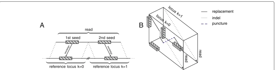
The algorithmic strategy for split-read alignments is sketched in Figure 5.
Chaining
A
1st seed 2nd seed
read
reference locus k=0 reference locus k=1 seeda
lign seeda
lign
B
locu s
k=1 locu
s k=0
read
read
replacement
[image:8.595.57.540.85.208.2]indel puncture
Figure 5A chain of seeds guides a local transition alignment across multiple genomic loci.High-quality seeds mapping to different genomic loci, strands or chromosomes(A)are chained. Subsequently, the order of the seeds within the chain guides a walk through the alignment cube (B). For each genomic locus, a local alignment with the read is performed. In addition to the regular Smith-Waterman recursions, the local transition alignment allows crossing between different reference loci.
in the genome. The start positions of each seed’s set of alignments in the reference genome are represented by the ESA interval [l,r]. In addition to the aforementioned E-value and maximum occurrence parameters, the align-ment seeds retrieved from the ESA are required to have a minimum Shannon entropy of 1.5. The Shannon entropy of a sequenceSis defined by
H(S)= −
n−1
i=0
p(si)log2p(si) (1)
where p(si) denotes the probability that the charactersi
occurs in S. This additional prerequisite is necessary to drop low-complexity seeds caused, e.g., by poly-A tails or repeats that bypass the maximum occurrence threshold due to sequencing errors. In general, it cannot be ruled out that the Shannon entropy filter affects the detection of splice events in repetitive elements (cf. [39]). However, our calculations show that the majority of 20-bp and 40-bp windows in human ALU repeats have a Shannon entropy well above our threshold of 1.5 (see Additional file 1). Therefore, this filter does not impede the split-read map-ping in ALU repeats per se. After passing the three filters, each alignment start of a seed in the reference genome is easily obtained in constant time using the suffix table of the ESA. Let S denote the set of seeds. In the chaining step, we aim to select an ordered chain of seedsc⊆Sthat optimally covers the read from start to end while at the same time maximizing the sum of alignment scores. Letψ denote a function to obtain the alignment score of a seed and letπsandπebe two functions to determine a seed’s
alignment start and end in the read, respectively. Finally, the score of a chain is evaluated using
σ(c)=
|c|
k=1
ψ(c[k])−
|c|
k=2
|πe(c[k−1])−πs(c[k])| (2)
where c[k]∈ S denotes the kth seed in chain c. In our implementation ψ(c[k]) is the number of correctly
matched nucleotides of fragmentiminus the sum of mis-matches, insertions and deletions in this fragment. A set of chains is obtained using a greedy chaining algorithm (Algorithm 1).
Algorithm 1 C=sort(S,πs)
fori := 1 to|C|do c=
forj := 1 toi−1do
ifσ(g(Cj,Ci)) > σ(Ci)then
c=Cj
end if end for ifc=then
Ci =g(c,Ci)
end if end for
Initially, seeds are sorted with respect to πs and the
sorted list is stored inC. In the first step, each single seed is a chain of its own. The computation proceeds by iter-ating over all chains in the listC. For each chainCi, the
best preceding chaincis identified and concatenated with it. For two chains,candc, the concatenation operator is denoted byg(c,c).
It is easy to see that algorithm terminates after (|C| · (|C| −1))/2 iterations. Since there are at most 2(m−) seeds, the algorithm is of complexityO(m2).
Local transition alignment
segemehl selects those alignments that minimize the distance on the genome and, if possible, are on the same strand. Finally, for each seed we obtain exactly one posi-tion in the reference.
Guided by the chain of seeds, the local transition alignment algorithm maps the reads across multiple loci (Algorithm 2). A similar idea was independently pro-posed by [14]. Unlike McPherson et al., we devised an algorithm that fully integrates the transition between mul-tiple matrices to obtain an optimal local split alignment across different genomic loci. The local transition align-ment method is a modification of the Smith-Waterman alignment.
Algorithm 2 fork := 1 to|c|do
fori := 1 tomdo
lms[k,i]=lms[k,i−1] * forj := 1 to|γ (c[k])|do
a = getchar_read(r,i,κ(c[k])) b = getchar_ref(γ (c[k]),j) M[i,k,j]=
max
⎧ ⎨ ⎩
M[i−1,k,j]+δ M[i,k,j−1]+δ
M[i−1,k,j−1]+s(a,b) forq := 1 tok−1do
M[i,k,j]=
max
M[i,k,j]
lms[q,i−1]+s(a,b) * end for
lms[k,i]=max(M[i,k,j] ,lms[k,i−1])* end for
end for end for
The algorithmic parts that realize the transition to other loci are marked by an asterisk. Note, that we have imple-mented the algorithm using lazy evaluation schemes. Furthermore, a penalty is applied to each transition in practice (not shown). Letγ (c[k])andκ(c[k])be functions that return the sequence and the strandedness for the reference locus to which the seedc[k] was aligned, respec-tively. Note, that in practice the sequence returned byγ extends the exact alignment boundaries ofc[k] by several nucleotides to account for inaccuracies in segemehl’s seed-finding heuristics. The algorithm iterates over all
seeds in the chain c and performs local alignments of
the read r with their respective reference locus. Inser-tions and deleInser-tions are penalized with δ. s(a,b) scores matches and mismatches. The resulting alignment scores are stored in a three-dimensional matrixM. During the local alignment at γ (c[k]), the algorithm keeps track of the last maximum score lms[k,i] seen prior to the
alignment of the ith character of the read. This addi-tional table is the key to the local transition alignment algorithm. In addition to the local alignment recursions that maximize the score ofM[i,k,j], allk−1 preceding loci are checked for a possible transition using thelms table.
Simulations and tools
To simulate both regular and irregular splice junctions, a sample of 10,000 isoforms was drawn from the ASTD database [40]. For the non-regular data set, 20% of the exons were either flipped to the opposite strand or sub-stituted by a distant exon from the ASTD database. Any exon with a distance > 200 kB from the isoform or on a different chromosome was denoted as long-range splic-ing. The isoforms of each data set were extracted from the human genome (hg19) by concatenating their exon sequences. Usingmason v.0.1.1 [41], we simulated Illumina and 454-like single-end reads of length 100 nucleotides and 400 nucleotides, respectively, from the isoforms of each data set with 10-fold, 15-fold and 20-fold cover-age. The parameter values of the Illumina and 454 error
model in mason were specified in accordance with the
Bowtie2 paper [42]. For simulated Illumina reads, we used
the default parameters; for 454, we used -k 0.3 -bm
0.4 -bs 0.1. Recall and false positive rates were cal-culated using the splice junctions predicted by each tool. Thus, the recall was calculated as the fraction of simulated junctions correctly identified by a tool. For each tool, the false positive rate was calculated by dividing the number of wrong junctions by the number of predicted
junc-tions. For the comparisons we used TopHat2(version
2.0.4),RUM(version 1.12_01),STAR(version 2.1.3e_r157), SOAPsplice(version 1.9),MapSplice(version 2.1.2), GSNAP (version 2013-11-27) and SpliceMap (version 3.3.5.2 (55)). All programs were run with default parame-ters for split-read mapping. With the exception ofRUM, no additional annotation information was provided to any of the tools. Further details are given in Additional file 1.
Split and isoform validation
To validate the newly identified canonical splice junc-tions inp53, we used RNA from venous fibroblasts. The RNA was isolated with TRIzol reagent (Life Technologies, Carlsbad, USA) and treated with RNase-free DNase (Qiagen, Hilden, Germany), according to the manufac-turers’ instructions. The RNA samples were prepared in the context of another study recently published in PLoS Genetics [43]. The protocols for reverse transcription into cDNA are described therein.
PCR reactions were prepared in a final volume
of 25 μl using AmpliTaq Gold(R) 360 DNA
co-ampli-fication of genomic DNA: common forward primer
(5-CCGAGAGCTGAATGAGGCCTTG-3, 300 nM)
and isoform-specific reverse primers (isoform (v) 5
-CATCACACTGCATACCTTGAATGTATGC-3; isoform
(vi) 5-CAGGCTAGAGTGCAATGGCGC-3; isoform
(vii) 5-GGCTCACGCCTGTAATCCCAGTAC-3; 300
nM each). The expected PCR product sizes were 322 bp, 213 bp and 178 bp for isoforms (v) to (vii), respectively. Cycling conditions were 95°C for 10 min and 40 three-step cycles of 95° for 20 s, 60°C (isoform (vi)) or 62°C (isoforms (v) and (vii)) for 30 s, and 72°C for 30 s. PCR products were subcloned using the TOPO TA Cloning Kit (Life Technologies, Carlsbad, USA) and sequencing reactions were performed with forward and reverse M13 primers (5μM, Life Technologies, Carlsbad, USA) and BigDye(R) Terminator v 3.1 chemistry (Life Technologies, Carlsbad, USA) according to the manufacturer’s instructions using an Applied Biosystems 3730xl DNA Analyzer.
Data sets
Publicly available Illumina RNA-seq data sets of D.
melanogaster[SRA:SRR166809] and a human melanoma sample [SRA:SRR018261-62] were downloaded from the National Center for Biotechnology Information short read archive. The 454 sequencing data for a human umbil-ical vein RNA-seq experiment [27] and RNA capture sequencing experiments of human fibroblasts [24] were retrieved from the Gene Expression Omnibus under [GEO:GSM951482] and [GEO:GSE29040]. The RNA-seq sample for HEK293 cells investigated in [5] was retrieved under [GEO:GSE43574]. Finally, we applied our
algorithm to a C. elegans data set [SRA:SRX151602]
to investigate RNA leader trans-splicing. All of these data sets are non-strand specific. In addition, we ana-lyzed a strand-specific prostate cancer data set (see Additional file 1).
Additional file
Additional file 1: Supplementary benchmark.Supplementary data on
the parameters for all tools tested, the algorithms’ performance with real and simulated data and the results of wet-lab experiments.
Abbreviations
bp: Base pair; ESA: Enhanced suffix array; kb: Kilobase; Mb: Megabase; PCR: Polymerase chain reaction; RT: Reverse transcriptase; seq: Sequencing; SL: Spliced leader sequence; UTR: Untranslated region.
Competing interests
The authors declare that they have no competing interests.
Authors’ contributions
SH wrote the manuscript and developed the presented algorithm. CO co-developed the algorithm. SH, CO, GD, AT and DL performed the simulations and tested and verified the algorithm using simulated and real-life data. SC, JH, MK, LH and DT performed the wet-lab experiments. PFS designed and supervised the research. All authors read and approved the final manuscript.
Acknowledgements
This research was supported by LIFE (Leipzig Research Center for Civilization Diseases), Leipzig University. LIFE is funded by the European Union, by the European Regional Development Fund (ERDF), the European Social Fund (ESF) and by the Free State of Saxony within the excellence initiative. Additionally, this work was supported in part by the Initiative and Networking Fund of the Helmholtz Association (VH-NG-738) and the BMBF through ICGC MMML-Seq (01KU1002J). The authors are grateful to Stephan Schreiber for technical assistance. The authors would like to thank Roderic Guigó for stimulating discussions.
Author details
1Junior Research Group Transcriptome Bioinformatics, Leipzig University,
Haertelstrasse 16-18, Leipzig, Germany.2Interdisciplinary Center for Bioinformatics and Bioinformatics Group, University Leipzig, Haertelstrasse 16-18, Leipzig, Germany.3LIFE Research Center for Civilization Diseases, Leipzig University, Haertelstrasse 16-18, Leipzig, Germany.4Department of Theoretical Chemistry, University of Vienna, Währinger Strasse 17, Vienna, Austria. 5RNomics Group, Fraunhofer Institute for Cell Therapy and Immunology – IZI,
Perlickstrasse 1, Leipzig, Germany.6Department of Dermatology, Venerology and Allergology, Leipzig University, Philipp-Rosenthal-Strasse 23, Leipzig, Germany.7Institute of Laboratory Medicine, Ludwig Maximilian University, Marchioninistrasse 15, Munich, Germany.8Young Investigators Group Bioinformatics and Transcriptomics, Department of Proteomics, Helmholtz Centre for Environmental Research – UFZ, Permoserstrasse 15, Leipzig, Germany.9Max Planck Institute for Mathematics in the Sciences, Inselstrasse 22, Leipzig, Germany.10Center for non-coding RNA in Technology and Health, University of Copenhagen, Grønnegårdsvej 3, Frederiksberg, Denmark.11Santa Fe Institute, 1399 Hyde Park Road, Santa Fe, NM, USA.
Received: 22 April 2013 Accepted: 10 February 2014 Published: 10 February 2014
References
1. Dorn R, Reuter G, Loewendorf A:Transgene analysis proves mRNA
trans-splicing at the complexmod(mdg4)locus inDrosophila.
Proc Natl Acad Sci USA2001,98:9724–9729.
2. Frenkel-Morgenstern M, Lacroix V, Ezkurdia I, Levin Y, Gabashvili A, Prilusky J, del Pozo A, Tress M, Johnson R, Guigó R, Valencia A:Chimeras taking shape: potential functions of proteins encoded by chimeric
RNA transcripts.Genome Res2012,22:1231–1242.
3. Jeck WR, Sorrentino JA, Wang K, Slevin MK, Burd CE, Liu J, Marzluff WF, Sharpless NE:Circular RNAs are abundant, conserved, and associated
with ALU repeats.RNA2013,19:141–157.
4. Salzman J, Gawad C, Wang PL, Lacayo N, Brown PO:Circular RNAs are the predominant transcript isoform from hundreds of human genes
in diverse cell types.PLoS ONE2012,7:30733.
5. Memczak S, Jens M, Elefsinioti A, Torti F, Krueger J, Rybak A, Maier L, Mackowiak SD, Gregersen LH, Munschauer M, Loewer A, Ziebold U, Landthaler M, Kocks C, le Noble F, Rajewsky N:Circular RNAs are a large
class of animal RNAs with regulatory potency.Nature2013,
495:333–338.
6. Hansen TB, Jensen TI, Clausen BH, Bramsen JB, Finsen B, Damgaard CK, Kjems J:Natural RNA circles function as efficient microRNA sponges. Nature2013,495:384–388.
7. Berger MF, Levin JZ, Vijayendran K, Sivachenko A, Adiconis X, Maguire J, Johnson LA, Robinson J, Verhaak RG, Sougnez C, Onofrio RC, Ziaugra L, Cibulskis K, Laine E, Barretina J, Winckler W, Fisher DE, Getz G, Meyerson M, Jaffe DB, Gabriel SB, Lander ES, Dummer R, Gnirke A, Nusbaum C, Garraway LA:Integrative analysis of the melanoma transcriptome. Genome Res2010,20:413–427.
8. Edgren H, Murumagi A, Kangaspeska S, Nicorici D, Hongisto V, Kleivi K, Rye IH, Nyberg S, Wolf M, Borresen-Dale A-L, Kallioniemi O:Identification of fusion genes in breast cancer by paired-end RNA-sequencing. Genome Biol2011,12:6.
9. Trapnell C, Pachter L, Salzberg SL:TopHat: discovering splice junctions
with RNA-Seq.Bioinformatics2009,25:1105–1111.
10. Kim D, Salzberg S:TopHat-Fusion: an algorithm for discovery of novel
11. Au KF, Jiang H, Lin L, Xing Y, Wong WH:Detection of splice junctions
from paired-end RNA-seq data by SpliceMap.Nucleic Acids Res2010,
38:4570–4578.
12. Wang K, Singh D, Zeng Z, Coleman SJ, Huang Y, Savich GL, He X, Mieczkowski P, Grimm SA, Perou CM, MacLeod JN, Chiang DY, Prins JF, Liu J:MapSplice: accurate mapping of RNA-seq reads for splice
junction discovery.Nucleic Acids Res2010,38:178.
13. Ameur A, Wetterbom A, Feuk L, Gyllensten U:Global and unbiased
detection of splice junctions from RNA-seq data.Genome Biol2010,
11:34.
14. McPherson A, Hormozdiari F, Zayed A, Giuliany R, Ha G, Sun MGF, Griffith M, Heravi Moussavi A, Senz J, Melnyk N, Pacheco M, Marra MA, Hirst M, Nielsen TO, Sahinalp SC, Huntsman D, Shah SP:deFuse: an algorithm for gene fusion discovery in tumor RNA-Seq data. PLoS Comput Biol1001,7:138.
15. Huang S, Zhang J, Li R, Zhang W, He Z, Lam T-W, Peng Z, Yiu S-M:
SOAPsplice: genome-wideab initiodetection of splice junctions
from RNA-Seq data.Fron Genet2011,2:46.
16. Wu T, Nacu S:Fast and SNP-tolerant detection of complex variants
and splicing in short reads.Bioinformatics2010,26:873–881.
17. Grant GR, Farkas MH, Pizarro AD, Lahens NF, Schug J, Brunk BP, Stoeckert CJ, Hogenesch JB, Pierce EA:Comparative analysis of RNA-Seq alignment algorithms and the RNA-Seq unified mapper
(RUM).Bioinformatics2011,27:2518–2528.
18. Langmead B, Trapnell C, Pop M, Salzberg S:Ultrafast and
memory-efficient alignment of short DNA sequences to the human
genome.Genome Biol2009,10:25.
19. Kent WJ:BLAT – the BLAST-like alignment tool.Genome Res2002,
12:656–664.
20. Dobin A, Davis CA, Schlesinger F, Drenkow J, Zaleski C, Jha S, Batut P, Chaisson M, Gingeras TR:STAR: ultrafast universal RNA-seq aligner. Bioinformatics2013,29:15–21.
21. Hoffmann S, Otto C, Kurtz S, Sharma CM, Khaitovich P, Vogel J, Stadler PF, Hackermüller J:Fast mapping of short sequences with mismatches,
insertions and deletions using index structures.PLoS Comput Biol
1000,5:502.
22. Myers G:A fast bit-vector algorithm for approximate string matching
based on dynamic programming.J ACM1999,46:395–415.
23. Danan M, Schwartz S, Edelheit S, Sorek R:Transcriptome-wide discovery
of circular RNAs in Archaea.Nucleic Acids Res2012,40:3131–3142.
24. Mercer TR, Gerhardt DJ, Dinger ME, Crawford J, Trapnell C, Jeddeloh JA, Mattick JS, Rinn JL:Targeted RNA sequencing reveals the deep
complexity of the human transcriptome.Nature Biotechnol2012,
30:99–104.
25. Marcel V, Olivier M, Mollereau B, Hainaut P, Bourdon J-C:First
Internationalp53Isoforms Meeting:p53isoforms through
evolution: from identification to biological function.Cell Death
Different2011,18:563–564.
26. Camus S, Ménendez S, Fernandes K, Kua N, Liu G, Xirodimas DP, Lane DP, Bourdon JC:Thep53isoforms are differentially modified by Mdm2. Cell Cycle2012,11:1646–1655.
27. Djebali S, Davis CA, Merkel A, Dobin A, Lassmann T, Mortazavi AM, Tanzer A, Lagarde J, Lin W, Schlesinger F, Xue C, Marinov GK, Khatun J, Williams BA, Zaleski C, Rozowsky J, Röder M, Kokocinski F, Abdelhamid RF, Alioto T, Antoshechkin I, Baer MT, Batut P, Bell K, Bell I, Chakrabortty S, Chen X, Chrast J, Curado J, Derrien T, et al:Landscape of transcription in
human cells.Nature2012,489:101–108.
28. Kim S-J, Choi H, Park S-S, Chang C, Kim E:Stearoyl CoA desaturase (SCD) facilitates proliferation of prostate cancer cells through
enhancement of androgen receptor transactivation.Mol Cells2011,
31:371–377.
29. Blumenthal T:Trans-splicing and operons inC. elegans.WormBook
2005. http://www.wormbook.org
30. Hillier LW, Reinke V, Green P, Hirst M, Marra MA, Waterston RH:Massively
parallel sequencing of the polyadenylated transcriptome ofC.
elegans.Genome Res2009,19:657–666.
31. Allen MA, Hillier LW, Waterston RH, Blumenthal T:A global analysis of
C. eleganstrans-splicing.Genome Res2011,21:255–264.
32. McManus CJ, Duff MO, Eipper-Mains J, Graveley BR:Global analysis of
trans-splicing inDrosophila.Proc Natl Acad Sci USA1297,107:5–12979.
33. Salzman J, Chen RE, Olsen MN, Wang PL, Brown PO:Cell-type specific
features of circular RNA expression.PLoS Genet1003,9:777.
34. Zhang Y, Zhang X-OO, Chen T, Xiang J-FF, Yin Q-FF, Xing Y-HH, Zhu S, Yang L, Chen L-LL:Circular intronic long noncoding RNAs.Mol Cell
2013,51:792–806.
35. Burd CE, Jeck WR, Liu Y, Sanoff HK, Wang Z, Sharpless NE:Expression of
linear and novel circular forms of anINK4/ARF-associated
non-coding RNA correlates with atherosclerosis risk.PLoS Genet1001,
6:233.
36. Ni M-J, Hu Z-H, Liu Q, Liu M-F, Lu M-H, Zhang J-S, Zhang L, Yong-Lian Z:
Identification and characterization of a novel non-coding RNA
involved in sperm maturation.PLoS ONE2605,6:3.
37. Frenkel-Morgenstern M, Valencia A:Novel domain combinations in
proteins encoded by chimeric transcripts.Bioinformatics2012,
28:67–74.
38. Gingeras TR:Implications of chimaeric non-co-linear transcripts. Nature2009,461:206–211.
39. Lin L, Shen S, Tye A, Cai JJ, Jiang P, Davidson BL, Xing Y:Diverse splicing
patterns of exonized Alu elements in human tissues.PLoS Genet1000,
4:225.
40. Koscielny G, Le Texier V, Gopalakrishnan C, Kumanduri V, Riethoven J-J, Nardone F, Stanley E, Fallsehr C, Hofmann O, Kull M, Harrington E, Boué S, Eyras E, Plass M, Lopez F, Ritchie W, Moucadel V, Ara T, Pospisil H, Herrmann A, Reich JG, Guigó R, Bork P, von Knebel Doeberitz M, Vilo J, Hide W, Apweiler R, Thanaraj TA, Gautheret D:ASTD: the alternative
splicing and transcript diversity database.Genomics2009,93:213–220.
41. Holtgrewe M, Emde A-K, Weese D, Reinert K:A novel and well-defined benchmarking method for second generation read mapping. BMC Bioinformatics2011,12:210.
42. Langmead B, Salzberg S:Fast gapped-read alignment with Bowtie 2. Nat Methods2012,9:357–359.
43. Holdt LM, Hoffmann S, Sass K, Langenberger D, Scholz M, Krohn K, Finstermeier K, Stahringer A, Wilfert W, Beutner F, Gielen S, Schuler G, Gäbel G, Bergert H, Bechmann I, Stadler PF, Thiery J, Teupser D:Alu
elements inANRILnon-coding RNA at chromosome 9p21 modulate
atherogenic cell functions through trans-regulation of gene
networks.PLoS Genet1003,9:588.
doi:10.1186/gb-2014-15-2-r34
Cite this article as:Hoffmannet al.:A multi-split mapping algorithm for circular RNA, splicing, trans-splicing and fusion detection.Genome Biology 201415:R34.
Submit your next manuscript to BioMed Central and take full advantage of:
• Convenient online submission
• Thorough peer review
• No space constraints or color figure charges
• Immediate publication on acceptance
• Inclusion in PubMed, CAS, Scopus and Google Scholar
• Research which is freely available for redistribution