Copyright © 1999, American Society for Microbiology. All Rights Reserved.
Development of a PCR Assay for Rapid Detection
of Enterococci
DANBING KE,1,2FRANC¸OIS J. PICARD,1FRANCIS MARTINEAU,1,2CHRISTIAN ME´NARD,1 PAUL H. ROY,1,3MARC OUELLETTE,1,2ANDMICHEL G. BERGERON1,2*
Centre de Recherche en Infectiologie de l’Universite´ Laval, Sainte-Foy, Que´bec, Canada G1V 4G2,1and Division de Microbiologie, Faculte´ de Medicine,2and De´partement de Biochimie, Faculte´ des Sciences et de Ge´nie,3
Universite´ Laval, Sainte-Foy, Que´bec, Canada G1K 7P4
Received 22 March 1999/Returned for modification 4 June 1999/Accepted 23 July 1999
Enterococci are becoming major nosocomial pathogens, and increasing resistance to vancomycin has been well documented. Conventional identification methods, which are based on culturing, require 2 to 3 days to provide results. PCR has provided a means for the culture-independent detection of enterococci in a variety of clinical specimens and is capable of yielding results in just a few hours. However, all PCR-based assays developed so far are species specific only for clinically important enterococci. We have developed a PCR-based assay which allows the detection of enterococci at the genus level by targeting thetuf gene, which encodes elongation factor EF-Tu. Initially, we compared the nucleotide sequences of thetufgene from several bacterial species (available in public databases) and designed degenerate PCR primers derived from conserved regions. These primers were used to amplify a target region of 803 bp from four enterococcal species (Enterococcus
avium,E. faecalis, E. faecium, andE. gallinarum). Subsequently, the complete nucleotide sequences of these
amplicons were determined. The analysis of a multiple alignment of these sequences revealed regions con-served among enterococci but distinct from those of other bacteria. PCR primers complementary to these regions allowed amplification of genomic DNAs from 14 of 15 species of enterococci tested (E. solitariusDNA could not be amplified). There was no amplification with a majority of 79 nonenterococcal bacterial species, except for 2Abiotrophiaspecies and severalListeriaspecies. Furthermore, this assay efficiently amplified all 159 clinical isolates of enterococci tested (61 E. faecium, 77 E. faecalis, 9 E. gallinarum, and 12 E. casseliflavus
isolates). Interestingly, the preliminary sequence comparison of the amplicons for four enterococcal species demonstrated that there were some sequence variations which may be used to generate species-specific internal probes. In conclusion, this rapid PCR-based assay is capable of detecting all clinically important enterococci and has potential for use in clinical microbiology laboratories.
Enterococci are members of the normal flora of the gastro-intestinal tract in humans and animals (24). The incidence of enterococcal infections has increased in recent years because of widespread multiresistant enterococcal strains and increas-ing numbers of immunosuppressed patients and catheter-re-lated infections. In fact, enterococci are now the second most common nosocomial pathogens in the United States (4, 31). There are two major pathogenic species in humans, Enterococ-cus faecalisand E. faecium, with occasional infections being caused byE. durans,E. gallinarum,E. casseliflavus,E. avium,E. hirae,E. mundtii, andE. raffinosus(24, 38). Devriese et al. (8) suggested that the extensive agricultural use of glycopeptides has created an animal reservoir of resistant enterococci which may lead to more enterococcal species resistant to glycopep-tides in animal sources and complicate the control of such infections. Enterococci resistant to glycopeptides have been isolated with increasing frequency and have become a major concern worldwide. Rapid identification of enterococci is im-portant in reducing the spread of multiresistant enterococci (2, 16).
Identification of enterococci through conventional methods, i.e., by determining phenotypic characters, is complicated and often requires 24 to 48 h (7, 11, 28). The automated methods
currently used are unable to reliably identify enterococci other than E. faecalis and E. faecium (29, 32, 34). Furthermore, phenotypic identification of some enterococcal species may be occasionally difficult or even impossible because these species lack typical characteristics. More rapid and accurate methods would be helpful for microbiology laboratories. Several DNA-based methods for the specific detection of E. faecalisorE. faeciumhave been reported (5, 10, 27, 30). Other molecular methods, such as contour-clamped homogeneous electric field electrophoresis patterns, amplified ribosomal DNA spacer polymorphisms, and randomly amplified polymorphic DNA analysis, have been used to identify enterococci at the species level (3, 9, 23, 26, 34). However, it is difficult to adapt these tests for use in clinical microbiology laboratories because of their complexity. AnEnterococcussp. assay based on the hy-bridization of rRNA genes (Gen-Probe, San Diego, Calif.) is commercially available for culture confirmation (6). The sen-sitivity of this assay is unsatisfactory for direct detection from clinical specimens.
A variety of conserved genes, including rRNA genes (3, 19, 22), the heat shock protein 60 (HSP60 or CPN60) gene (13, 14), the major cold shock protein gene (12), and thesodgene (39), have been exploited for the detection of bacteria. Thetuf gene, encoding elongation factor EF-Tu, is involved in peptide chain formation and is an essential constituent of the bacterial genome (15). These characteristics make it a target of choice for diagnostic purposes. PCR-based assays in which the tuf gene serves as the target sequence have been developed for Mycoplasma fermentans (1) andM. pneumoniae (20). We re-* Corresponding author. Mailing address: Centre de Recherche en
Infectiologie, Centre Hospitalier Universitaire de Que´bec, Pavillon CHUL, 2705 Boul. Laurier, Sainte-Foy, Que´bec, Canada G1V 4G2. Phone: (418) 654-2705. Fax: (418) 654-2715. E-mail: Michel [email protected].
3497
on May 15, 2020 by guest
http://jcm.asm.org/
port here the development of a PCR-based assay that targets the tufgene, that can detect most enterococcal species with excellent sensitivity and acceptable specificity, and that has potential for the development of species-specific internal probes.
(This study was presented in part at the 98th General Meet-ing of the American Society for Microbiology 1998, Atlanta, Ga., 17 to 21 May 1998.)
MATERIALS AND METHODS
Bacterial strains.Twenty enterococcal strains obtained from the American
Type Culture Collection (ATCC), Manassas, Va., were used in this study. These strains represent the following species:E. avium(ATCC 14025),E. casseliflavus
(ATCC 25788),E. cecorum(ATCC 43198),E. dispar(ATCC 51266),E. durans
(ATCC 19432),E. faecalis(ATCC 19433, ATCC 29212, ATCC 33186, ATCC 49533, and ATCC 51299), E. faecium(ATCC 19434 and ATCC 51559),E. flavescens(ATCC 49996),E. gallinarum(ATCC 49573),E. hirae(ATCC 8043),
E. mundtii(ATCC 43186),E. pseudoavium(ATCC 49372),E. raffinosus(ATCC 49427),E. saccharolyticus(ATCC 43076), andE. solitarius(ATCC 49428). An additional 159 clinical isolates of enterococci obtained from various sources were also used in this study (Table 1).
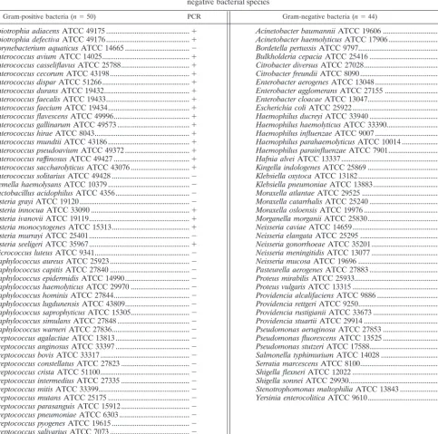
The specificity of the PCR-based assay was verified by use of a battery of ATCC reference strains consisting of 44 gram-negative and 50 gram-positive bacterial species (Table 2). The 159 clinical isolates of enterococci (61E. fae-cium, 77E. faecalis, 9E. gallinarum, and 12E. casseliflavus) from various origins (Table 1) were also tested to further validate theEnterococcus-specific PCR-based assay. The reference strains as well as the clinical isolates were all iden-tified by conventional methods or with an automated MicroScan Autoscan-4 system equipped with a Positive BP Combo Panel Type 6 (Dade Diagnostics, Mississauga, Ontario, Canada). Bacterial strains were grown from frozen stocks kept at⫺80°C in brain heart infusion medium containing 10% glycerol and were cultured on sheep blood agar or in brain heart infusion broth.
PCR primers.Thetufgene sequences available from public databases were
analyzed with GCG programs (version 8.0) (Genetics Computer Group, Madi-son, Wis.). Based on multiple sequence alignments, regions of thetufgene highly conserved among eubacteria were chosen, and PCR primers were derived from these regions with Oligo primer analysis software (version 5.0) (National Bio-sciences, Plymouth, Minn.). When required, the primers contained inosines or degeneracies at one or more variable positions. Oligonucleotide primers were synthesized with a model 391 DNA synthesizer (Perkin-Elmer Corp., Applied Biosystems Division, Mississauga, Ontario, Canada). PCR primers used in this study are listed in Table 3.
DNA sequencing.An 803-bp portion of thetufgene was sequenced forE.
avium,E. faecalis,E. faecium, andE. gallinarum. Amplification was performed with 1 ng of genomic DNA prepared by use of a G NOME DNA kit (Bio 101, Vista, Calif.). The 20-l PCR mixtures used to generate PCR products for sequencing contained 1.0M each universal primer (U1 and U2; Table 3), 200
M each deoxyribonucleoside triphosphate (Pharmacia Biotech Inc., Baie d’Urfe´, Que´bec, Canada), 10 mM Tris-HCl (pH 9.0), 50 mM KCl, 0.1% Triton X-100, 2.5 mM MgCl2, 0.5 U ofTaqpolymerase (Promega Corp., Madison,
Wis.), and TaqStart antibody (Clontech Laboratories Inc., Palo Alto, Calif.). The TaqStart antibody, which is a neutralizing monoclonal antibody forTaqDNA polymerase, was added to all PCR mixtures to enhance the efficiency of the amplifications (18).
The PCR mixtures were subjected to thermal cycling (3 min at 95°C and then
35 cycles of 30 s at 95°C, 30 s at 55°C, and 1 min at 72°C, with a 7-min final extension at 72°C) with a PTC-200 DNA Engine thermocycler (MJ Research Inc., Watertown, Mass.). The amplified PCR mixtures were resolved by electro-phoresis through 1.5% agarose gels at 4 V/cm for 90 min; the gels were then stained with ethidium bromide and visualized under 312-nm UV light. Subse-quently, PCR products having the predicted sizes were recovered from the gels with a QIAquick gel extraction kit (QIAGEN Inc., Mississauga, Ontario, Can-ada).
The purified DNA fragments were cloned into pCR2.1 vector (Invitrogen Corp., Carlsbad, Calif.). Plasmids were isolated from transformedEscherichia coliwith a QIAGEN plasmid mini-kit. The presence of DNA inserts in the recombinant plasmids was confirmed by digesting purified plasmid DNA with
EcoRI (New England Biolabs, Ltd., Mississauga, Ontario, Canada), which al-lowed excision of the inserted fragments. Both strands of the DNA inserts for each of the selected recombinant plasmids were sequenced with a PRISM Ready Reaction DyeDeoxy Terminator cycle sequencing kit and an Applied Biosystems 373A sequencer (Perkin-Elmer). In order to exclude the possibility of sequencing errors attributable to misincorporations byTaqpolymerase, each strand of the insert was sequenced from three different clones.
PCR amplification.For all bacterial species, amplification was performed from
purified genomic DNA or from a bacterial suspension whose turbidity was adjusted to that of a 0.5 McFarland standard, which corresponds to approxi-mately 1.5⫻108bacteria per ml. One nanogram of genomic DNA or 1l of
standardized bacterial suspension was transferred directly to a 19-l PCR mix-ture containing 50 mM KCl, 10 mM Tris-HCl (pH 9.0), 0.1% Triton X-100, 2.5 mM MgCl2, 0.2M eachEnterococcus-specific primer (Ent1 and Ent2; Table 3),
200M each deoxynucleoside triphosphate (Pharmacia Biotech), 3.3g of bovine serum albumin (BSA) (Sigma-Aldrich Canada Ltd., Oakville, Ontario, Canada) perl, 0.5 U ofTaqpolymerase (Promega), and TaqStart antibody (Clontech). PCR amplification and agarose gel analysis of the amplified products were performed as previously described (21).
The Superlinker phagemid pSL1180 (Pharmacia Biotech) linearized by diges-tion withEcoRI (New England Biolabs) and the primers (IC1 and IC2; Table 3) derived from the multiple cloning sites of this plasmid were used to provide an internal control for allEnterococcus-specific PCR-based assays. These primers can amplify a 252-bp product. The internal control was integrated into the PCR-based assays to verify the efficiency of the amplifications and to ensure that significant PCR inhibition was absent. Four thousand copies of the linearized plasmid were added to each PCR. The concentrations of the internal control primers were adjusted to ensure that there was no detrimental effect on the
Enterococcus-specific amplification. We found that concentrations of 0.1 and 0.04
M were optimal for 30- and 40-cycle PCRs, respectively.
For determination of the sensitivities of the PCR-based assays, twofold dilu-tions of purified genomic DNA were used to determine the minimal number of genomes which can be detected.
Nucleotide sequence accession numbers.GenBank accession numbers for the
751-bp partial sequence of thetufgene (excluding the sequences of the two universal amplification primers) are as follows: AF124220 for E. avium, AF124221 forE. faecalis, AF124222 forE. faecium, AF124223 forE. gallinarum, AF124224 forAbiotrophia adiacens, and AF124225 forA. defectiva.
RESULTS
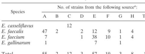
[image:2.612.53.293.92.182.2]Sequencing of a portion of thetufgene from four enterococ-cal species. The tuf sequences from a number of selected bacterial species, including E. coli (J01690), M. genitalium (U39732),Haemophilus influenzae (U32746),Neisseria gonor-rhoeae (L36380), Salmonella typhimurium(X55116), and Mi-crococcus luteus (M17788), were aligned and compared. Two highly conserved regions were identified, and a pair of primers (U1 and U2) amplifying a region of 803 bp was designed (Table 3). Several degeneracies and inosines were incorpo-rated into these two primers because some positions are vari-able among eubacteria. These primers allowed the amplifica-tion oftufsequences from a wide variety of bacteria, including 14 enterococcal species. By using these primers, we were able to amplify the 803-bp portion of tuf for four enterococcal species: E. avium, E. faecalis, E. faecium, and E. gallinarum. After purification from agarose gels, the 803-bp PCR product was cloned into a TA cloning vector. Subsequently, the se-quence of the inserted DNA fragment was determined by sequencing of three randomly selected clones for each entero-coccal species to ensure that no sequencing errors were attrib-utable to misincorporation by theTaqpolymerase. In order to facilitate the selection of Enterococcus-specific primers, we conducted a multiple sequence analysis using the sequences TABLE 1. Sources of the 159 enterococcal clinical isolates used in
this study
Species No. of strains from the following source a:
A B C D E F G H Total
E. casseliflavus 12 12
E. faecalis 47 2 2 12 9 1 4 77 E. faecium 7 1 38 10 1 4 61
E. gallinarum 1 7 1 9
Total 55 2 12 3 57 19 3 8 159
aA, Centre Hospitalier Universitaire de Que´bec (Pavillon Centre Hospitalier de l’Universite´ Laval), Sainte-Foy, Que´bec, Canada; B, Centers for Disease Control and Prevention, Atlanta, Ga; C, Hoˆpital Charles Lemoyne, Montre´al, Que´bec, Canada; D, Huashan Hospital, Shanghai, China; E, Laboratoire de Sante´ Publique du Que´bec, Sainte-Anne-de-Bellevue, Que´bec, Canada; F, Mount Sinai Hospital, Toronto, Ontario, Canada; G, Universidad de Buenos Aires, Buenos Aires, Argentina; H, University of Texas, Houston.
on May 15, 2020 by guest
http://jcm.asm.org/
mentioned above (available in public databases) as well as partialtufsequences from staphylococci, streptococci, entero-cocci,Listeriaspp., andAbiotrophiaspp. (all obtained from our laboratory; unpublished data). By using this approach, we were able to identify regions conserved in the four enterococcal species but variable in the other bacterial species. The Entero-coccus-specific PCR primers were derived from these regions. A similarity comparison of the tuf sequences for the entero-coccal speciesE. avium,E. faecalis,E. faecium, andE. gallina-rumas well as for twoAbiotrophiaspecies is given in Table 4. The sequence similarities for the 803-bp fragment in these species ranged from 85 to 91%.
The selected primers revealed no more than two mismatches within thetufsequences of the enterococcal species, except for
[image:3.612.64.547.86.564.2]E. faecium and twoAbiotrophia species, in which three mis-matches were present at the 5⬘end of the 5⬘primer. Impor-tantly, more than five mismatches were found in the corre-sponding regions of the other bacterial species mentioned above. Since the mismatches in the other bacteria were clus-tered at the 3⬘end of the primers, a position critical for dis-criminatory PCR amplification, the amplification of bacterial species other than enterococci could be efficiently prevented. Amplifications with the Enterococcus-specific PCR assay. The specificity of the assay was assessed by performing 30-cycle and 40-cycle PCR amplifications with the panel of gram-posi-tive (50 species from 9 genera) and gram-negagram-posi-tive (44 species from 21 genera) bacterial species listed in Table 2. The PCR assay was able to detect 14 of 15 enterococcal species tested in TABLE 2. Specificity test performed with theEnterococcus-specific 40-cycle PCR assay and DNA from a variety of positive and
gram-negative bacterial species
Gram-positive bacteria (n⫽50) PCR Gram-negative bacteria (n⫽44) PCR Abiotrophia adiacensATCC 49175 ...⫹ Acinetobacter baumanniiATCC 19606 ...⫺ Abiotrophia defectivaATCC 49176...⫹ Acinetobacter haemolyticusATCC 17906 ...⫺ Corynebacterium aquaticusATCC 14665 ...⫺ Bordetella pertussisATCC 9797...⫺ Enterococcus aviumATCC 14025...⫹ Bulkholderia cepaciaATCC 25416 ...⫺ Enterococcus casseliflavusATCC 25788...⫹ Citrobacter diversusATCC 27028...⫺ Enterococcus cecorumATCC 43198...⫹ Citrobacter freundiiATCC 8090...⫺ Enterococcus disparATCC 51266...⫹ Enterobacter aerogenesATCC 13048 ...⫺ Enterococcus duransATCC 19432...⫹ Enterobacter agglomeransATCC 27155 ...⫺ Enterococcus faecalisATCC 19433...⫹ Enterobacter cloacaeATCC 13047...⫺ Enterococcus faeciumATCC 19434...⫹ Escherichia coliATCC 25922...⫺ Enterococcus flavescensATCC 49996...⫹ Haemophilus ducreyiATCC 33940 ... ⫺ Enterococcus gallinarumATCC 49573 ...⫹ Haemophilus haemolyticusATCC 33390...⫺ Enterococcus hiraeATCC 8043...⫹ Haemophilus influenzaeATCC 9007 ...⫺ Enterococcus mundtiiATCC 43186 ...⫹ Haemophilus parahaemolyticusATCC 10014 ...⫺ Enterococcus pseudoaviumATCC 49372 ...⫹ Haemophilus parainfluenzaeATCC 7901...⫺ Enterococcus raffinosusATCC 49427 ...⫹ Hafnia alveiATCC 13337...⫺ Enterococcus saccharolyticusATCC 43076 ...⫹ Kingella indologenesATCC 25869 ...⫺ Enterococcus solitariusATCC 49428 ...⫺ Klebsiella oxytocaATCC 13182...⫺ Gemella haemolysansATCC 10379 ...⫺ Klebsiella pneumoniaeATCC 13883... ⫺ Lactobacillus acidophilusATCC 4356...⫺ Moraxella atlantaeATCC 29525 ...⫺ Listeria grayiATCC 19120...⫺ Moraxella catarrhalisATCC 25240 ...⫺ Listeria innocuaATCC 33090 ...⫹ Moraxella osloensisATCC 19976 ...⫺ Listeria ivanoviiATCC 19119...⫹ Morganella morganiiATCC 25830...⫺ Listeria monocytogenesATCC 15313...⫹ Neisseria caviaeATCC 14659... ⫺ Listeria murrayiATCC 25401...⫺ Neisseria elangataATCC 25295 ...⫺ Listeria seeligeriATCC 35967...⫹ Neisseria gonorrhoeaeATCC 35201...⫺ Micrococcus luteusATCC 9341...⫺ Neisseria meningitidisATCC 13077 ...⫺ Staphylococcus aureusATCC 25923...⫺ Neisseria mucosaATCC 19696 ...⫺ Staphylococcus capitisATCC 27840 ...⫺ Pasteurella aerogenesATCC 27883 ...⫺ Staphylococcus epidermidisATCC 14990...⫺ Proteus mirabilisATCC 25933...⫺ Staphylococcus haemolyticusATCC 29970 ...⫺ Proteus vulgarisATCC 13315 ... ⫺ Staphylococcus hominisATCC 27844...⫺ Providencia alcalifaciensATCC 9886 ... ⫺ Staphylococcus lugdunensisATCC 43809...⫺ Providencia rettgeriATCC 9250...⫺ Staphylococcus saprophyticusATCC 15305...⫺ Providencia rustigianiiATCC 33673 ...⫺ Staphylococcus simulansATCC 27848 ...⫺ Providencia stuartiiATCC 29914 ... ⫺ Staphylococcus warneriATCC 27836...⫺ Pseudomonas aeruginosaATCC 27853 ...⫺ Streptococcus agalactiaeATCC 13813...⫺ Pseudomonas fluorescensATCC 13525 ...⫺ Streptococcus anginosusATCC 33397 ...⫺ Pseudomonas stutzeriATCC 17588...⫺ Streptococcus bovisATCC 33317 ...⫺ Salmonella typhimuriumATCC 14028 ...⫺ Streptococcus constellatusATCC 27823 ...⫺ Serratia marcescensATCC 8100... ⫺ Streptococcus cristaATCC 51100...⫺ Shigella flexneriATCC 12022 ...⫺ Streptococcus intermediusATCC 27335 ...⫺ Shigella sonneiATCC 29930...⫺ Streptococcus mitisATCC 33399...⫺ Stenotrophomonas maltophiliaATCC 13843 ...⫺ Streptococcus mutansATCC 25175 ...⫺ Yersinia enterocoliticaATCC 9610...⫺ Streptococcus parasanguisATCC 15912...⫺
Streptococcus pneumoniaeATCC 6303 ...⫺ Streptococcus pyogenesATCC 19615 ...⫺ Streptococcus salivariusATCC 7073 ...⫺ Streptococcus sanguisATCC 10556 ...⫺ Streptococcus suisATCC 43765 ...⫺
on May 15, 2020 by guest
http://jcm.asm.org/
both 30-cycle and 40-cycle regimens.E. solitariuswas the only enterococcal species tested that was not amplified by the En-terococcus-specific assay (Fig. 1). For 30-cycle PCR, all bacte-rial species tested other than enterococci were negative, except for twoAbiotrophiaspecies,A. adiacensandA. defectiva. For 40-cycle PCR, fourListeriaspecies,Listeria innocua,L. ivano-vii, L. monocytogenes, and L. seeligeri, were also positive in addition to the enterococci andAbiotrophiaspecies. The other species tested remained negative. The internal control was always efficiently amplified when no target DNA was present, thereby showing the absence of PCR inhibitors. On the con-trary, the internal control was not amplified when target DNA was present in a sample (Fig. 1). This result is explained by the fact that the concentrations of the internal control primers were limited in order to favor the amplification of the target DNA. Tests of a collection of clinical isolates comprising E. faecalis(n⫽77),E. faecium(n⫽61),E. gallinarum(n⫽9), and E. casseliflavus(n ⫽12) showed a uniform amplification signal with both the 30-cycle and the 40-cycle PCR assays and a perfect relationship between the genotype and classical means of identification.
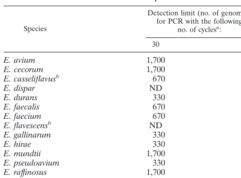
The sensitivity of our Enterococcus-specific assay with 30-cycle and 40-30-cycle PCR protocols was determined by using purified genomic DNA from 14 enterococcal species. For PCR with 30 cycles, we found a detection limit ranging from 330 to 1,700 copies of genomic DNA, depending on the enterococcal species tested (Table 5). In order to enhance the sensitivity of the assay, we increased the number of cycles. For PCR with 40
cycles, the detection limit was lowered to two to eight genome copies (Table 5).
DISCUSSION
[image:4.612.52.553.83.212.2]Enterococci can be encountered throughout the environ-ment, from human, animal, and food sources. The emergence and dissemination of vancomycin-resistant enterococci (VRE) underscore the importance of the rapid detection of these organisms (16). Conventional identification of some entero-cocci is difficult because no phenotypic criteria are available to unequivocally separate the genusEnterococcusfrom the other genera of gram-positive cocci. A number of tests, such as Lancefield group D antigen, the ability to grow in 6.5% NaCl broth, the pyrolidonylarylamidase test, the leucinearylamidase test, and the bile esculin test, are all valuable for the identifi-cation of enterococci. Unfortunately, none of these tests alone or in combinations provides a phenotype unique to the entero-cocci (7). Furthermore, such tests require 24 to 48 h to provide results after pure cultures are obtained. Even with automated systems, incubation for up to 24 h is routinely required, and some additional tests may have to be carried out for species differentiation (29, 32). Since the vancomycin resistance genes are transferable among different enterococcal species or even
FIG. 1. Example of multiplex PCR amplifications with theEnterococcus -specific PCR assay and the internal control. PCR assays (40 cycles) were per-formed with 1l of genomic DNA (1 ng/l) from various bacteria and 4,000 copies of linearized pSL1180 as an internal control. The internal control was not amplified when target DNA was present due to competitive inhibition by am-plification ofEnterococcusDNA. Lanes: 2,E. aviumATCC 14025; 3,E. cas-seliflavusATCC 25788; 4,E. cecorumATCC 43198; 5,E. duransATCC 19432; 6,
E. disparATCC 51266; 7,E. faecalisATCC 19433; 8,E. faeciumATCC 19434; 9,E. flavescensATCC 49996; 10,E. gallinarumATCC 49573; 11,E. hiraeATCC 8043; 12,E. mundtiiATCC 43186; 13,E. pseudoaviumATCC 49372; 14,E. raffinosus ATCC 49427; 15,E. saccharolyticus ATCC 43076; 16,E. solitarius
[image:4.612.320.544.515.619.2]ATCC 49428; 17,E. coliATCC 25922; 1 and 18, controls to which no DNA was added; M, 100-bp molecular size standard ladder.
TABLE 4. Sequence homology for an 803-bp portion of thetuf gene andEnterococcus-specific PCR products for four enterococcal
species and twoAbiotrophiaspecies
Species
% of sequence similarityafor: A.
adiacens defectivaA. aviumE. faecalisE. faeciumE. gallinarumE.
A. adiacens 85 89 89 89 87 A. defectiva 89.8 87 85 86 85 E. avium 92.7 97.1 91 90 90 E. faecalis 92.7 91.3 92.7 89 90 E. faecium 95.7 89.8 91.3 91.3 90 E. gallinarum 94.2 95.7 95.7 95.7 94.2
aThe numbers in the upper right triangle are the percentage of similarity for the sequenced 803-bp fragment, and those in the lower left triangle represent the percentage of similarity for the 69-bp sequences flanked by the twoEnterococcus -specific PCR primers. The similarity scores were obtained by use of the PILEUP program of the GCG software package.
TABLE 3. PCR primers used in this study
Primer Sequence Nucleotidepositionsa length (bp)Product
Universal amplification 803
U1 5⬘-AAYATGATIACIGGIGCIGCICARATGGA-3⬘ 271–300
U2 5⬘-AYRTTITCICCIGGCATIACCAT-3⬘ 1051–1073
Enterococcusspecific 112
Ent1 5⬘-TACTGACAAACCATTCATGATG-3⬘ 618–639
Ent2 5⬘-AACTTCGTCACCAACGCGAAC-3⬘ 708–729
Internal control 252
IC1 5⬘-TCTCGAGCTCTGTACATGTCC-3⬘
IC2 5⬘-GTTCTAGAGGTACCGGTTGTT-3⬘
aThe nucleotide positions given are for theE. coli tufsequence (GenBank accession no. J01690).
on May 15, 2020 by guest
http://jcm.asm.org/
[image:4.612.52.293.588.685.2]among different genera (38), the inability to detect enterococci promptly may cause delays in reporting VRE; this situation may lead to complex and costly containment efforts to elimi-nate VRE colonization and infection.
The development of rapid and sensitive DNA-based assays which are applicable for the direct detection of enterococci from clinical specimens may improve the rapidity and the ac-curacy of the diagnosis of enterococcal infections. The validity and versatility of PCR in applications for the detection of nucleic acids from a variety of infectious agents have been well reviewed by Whelen and Persing (36). In the present study, we have developed a rapid PCR-based assay to improve the diag-nosis of enterococcal infections. Initially, PCR primers com-plementary to highly conserved regions of thetufgene among eubacteria were designed and used to amplify an 803-bp por-tion of thetufgene from 14 enterococcal species. By sequenc-ing four enterococcal species, which represented four different enterococcal subgroups based on 16S rRNA gene analysis, we have obtained adequate sequence information to design a pair ofEnterococcus-specific PCR primers. Subsequently, a PCR-based assay was set up and optimized to be simple and rapid. Among 50 species of gram-positive (representing 9 genera) and 44 species of gram-negative (representing 21 genera) clin-ically important bacteria tested, this PCR-based assay was able to detect all enterococcal species tested, except forE. solitarius, which in fact does not appear to be a member of the genus Enterococcusbased on a phylogenetic analysis (25, 37). Two Abiotrophiaspecies were also amplified efficiently under the same amplification conditions. Moreover, amplification of four Listeriaspecies was also observed when the number of PCR cycles was increased from 30 to 40. It should be noted that DNA fromListeriaspecies was amplified about 100 times less efficiently than enterococcal DNA. However, most clinically important bacteria can be easily differentiated from entero-cocci by this assay, indicating its usefulness for the detection of enterococci. The concomitant use of species-specific internal probes should allow exclusion ofAbiotrophiaandListeria spe-cies by increasing the specificity of theEnterococcus-specific PCR-based assay.
To elucidate the fact that the developed PCR-based assay failed to detectE. solitarius, which rarely causes infections in humans, we amplified and sequenced an 890-bp portion oftuf encompassing the 803-bp region by using another pair of prim-ers (unpublished data). The sequence similarity between E. solitariusand other enterococci ranged from 79 to 81%, while that between the other enterococci ranged from 89 to 91%. This finding supports 16S rRNA data reported by others (25, 37) suggesting thatE. solitariusis not a member of the genus Enterococcus(93.0 to 94.8% homology) but is phylogenetically more closely related to the genusTetragenococcus(97.8% ho-mology). Sequence data revealed that there were six mis-matches at the Enterococcus-specific 3⬘-primer binding site which led to a failure in the amplification ofE. solitariusby the developed PCR-based assay.
The finding that A. adiacensand A. defectiva, formerly re-ferred to as nutritionally variant streptococci (17), were also positive in our Enterococcus-specific PCR assay indicates a high level of similarity of thetufgenes at the primer binding sites in the genus Abiotrophia and the genus Enterococcus. Sequencing of the 803-bp region of tuf showed that the se-quence similarities between the enterococcal andAbiotrophia spp. ranged from 85 to 89%, values which are quite high. For comparison, the tuf sequences among enterococci are 89 to 91% similar. Therefore, it is not surprising that PCR primers derived from thetufsequence amplify DNA fromAbiotrophia spp. as well. In fact, there are only two mismatches at the 5⬘ end of theEnterococcus-specific primers compared to the se-quences of A. defectiva and A. adiacens. Consequently, the sensitivity level achieved for the twoAbiotrophiaspecies was similar to that obtained for the enterococcal species. In addi-tion, several Listeria species were detectable when 40-cycle PCR was performed, even though three mismatches located near the 3⬘end of the upper primer were found between the four Listeriaspecies and enterococci. However, it should be noted that Listeria DNA was amplified approximately 100 times less efficiently than enterococcal DNA. The fourListeria species, L. monocytogenes, L. innocua, L. ivanovii, and L. seelegeri, showed sequence similarities to enterococci ranging from 82 to 83% (data not shown). Based on the level of se-quence divergence for thetuf gene, it should be possible to develop enterococcal species-specific internal probes which al-low discrimination of members of the generaAbiotrophiaand Listeriafrom those of the genusEnterococcus.
Others have developed E. faecalis-specific andE. faecium -specific PCR-based assays by targeting various genes. The tar-get genes include (i)ddl(coding forD-alanine-D-alanine ligase) for the species-specific detection ofE. faecalisandE. faecium (10, 30) and (ii)PBP5(coding for penicillin binding protein) for the species-specific detection ofE. faecalis (27). A PCR-based assay amplifying differentvanCgenes (coding for intrin-sic vancomycin resistance inE. gallinarum,E. casseliflavus, and E. flavescens) has also been developed to specifically detectE. gallinarum,E. casseliflavus, andE. flavescens(10). A sequence of unknown coding potential, selected from an E. faecium genomic library, has been used to develop an E. faecium -specific PCR-based assay (5). AlthoughE. faecalisandE. fae-ciumaccount for a majority of enterococcal infections, other enterococci may be associated with infections. However, cur-rently available systems based on culturing for the identifica-tion of gram-positive cocci are often unable to correctly iden-tify these less frequently encountered enterococci (29, 32, 34). Tyrrell et al. (35) reported using an internally transcribed spacer region PCR to identify enterococcal species based on characteristic amplicon profiles and the different patterns of Sau3A-digested PCR amplicons. Donabedian et al. (9) dem-TABLE 5. Detection limits of theEnterococcus-specific PCR assays
for 14 enterococcal species
Species
Detection limit (no. of genomes) for PCR with the following
no. of cyclesa:
30 40
E. avium 1,700 8
E. cecorum 1,700 2
E. casseliflavusb 670 4
E. dispar ND 8
E. durans 330 4
E. faecalis 670 2
E. faecium 670 8
E. flavescensb ND 8
E. gallinarum 330 8
E. hirae 330 4
E. mundtii 1,700 8
E. pseudoavium 330 2
E. raffinosus 1,700 8
E. saccharolyticus 330 2
aThe genome size for all enterococcal species was considered the same as that ofE. faecalis(3.0 Mb). Serial twofold dilutions of genomic DNA from each enterococcal species were tested to determine the detection limit of the assay for each species. ND, not determined.
bE. casseliflavusandE. flavescensare now considered the same species (33).
on May 15, 2020 by guest
http://jcm.asm.org/
[image:5.612.52.294.91.270.2]onstrated that contour-clamped homogeneous electric field electrophoresis patterns and DNA-DNA hybridization with biotin-labeled genomic DNAs from type strains of enterococci used as probes may be suitable for species differentiation of some enterococci. However, neither method is feasible for routine use in the clinical laboratory because of high complex-ity and must be used in conjunction with phenotypic tests to provide reliable results (9, 35). Recently, Quednau et al. (26) and Monstein et al. (23) identified enterococci to the species or species group level by using randomly amplified polymorphic DNA methods. A simple test for the detection ofEnterococcus spp. has been developed by Gen-Probe for use as a culture confirmation assay; this test was 100% accurate in identifying enterococcal isolates from cultures by hybridization to rRNA (6). However, this assay does not offer potential for enterococ-cal species identification. Moreover, the sensitivity of the Gen-Probe assay is not sufficient for direct detection of enterococci from clinical specimens.
Besides identification of bacterial species by species-specific PCR-based assays, identification may also be performed by coupling the amplification of a highly conserved gene with hybridization to internal probes or DNA sequencing (1, 14, 20, 22). Many conserved genes have been selected as targets for this purpose. Among them, the 16S rRNA gene has been used to detect a wide variety of eubacteria because the presence of conserved regions and variable regions in this gene provides the possibility of developing PCR-based assays suitable for detecting and identifying bacteria at the species level or higher taxonomic levels (22). Other genes have been exploited for similar purposes. The sod gene, coding for superoxide dis-mutase, has been used as a target to amplify 28 species of mycobacteria and to differentiate one from another with probes recognizing species-specific regions (39). A similar ap-proach has been used for the detection of staphylococci by targeting the chaperonin 60 (cpn60) gene (13, 14). These meth-ods are specific, but their sensitivity remains unclear. The latter is critical when such tests are used to detect bacteria directly from clinical specimens. Berg et al. (1) have devel-oped a PCR-based detection system forM. fermentansby tar-geting thetufgene; the system has a high sensitivity. A similar assay has also been used for the identification of M. pneu-moniae(20).
The tuf gene acts in translation to bring aminoacylated tRNA molecules to the ribosome. This gene represents an ideal candidate target for diagnostic purposes because it is highly conserved at the nucleotide level and ubiquitous in bacteria. By analyzing a rather small tuf sequence data set available in public databases, we were able to design PCR primers which could amplify an 803-bp portion of thetufgene from a variety of bacteria, including members of the genera Enterococcus, Streptococcus, and Staphylococcus. We found that there were more nucleotide sequence variations in thetuf gene sequences than in the corresponding 16S rRNA gene sequences for four enterococcal species (i.e., E. faecalis, E. faecium, E. avium, andE. gallinarum). An analysis of thetuf sequences from gram-positive bacteria allowed the develop-ment of a PCR-based assay amplifying 14 of 15 enterococcal species tested. The PCR-based assay described here differen-tiates enterococci from most clinically relevant bacteria, indi-cating that thetufgene is a target of choice for the molecular detection of enterococci. Furthermore, the amplicon sequence polymorphism should be sufficient to provide discriminatory internal probes specific for clinically important enterococcal species. The sensitivity of the 30-cycle PCR-based assay varies from 330 to 1,700 copies of enterococcal genomes for the 14 enterococcal species detected. This sensitivity level is sufficient
for culture confirmation assays. It is possible to efficiently in-crease the sensitivity of the assay. For example, in sensitivity assays performed with 40-cycle PCR, the detection limit was reduced to about two to eight copies of enterococcal genomes for the same 14 enterococcal species. This sensitivity level should be sufficient for the direct detection of enterococci in clinical specimens, such as fecal or urine samples. The use of this PCR-based assay coupled with species-specific internal probes specific for clinically important enterococci and other PCR assays targeting vancomycin resistance genes should pro-vide a useful screening test for VRE from rectal swabs. Such an assay is currently under development in our laboratory.
In conclusion, we have developed a PCR-based diagnostic assay which is simple to conduct and reliable for the detection of enterococci. This assay offers an alternative to currently used methods and should allow the identification of clinically important enterococcal species if coupled with species-specific probes complementary to internal regions of the amplicons. This new diagnostic tool may lead to the early diagnosis of enterococcal infections, which is essential for the prevention and control of transmission of the infections.
ACKNOWLEDGMENTS
We thank Louise Cote´, director of the microbiology laboratory of CHUQ, Pavillon CHUL, for free access to the laboratory and for providing enterococcal and other clinical isolates. We thank Jean-Luc Simard, Martin Gagnon, Marie-Jose´e Boily, Caroline Paquet, Nicolas Lansac, Marie-Claude Bergeron, and Gise`le Chasse´ for help. We also thank Louise Jette´ (Laboratoire de Sante´ Publique du Que´bec), Donald E. Low (Mount Sinai Hospital), Barbara E. Murray (Univer-sity of Texas, Houston), Fred C. Tenover (Centers for Disease Control and Prevention), Wang Fu (Huashan Hospital), and Daniela Centron-Garcia (Universidad de Buenos Aires) for providing enterococcal strains. We thank Maurice Boissinot for critical comments regarding the manuscript.
Marc Ouellette is an MRC scientist and a recipient of the Burroughs Wellcome Fund new investigator award in molecular parasitology. This study was supported by grant PA-15586 from the Medical Re-search Council of Canada and by Infectio Diagnostic (I.D.I) Inc., Sainte-Foy, Que´bec, Canada.
REFERENCES
1.Berg, S., E. Luneberg, and M. Frosch.1996. Development of an
amplifica-tion and hybridizaamplifica-tion assay for the specific and sensitive detecamplifica-tion of My-coplasma fermentansDNA. Mol. Cell. Probes10:7–14.
2.Bergeron, M. G., and M. Ouellette.1998. Preventing antibiotic resistance
through rapid genotypic identification of bacteria and of their antibiotic resistance genes in the clinical microbiology laboratory. J. Clin. Microbiol.
36:2169–2172.
3.Betzl, D., W. Ludwig, and K. H. Schleifer.1990. Identification of lactococci
and enterococci by colony hybridization with 23S rRNA-targeted oligonu-cleotide probes. Appl. Environ. Microbiol.56:2927–2929.
4.Centers for Disease Control and Prevention.1993. Nosocomial enterococci
resistant to vancomycin—United States, 1989–1993. Morbid. Mortal. Weekly Rep.42:597–599.
5.Cheng, S., F. K. McCleskey, M. J. Gress, J. M. Petroziello, R. Liu, H.
Namdari, K. Beninga, A. Salmen, and V. G. Del Vecchio.1997. A PCR assay
for identification ofEnterococcus faecium. J. Clin. Microbiol.35:1248–1250.
6.Daly, J. A., N. L. Clifton, K. C. Seskin, and W. M. Gooch.1991. Use of rapid,
nonradioactive DNA probes in culture confirmation tests to detect Strepto-coccus agalactiae,Haemophilus influenzae, andEnterococcusspp. from pedi-atric patients with significant infections. J. Clin. Microbiol.29:80–82.
7.Devriese, L. A., B. Pot, and M. D. Collins.1993. Phenotypic identification of
the genusEnterococcusand differentiation of phylogenetically distinct en-terococcal species and species groups. J. Appl. Bacteriol.75:399–408.
8.Devriese, L. A., M. Ieven, H. Goossens, P. Vandamme, B. Pot, J. Hommez,
and F. Haesebrouck.1996. Presence of vancomycin-resistant enterococci in
farm and pet animals. Antimicrob. Agents Chemother.40:2285–2287.
9.Donabedian, S., J. W. Chow, D. M. Shlaes, M. Green, and M. J. Zervos.1995.
DNA hybridization and contour-clamped homogeneous electric field elec-trophoresis for identification of enterococci to the species level. J. Clin. Microbiol.33:141–145.
10. Dutka-Malen, S., S. Evers, and P. Courvalin.1995. Detection of
on May 15, 2020 by guest
http://jcm.asm.org/
tide resistance genotypes and identification to the species level of clinically relevant enterococci by PCR. J. Clin. Microbiol.33:24–27.
11.Facklam, R. R., and M. D. Collins.1989. Identification ofEnterococcus
species isolated from human infections by a conventional test scheme. J. Clin. Microbiol.27:731–734.
12.Francis, K. P., and G. S. Stewart.1997. Detection and speciation of bacteria
through PCR using universal major cold-shock protein primer oligomers. J. Ind. Microbiol. Biotechnol.19:286–293.
13. Goh, S. H., S. Potter, J. O. Wood, S. M. Hemmingsen, R. P. Reynolds, and
A. W. Chow.1996. HSP60 gene sequences as universal targets for microbial
species identification: studies with coagulase-negative staphylococci. J. Clin. Microbiol.34:818–823.
14. Goh, S. H., Z. Santucci, W. E. Kloos, M. Faltyn, C. G. George, D. Driedger,
and S. M. Hemmingsen.1997. Identification ofStaphylococcusspecies and
subspecies by the chaperonin 60 gene identification method and reverse checkerboard hybridization. J. Clin. Microbiol.35:3116–3121.
15. Grunberg-Manago, M.1996. Regulation of the expression of
aminoacyl-tRNA synthetases and translation factors, p. 1432–1457.InF. C. Neidhardt, R. Curtiss III, J. L. Ingraham, E. C. C. Lin, K. B. Low, B. Magasanik, W. S. Reznikoff, M. Riley, M. Schaechter, and H. E. Umbarger (ed.),Escherichia coliandSalmonella: cellular and molecular biology, 2nd ed., vol. 2. ASM Press, Washington, D.C.
16. Hospital Infection Control Practices Advisory Committee.1995.
Recom-mendations for preventing the spread of vancomycin resistance. Infect. Con-trol Hosp. Epidemiol.16:105–113.
17. Kawamura, Y., X. G. Hou, F. Sultana, S. J. Liu, H. Yamamoto, and T. Ezaki.
1995. Transfer ofStreptococcus adjacens andStreptococcus defectivus to
Abiotrophiagen. nov. asAbiotrophia adiacenscomb. nov. andAbiotrophia defectivacomb. nov., respectively. Int. J. Syst. Bacteriol.45:798–803.
18. Kellogg, D. E., I. Rybalkin, S. Chen, N. Mukhamedova, T. Vlasik, P. D.
Siebert, and A. Chenchik.1994. TaqStart Antibody™: “hot start” PCR
fa-cilitated by a neutralizing monoclonal antibody directed againstTaqDNA polymerase. BioTechniques16:2888–2893.
19. Ludwig, W., S. Dorn, N. Springer, G. Kirchhof, and K.-H. Schleifer.1994.
PCR-based preparation of 23S rRNA-targeted group-specific polynucleotide probes. Appl. Environ. Microbiol.60:3236–3244.
20. Luneberg, E., J. S. Jensen, and M. Frosch.1993. Detection ofMycoplasma
pneumoniaeby polymerase chain reaction and nonradioactive hybridization in microtiter plates. J. Clin. Microbiol.31:1088–1094.
21. Martineau, F., F. J. Picard, P. H. Roy, M. Ouellette, and M. G. Bergeron.
1998. Species-specific and ubiquitous DNA-based assays for rapid identifi-cation ofStaphylococcus aureus. J. Clin. Microbiol.36:618–623.
22. McCabe, K. M., G. Khan, Y.-H. Zhang, E. O. Mason, and E. R. B. McCabe.
1995. Amplification of bacterial DNA using highly conserved sequences: automated analysis and potential for molecular triage of sepsis. Pediatrics
95:165–169.
23. Monstein, H.-J., M. Quednau, A. Samuelsson, S. Ahrne´, B. Isaksson, and J.
Jonasson.1998. Division of the genusEnterococcusinto species groups using
PCR-based molecular typing methods. Microbiology144:1171–1179.
24. Murray, B. E.1990. The life and times of theEnterococcus. Clin. Microbiol.
Rev.3:46–65.
25. Patel, R., K. E. Piper, M. S. Rouse, J. M. Steckelberg, J. R. Uhl, P. Kohner,
M. K. Hopkins, F. R. Cockerill III, and B. C. Kline.1998. Determination of
16S rRNA sequences of enterococci and application to species identification of nonmotileEnterococcus gallinarumisolates. J. Clin. Microbiol.36:3399– 3407.
26. Quednau, M., S. Ahrne´, A. C. Petersson, and G. Molin.1998. Identification
of clinically important species ofEnterococcuswithin 1 day with randomly amplified polymorphic DNA (RAPD). Curr. Microbiol.36:332–336.
27. Robbi, C., C. Signoretto, M. Boaretti, and P. Canepari.1996. The gene
coding for penicillin-binding protein 5 ofEnterococcus faecalisis useful for the development of a species-specific DNA probe. Microb. Drug Resist.
2:215–218.
28. Ruoff, K. L., L. De La Maza, M. J. Murtagh, J. D. Spargo, and M. J. Ferraro.
1990. Species identification of enterococci isolated from clinical specimens. J. Clin. Microbiol.28:435–437.
29. Sader, H. S., D. Biedenbach, and R. N. Jones.1995. Evaluation of Vitek and
API20S for species identification of enterococci. Diagn. Microbiol. Infect. Dis.22:315–319.
30. Satake, S., N. Clark, D. Rimland, F. S. Nolte, and F. C. Tenover.1997.
Detection of vancomycin-resistant enterococci in fecal samples by PCR. J. Clin. Microbiol.35:2325–2330.
31. Schaberg, D. R., D. H. Culver, and R. P. Gaynes.1991. Major trends in the
microbial etiology of nocosomial infection. Am. J. Med.91:72S–75S.
32. Singer, D. A., E. M. Jochimsen, P. Gielerak, and W. R. Jarvis.1996.
Pseudo-outbreak ofEnterococcus duransinfections and colonization associated with introduction of an automated identification system software update. J. Clin. Microbiol.34:2685–2687.
33. Teixeira, L. M., M. G. S. Carvalho, V. L. C. Merquior, A. G. Steigerwalt,
M. G. M. Teixeira, D. J. Brenner, and R. R. Facklam.1997. Recent
ap-proaches on the taxonomy of the enterococci and some related microorgan-isms. Adv. Exp. Med. Biol.418:397–400.
34. Tritz, D. M., P. C. Iwen, and G. L. Woods.1990. Evaluation of MicroScan for
identification ofEnterococcusspecies. J. Clin. Microbiol.28:1477–1478.
35. Tyrrell, G. J., R. N. Bethune, B. Willey, and D. E. Low.1997. Species
identification of enterococci via intergenic ribosomal PCR. J. Clin. Micro-biol.35:1054–1060.
36. Whelen, A. C., and D. H. Persing.1996. The role of nucleic acid amplification
and detection in the clinical microbiology laboratory. Annu. Rev. Microbiol.
50:349–373.
37. Williams, A. M., U. M. Rodrigues, and M. D. Collins.1991. Intragenic
relationships of enterococci as determined by reverse transcriptase sequenc-ing of small-subunit rRNA. Res. Microbiol.142:67–74.
38. Woodford, N. J., A. P. Morrison, D. Speller, and C. E. David.1995. Current
perspectives on glycopeptide resistance. Clin. Microbiol. Rev.8:585–615.
39. Zolg, J. W., and S. Philippi-Schulz.1994. The superoxide dismutase gene, a
target for detection and identification of mycobacteria by PCR. J. Clin. Microbiol.32:2801–2812.