0095-1137/06/$08.00⫹0 doi:10.1128/JCM.00542-06
Sensitive, Seminested PCR Amplification of VP1 Sequences for
Direct Identification of All Enterovirus Serotypes from
Original Clinical Specimens
W. Allan Nix, M. Steven Oberste,* and Mark A. Pallansch
Polio and Picornavirus Laboratory Branch, Division of Viral Diseases, National Center for Immunization and Respiratory Diseases, Centers for Disease Control and Prevention, Atlanta, Georgia 30333
Received 13 March 2006/Returned for modification 24 April 2006/Accepted 24 May 2006
A reverse transcription-seminested PCR (RT-snPCR) assay was developed for the detection and identifica-tion of enterovirus (EV) RNA in clinical specimens. Three conserved protein motifs were identified by aligning the VP3 and VP1 sequences of prototype EV strains. Consensus degenerate primers were designed from a conserved VP3 motif and a distal VP1 motif for the first PCR. Consensus-degenerate hybrid oligonucleotide primers were designed from an internal VP1 motif and used with the same distal VP1 motif for the second, seminested PCR step. The primers were designed for broad target specificity and amplified all recognized and proposed EV serotypes and other antigenic variant strains tested. The VP1 RT-snPCR assay was slightly more sensitive than our in-house EV 5ⴕnontranslated region RT-snPCR assay, detecting as few as 10 RNA copies per reaction. As an example application, the VP1 RT-snPCR assay was used to identify EVs in clinical specimens. A product of the expected size was successfully amplified and sequenced from cerebrospinal fluid; serum; stool suspensions; and nasopharyngeal, eye, and rectal swab specimens, allowing unambiguous identification of the infecting virus in all cases. The VP1 sequences derived from the RT-snPCR products allow rapid phylogenetic and molecular epidemiologic analysis of strains circulating during the EV season and comparison with EV sequences from past seasons or from different locations around the world.
Enteroviruses (EVs) (genus Enterovirus, family
Picornaviri-dae) are among the most common viruses infecting humans. Most infections are asymptomatic or result in only mild symp-toms, such as nonspecific febrile illness or mild upper respira-tory symptoms (common cold). However, enteroviruses can also cause a wide variety of other clinical illnesses, including acute hemorrhagic conjunctivitis, aseptic meningitis, undiffer-entiated rash, acute flaccid paralysis, myocarditis, and neonatal sepsis-like disease (23).
Molecular diagnostic tests for the detection of EVs in
clin-ical specimens usually target highly conserved sites in the 5⬘
nontranslated region (5⬘-NTR), allowing detection of all
mem-bers of the genus (27). Many enteroviruses do not grow readily in cell culture; hence, these diagnostic tests based on PCR are frequently more sensitive than traditional methods. Because it is more sensitive and much faster than culture, providing reli-able results in a clinically relevant time frame, PCR is rapidly becoming the new “gold standard” for enterovirus detection (5, 25, 26, 32). Despite their generally better detection sensi-tivities, however, these tests are genus specific and provide an EV-positive or EV-negative result, but they cannot be used to identify the serotype.
The sequence of the EV VP1 capsid gene correlates with the serotype determined by antigenic methods (14) and is an ideal target for EV detection tests that can both detect and identify EVs. To take advantage of the excellent correlation between the VP1 sequence and the enterovirus serotype, we developed
prim-ers with broad target specificities to facilitate sequencing of a portion of VP1 as a molecular typing system (12, 13, 19). Since then, other investigators have developed analogous methods tar-geting different portions of VP1 (3, 4, 11, 31). In this system, nucleotide sequences which are at least 75% identical (85% amino acid identity) are considered to represent strains of the same serotype (12–14, 19, 20). As a result, this approach has been widely accepted as a surrogate for antigenic typing for the iden-tification of EV isolates (1, 2, 6–9, 15–18, 21, 22, 24).
Even though the detection of virus from original clinical
specimens by targeting the 5⬘-NTR has achieved very high
sensitivities and sequencing of the VP1 region provides a prac-tical means of establishing the identity of an enterovirus, iden-tification of the serotype directly from clinical specimens has been considerably more problematic. This is primarily because the virus titer is typically very low in original specimens; as a result, nonspecific amplification may outcompete virus-specific amplification. Several reverse transcription (RT)-nested or RT-seminested PCR (RT-snPCR) methods have been devel-oped to address these issues (4, 10, 31). However, these meth-ods either target only a subset of EV serotypes (31) or rely on highly degenerate, inosine-containing primers in both PCR steps to broaden the target specificity to include all serotypes (4, 10), which can adversely affect sensitivity. The use of de-generate, inosine-containing primers often results in nonspe-cific amplification of host cell nucleic acids and may obscure the virus-specific product (30). We have adapted a primer design strategy that reduces nonspecific amplification and that was originally applied to the amplification and identification of new homologs of cellular genes and to the discovery of novel herpesviruses (28, 30). Our RT-snPCR method achieves high sensitivity, amplifies all known human EV serotypes, and
per-* Corresponding author. Mailing address: Enterovirus Section, Cen-ters for Disease Control and Prevention, 1600 Clifton Road NE, Mail-stop G-17, Atlanta, GA 30333. Phone: (404) 639-5497. Fax: (404) 639-4011. E-mail: [email protected].
2698
on May 16, 2020 by guest
http://jcm.asm.org/
mits the detection of EVs in clinical specimens as well as the identification of the serotype by amplicon sequencing.
MATERIALS AND METHODS
Virus isolates and model templates.To evaluate the specificity of the VP1 RT-snPCR, we assembled panels of virus isolates that included reference strains of all 64 recognized EV serotypes (see Table 2), reference strains of 22 proposed new serotypes (see Table 2), and 87 recent clinical isolates (see Table 3).
RNA extraction.Stool suspensions were prepared by adding 5 ml of phos-phate-buffered saline, 1 g of glass beads (Corning Inc., Corning, NY), and 0.5 ml of chloroform to 1 g of stool sample, shaking the mixture vigorously for 20 min in a mechanical shaker, and centrifuging at 1,500⫻gfor 20 min at 4°C (33). For rectal swab samples, the fluid was centrifuged at 13,000⫻gfor 1 min at room temperature to remove the solids, and the supernatant was transferred to a fresh tube. For fecal specimens (10% stool suspensions or clarified rectal swab super-natants), 140l of the specimen extract was combined with an equal volume of Vertrel XF (Miller-Stephenson Chemical Co., Danbury, CT), shaken vigorously, and then centrifuged at 13,000⫻gfor 1 min at room temperature. The aqueous phase was transferred to a fresh tube. Other specimen types (including cerebro-spinal fluid; serum; virus isolates; and supernatants from nasopharyngeal, oro-pharyngeal, and conjunctival swab samples) were processed without pretreat-ment. Twenty micrograms of proteinase K (Roche Applied Science, Indianapolis, IN) was added to 140l of each liquid specimen or fecal extract, and the mixture was then incubated for 30 min at 37°C. Nucleic acid was extracted from the digested specimen with a QIAamp Viral RNA mini kit (QIAGEN, Inc., Valencia, CA), which was used according to the manufacturer’s instructions. The eluted RNAs were passively dried in a benchtop desiccator under vacuum. The dried RNA was resuspended in 16l of sterile nuclease-free water and stored at⫺20°C until use.
RT-PCR and sequencing.Synthesis of cDNA was carried out in a 10-l reaction mixture containing 5l of RNA, 100M each deoxynucleoside triphos-phate (dNTP; Amersham Biosciences, Piscataway, NJ), 2l of 5⫻reaction buffer (Invitrogen, Carlsbad, CA), 0.01 M dithiothreitol, 1 pmol each cDNA primer (primers AN32, AN33, AN34, and AN35; Table 1), 20 U of RNasin (Promega Corp., Madison, WI), and 100 U of SuperScript II reverse transcrip-tase (Invitrogen). Following incubation at 22°C for 10 min, 45°C for 45 min, and 95°C for 5 min, the entire 10l RT reaction mixture was then used in the first PCR (final volume, 50l) (PCR1), consisting of 5l of 10⫻PCR buffer (Roche Applied Science), 200M each dNTP, 50 pmol each of primers 224 and 222 (Table 1), and 2.5 U ofTaqDNA polymerase (Roche Applied Science), with 40 cycles of amplification (95°C for 30 s, 42°C for 30 s, 60°C for 45 s). One microliter of the first PCR was added to a second PCR (PCR2) for seminested amplifica-tion. PCR2 contained 40 pmol each of primers AN89 and AN88 (Table 1), 200 M each dNTP, 5l of 10⫻FastStartTaqbuffer (Roche Applied Science), and 2.5 U of FastStartTaqDNA polymerase (Roche Applied Science) in a final volume of 50l. The FastStartTaqpolymerase was activated by incubation at
95°C for 6 min prior to 40 amplification cycles of 95°C for 30 s, 60°C for 20 s, and 72°C for 15 s. The reaction products were separated and visualized on 1.2% agarose gels containing 0.5g ethidium bromide per ml and were purified from the gel by using a QIAquick gel extraction kit (QIAGEN). Slight variations in the sizes of the PCR products (⬃350 to 400 bp) were observed due to VP1 gene length differences in the different serotypes, as described previously (12–14, 19). The resulting DNA templates were sequenced with a BigDye Terminator v1.1 ready reaction cycle sequencing kit on an ABI Prism 3100 automated sequencer (both from Applied Biosystems, Foster City, CA) by using primers AN89 and AN88 or primers AN232 and AN233 (Table 1).
Sequence analysis. The amplicon sequences were compared with the VP1 sequences of EV reference strains, including at least one representative of each recognized serotype, by script-driven sequential pairwise comparison with the program Gap (Wisconsin Sequence Analysis Package, version 10.2; Accelrys, Inc., San Diego, CA), as described previously (15, 18, 19). In cases where the result was not unequivocal (highest score less than 75% or second-highest score greater than 70%), the deduced amino acid sequences were compared by a similar method.
Assay sensitivity.Sensitivity was tested by two methods. The sensitivity relative to the results of cell culture infectivity was measured by using a stock of the EV serotype 68 (EV68) prototype strain Fermon whose titer had been determined. Serial 10-fold dilutions of the EV68-Fermon stock were made in Hank’s bal-anced salt solution, and RNA from 100l of each dilution was extracted with the QIAamp Viral RNA mini kit. RNAs representing from 104cell culture infectious
dose 50% end-point units (CCID50) to 10⫺3CCID50per 5l were tested with
the VP1 RT-snPCR assay.
Absolute sensitivity was measured by using an in vitro-transcribed synthetic RNA standard derived from EV68-Fermon. To make the synthetic RNA stan-dard, RT-PCR primers were designed to flank the VP3-VP1 RT-snPCR assay cDNA product. The sense primer AN230 contains the 23-base T3 RNA poly-merase promoter at the 5⬘end, and it was used with the antisense primer AN231 (Table 1) in a two-step RT-PCR. cDNA was made with SuperScript II reverse transcriptase (Invitrogen), according to the instructions provided with the kit, by using 10 pmol AN231 to prime the cDNA. PCR was performed with FastStart
Taq(Roche Applied Science) by using the manufacturer’s 10⫻buffer with MgCl2, 2l of cDNA, 200M each dNTP, and 20 pmol each of primers AN230
and AN231 in a final reaction volume of 50l. The thermocycler program consisted of 40 cycles of 95°C for 30 s, 55°C for 40 s, and 72°C for 40 s. The PCR product was purified by using a High Pure PCR product purification kit (Roche Applied Science), according to the manufacturer’s instructions. The purified PCR product was quantitated spectrophotometrically, and 1g of PCR product was used as the template for in vitro RNA transcription with a MEGAscript high yield transcription kit (Ambion, Inc., Austin, TX), according to the manufactur-er’s protocol. The VP3-VP1 single-stranded, positive-sense standard RNA prod-uct (VP3-VP1 sRNA; 1,082 nucleotides) was digested with DNase I to remove the template DNA and was then purified with the QIAamp Viral RNA mini kit (QIAGEN). The manufacturer’s instructions were followed, except that no car-TABLE 1. Primers used for cDNA synthesis, PCR amplification, and sequencing
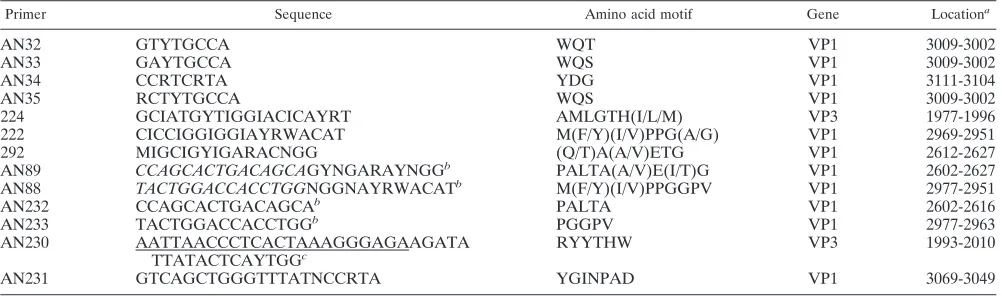
Primer Sequence Amino acid motif Gene Locationa
AN32 GTYTGCCA WQT VP1 3009-3002
AN33 GAYTGCCA WQS VP1 3009-3002
AN34 CCRTCRTA YDG VP1 3111-3104
AN35 RCTYTGCCA WQS VP1 3009-3002
224 GCIATGYTIGGIACICAYRT AMLGTH(I/L/M) VP3 1977-1996
222 CICCIGGIGGIAYRWACAT M(F/Y)(I/V)PPG(A/G) VP1 2969-2951
292 MIGCIGYIGARACNGG (Q/T)A(A/V)ETG VP1 2612-2627
AN89 CCAGCACTGACAGCAGYNGARAYNGGb PALTA(A/V)E(I/T)G VP1 2602-2627
AN88 TACTGGACCACCTGGNGGNAYRWACATb M(F/Y)(I/V)PPGGPV VP1 2977-2951
AN232 CCAGCACTGACAGCAb PALTA VP1 2602-2616
AN233 TACTGGACCACCTGGb PGGPV VP1 2977-2963
AN230 AATTAACCCTCACTAAAGGGAGAAGATA
TTATACTCAYTGGc
RYYTHW VP3 1993-2010
AN231 GTCAGCTGGGTTTATNCCRTA YGINPAD VP1 3069-3049
aThe locations of all primers except AN230 and AN231 are those relative to the genome of PV1 Mahoney (GenBank accession number J02281); the locations for AN230 and AN231 are relative to those of the genome of EV68 Fermon (GenBank accession number AY426531).
bAN232 is the nondegenerate “clamp” portion of AN89, and AN233 is the nondegenerate clamp portion of AN88. Within the AN88 and AN89 sequences, these clamp regions are indicated in italic type.
cThe T3 RNA polymerase promoter sequence is underlined.
on May 16, 2020 by guest
http://jcm.asm.org/
[image:2.585.43.544.81.229.2]rier tRNA was added to the kit’s lysis buffer. The purified VP3-VP1 sRNA was quantitated spectrophotometrically, and the concentration was calculated in units of RNA molecules per microliter. Two separate lots of the VP3-VP1 sRNA were synthesized and diluted to contain from 104
copies to 1 copy per 5l and then tested in two separate experiments with the VP1 RT-snPCR assay.
The sensitivity of the VP1 RT-snPCR assay was also directly compared to that of our published 5⬘-NTR RT-snPCR assay (10) by serially diluting RNA ex-tracted from a recent EV68 clinical isolate and running both the VP1 and the 5⬘-NTR RT-snPCR assays in parallel by using the same diluted RNA prepara-tions.
Nucleotide sequence accession numbers.The sequences for the original clin-ical specimens described here have been deposited in the GenBank sequence database (accession numbers AY903638 to AY903644).
RESULTS
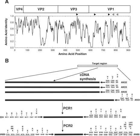
Primer design.To achieve the sensitivity necessary for the amplification and identification of EVs in original clinical spec-imens, we previously developed an RT-snPCR approach, using a primer (primer 224) in VP3 paired with a primer (primer 222) in VP1 for the first round of amplification and primers (primers 292 and 222) within VP1 (19) for the second, semin-ested PCR amplification (10) (Table 1; Fig. 1). This primer combination unfortunately sometimes results in an undesirable accumulation of nonspecific amplification products (data not shown), probably due to the use of highly degenerate inosine-containing primers, which require relatively low annealing temperatures for both PCR steps. To eliminate this problem, we designed a new set of internal primers to replace primers 292 and 222 using the consensus degenerate hybrid oligonucleotide primer (CODEHOP) approach (28, 30). CODEHOP primers contain a consensus degenerate core region of 9 to 12 bases at the
3⬘ end that is designed from conserved motifs within aligned
amino acid sequences. The 5⬘portion of a CODEHOP primer
comprises a nondegenerate consensus clamp region that is typi-cally 15 to 25 bases long. The degenerate core provides for broad target specificity, while the clamp increases the stability of the primer-template helix and allows the use of higher annealing temperatures to reduce nonspecific amplification. The optimal design strategy for the nondegenerate consensus clamp requires the analysis of codon usage for any conserved amino acid residues that occur within the clamp region. The predominant codon, if any, is incorporated into the primer sequence. For positions with multiple amino acid residues in the nondegenerate clamp, an arbitrary codon can be chosen or a specific codon can be selected
to manipulate the G⫹C content of the primer.
Consensus degenerate primers for the cDNA and PCR1 steps were designed from conserved amino acid motifs in the aligned capsid sequences of the 64 serotype prototype strains (Fig. 1). The four cDNA primers were designed to anneal to conserved sites downstream of the reverse PCR primer; these sites encode the motifs WQT (primer AN32), WQS (primers AN33 and AN35), and YDG (primer AN34) (Table 1; Fig. 1). PCR1 forward primer 224 was designed to target the site in VP3 encoding the highly conserved motif AMLGTH(I/L/M) (Table 1; Fig. 1), while PCR1 reverse primer 222 targets a conserved motif M(F/Y)(I/V)PPG( A/G), near the middle of VP1 (Table 1; Fig. 1) (12, 19). These two motifs are conserved among all EV serotypes and many human rhinoviruses. The degenerate primer design approach was used for the PCR1 primers to broaden the target specificity of ampli-fication, allowing amplification of all EV serotypes and increasing the absolute concentration of virus-specific product to be used as
the template for PCR2. The presence of inosine residues in po-sitions of fourfold codon degeneracy reduces the overall degen-eracy of the PCR1 primers but also results in the decreased thermostability of the primer-template helix. As a result, low-stringency annealing conditions are required for PCR1.
The internal primers used in PCR2 were designed without the use of inosine residues by using the CODEHOP strategy and with target sites encoding conserved motifs in VP1. Our original protocol used forward internal primer 292, which tar-gets the conserved motif (Q/T)A(A/V)ETG, paired with re-verse primer 222 (Table 1; Fig. 1). The new forward PCR2
primer, primer AN89, is 3⬘coterminal with primer 292, but it
targets a longer motif [PALTA(A/V)E(I/T)G] and
incorpo-rates an additional degeneracy at position⫺6 to allow
anneal-ing to codons that encode either isoleucine or threonine in the
penultimate codon of the motif. Primer AN88 is 3⬘coterminal
with primer 222 and targets the motif M(F/Y)(I/V)PPGGPV (Table 1; Fig. 1). The consensus clamp and increased length both probably contribute to the increased thermostabilities of primers AN88 and AN89 compared with those of primers 292 and 222.
Assay evaluation. To ensure that the new primers are broadly reactive and have the same ability to amplify VP1 sequences of all EV serotypes as our previous primers, we tested a panel of reference strains representing all serotypes (101 strains total), as well as a panel of 87 recent clinical isolates of 29 different types. All 64 EV serotype reference strains, the 22 proposed new serotypes, and 15 additional ref-erence strains for some serotypes were successfully amplified and sequenced by the CODEHOP VP1 RT-snPCR procedure (Table 2). All 87 clinical isolates tested were also successfully amplified, sequenced, and identified by comparing the nucleic acid sequence to the sequences in our EV reference strain VP1 sequence database (Table 3). In all cases, the serotype based on the VP1 RT-snPCR amplicon was identical to the serotype previously determined by neutralization or by VP1 sequencing with different primers and conventional PCR (12, 13, 19).
Assay sensitivity.Due to the absence of a gold standard for an analytical sensitivity measure that is applicable across all serotypes of enteroviruses, sensitivity was measured in two different ways so that the sensitivity could be compared to those obtained by previously described assays. The VP1 RT-snPCR assay detected RNA extracted from as little as 0.01
CCID50per 5l of EV68-Fermon (Fig. 2A), indicating that
the assay is at least 100-fold more sensitive than cell culture for
this strain of virus (since 1 CCID50defines the cell culture end
point). As few as 10 copies of the in vitro-transcribed VP3-VP1 sRNA produced a detectable gel band in two independent experiments, indicating a low limit of absolute sensitivity (Fig. 2B). The diluted EV68 clinical isolate RNA was amplified
from the 10⫺1to 10⫺7dilutions with the 5⬘-NTR RT-snPCR
assay and from the 10⫺1to 10⫺8dilutions with the VP1
RT-snPCR assay (Fig. 2C), indicating that the VP1 assay has sen-sitivity at least equal to or possibly 10-fold better than that of
the 5⬘-NTR assay.
Application to clinical specimens.To illustrate the applica-tion of the VP1 RT-snPCR method, RNA was extracted from original clinical specimens obtained from patients with a num-ber of different enteroviral illnesses and specimen types. The specimens (and associated illnesses) included cerebrospinal
on May 16, 2020 by guest
http://jcm.asm.org/
fluid (aseptic meningitis), stool (aseptic meningitis), rectal swab (febrile rash), nasopharyngeal swab (mild upper respi-ratory illness), conjunctival swab (acute hemorrhagic con-junctivitis), serum (febrile rash), and postmortem liver tis-sue (neonatal sepsis-like illness). A specific product was amplified by VP1 RT-snPCR from each of these extracted RNA templates (Fig. 3). In each assay, the tested RNA
represents the equivalent of approximately 45l of original
specimen fluid or 2 mg of stool sample. Following gel
[image:4.585.60.524.72.523.2]puri-fication, the EV present in each specimen was identified by amplicon sequencing and comparison to a database of EV VP1 sequences. All of the amplification products yielded clean, readable sequences, including those with weak or multiple bands (e.g., rectal swab and liver tissue specimens). In this example, the EVs identified were E30 (cerebrospinal fluid), CVA1 (stool), E9 (rectal swab), CVA9 (nasopharyn-geal swab), CVA24 (conjunctival swab), CVA10 (serum), and E11 (liver tissue).
FIG. 1. Schematic representation of the locations of the primers used in the CODEHOP VP1 RT-snPCR. (A) Similarity plot of the aligned capsid amino acid sequences of 64 enterovirus prototype strains. Sequence identity scores were calculated within each 6-residue window, and the window was progressively moved across the alignment in 1-residue increments, with the identity score plotted versus the position at the center of the window. The positions of the four mature EV capsid proteins, VP4, VP2, VP3, and VP1, are shown at the top. The orientations and approximate positions of the cDNA primers (open arrowheads) and PCR primers (filled arrowheads) are shown directly above the plot. (B) Amino acid motifs used in primer design and schematic representation of the steps in the CODEHOP VP1 RT-snPCR assay. Consensus amino acid motifs are shown. Asterisks indicate that the residue directly above the asterisk is present at that position in at least 90% of EV prototype strains; when only a single residue is shown, it is present in all prototype strains. Primer sequences are shown directly below the amino acid motif sequences. Ambiguity codes are as follows: R, A or G; Y, C or T; W, A or T; N, A, C, G, or T; I, inosine.
on May 16, 2020 by guest
http://jcm.asm.org/
DISCUSSION
Because of the degeneracy of the genetic code and the high degree of EV sequence diversity, it is more efficient to conceive of molecular diagnostic targets in the coding region as con-served amino acid sequences from which oligonucleotide primer sequences may be derived by back-translation. Codon degeneracy and sequence diversity, even within a serotype, necessitate the use of degenerate primers or multiple primer
[image:5.585.47.542.82.562.2]pairs to amplify all EV serotypes. Even with the use of multiple nondegenerate or minimally degenerate primers, it is difficult to design “pan-EV” primers that can be used for VP1 sequenc-ing and molecular serotypsequenc-ing. To address this problem, our strategy has been to design degenerate primers that contain deoxyinosine residues at positions of fourfold codon degener-acy (e.g., using GGI to specify a glycine codon, which is nor-mally GGN) (12, 13, 15, 16, 18, 19, 21). For example, the
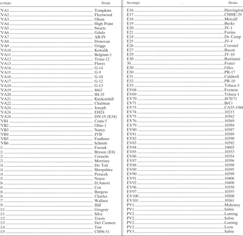
TABLE 2. Enterovirus reference strains amplified by VP1 RT-snPCRa
Serotype Strain Serotype Strain
CVA1 ... Tompkins CVA2 ... Fleetwood CVA3 ... Olson CVA4 ... High Point CVA5 ... Swartz CVA6 ... Gdula CVA7 ... AB-IV CVA8 ... Donovan CVA9 ... Griggs CVA10 ... Kowalik CVA11 ... Belgium-1 CVA12 ... Texas-12 CVA13 ... Flores CVA14 ... G-14 CVA15 ... G-9 CVA16 ... G-10 CVA17 ... G-12 CVA18 ... G-13
CVA19 ... 8663
CVA20 ... IH-35 CVA21 ... Kuykendall CVA22 ... Chulman CVA24 ... Joseph CVA24 ... EH24 CVA24 ... DN-19 (E34) CVB1 ... Conn-5 CVB2 ... Ohio-1 CVB3 ... Nancy CVB4 ... JVB CVB5 ... Faulkner CVB6 ... Schmitt E1 ... Farouk E1 ... Bryson (E8) E2 ... Cornelis E3 ... Morrisey E4 ... Du Toit E4 ... Shropshire E4 ... Pesacek E5 ... Noyce E6 ... D;Amori E6 ... Cox E6 ... Burgess E6 ... Charles E7 ... Wallace E9 ... Hill E11 ... Gregory E11 ... Silva E12 ... Travis E13 ... Del Carmen E14 ... Tow E15 ... CH96-51 a Reference strains for EV77 to EV78 and EV93 to EV95 were not tested because they are not publicly available. Some other numbers are missing due to reclassification (i.e., coxsackievirus A serotype 23关CVA23兴is a variant of echovirus 9 [E9], E8 is a variant of E1, E34 is a variant of CVA24, E10 is reovirus 1关genus Orthoreovirus, familyReoviridae兴, E28 is human rhinovirus 1A关genusRhinovirus, familyPicornaviridae兴, and EV72 is human hepatitis A virus关genusHepatovirus, family Picornaviridae兴). The “newer” serotypes (EV73 to EV76, EV79 to EV92, EV96, EV97, EV100, and EV101) were identified and classified by molecular means rather than by antigenic means (15, 18, 21; M. S. Oberste, unpublished data). PV, poliovirus. . E16 ...Harrington E17 ...CHHE-29 E18 ...Metcalf E19 ...Burke E20 ...JV-1 E21 ...Farina E24 ...De Camp E25 ...JV-4 E26 ...Coronel E27 ...Bacon E29 ...JV-10 E30 ...Bastianni 30 ...Frater E30 ...Giles E30 ...PR-17 E31 ...Caldwell E32 ...PR-10 E33 ...Toluca-3 EV68 ...Fermon EV69 ...Toluca-1 EV70 ...J670/71 EV71 ...BrCr EV73 ...CA55-1988 EV74 ...10213
EV75 ...10362
EV76 ...10369
EV79 ...10384
EV80 ...10387
EV81 ...10389
EV82 ...10390
EV83 ...10392
EV84 ...10603
EV85 ...10353
EV86 ...10354
EV87 ...10396
EV88 ...10398
EV89 ...10395
EV90 ...10399
EV91 ...10406
EV92 ...10408
EV96 ...10358
EV97 ...10355
EV100 ...10500
EV101 ...10361 PV1...Mahoney PV1...Sabin PV2...Lansing PV2...Sabin PV2...Lansing PV3...Leon PV3...Sabin
on May 16, 2020 by guest
http://jcm.asm.org/
primers that we currently use to identify EV cell culture iso-lates (primers 292 and 222) target the conserved VP1 amino acid motifs (QT)A(AV)ETG and M(FY)(IV)PPG(AG), re-spectively (19). These primers amplify RNA templates from all human EV serotypes, most nonhuman EVs, and some human rhinoviruses (12, 15, 16, 18, 19, 21). Casas et al. have used a similar approach, using degenerate, deoxyinosine-containing primers to target a different part of VP1 (4).
Several PCR assays have been developed to allow EV sero-type identification directly from original clinical specimens by nested amplification and sequencing of VP1 (4, 10, 31). The nested amplification format increases the sensitivity
substan-tially, rivaling that of nested 5⬘-NTR assays (4, 10), so that
nested VP1 assays can be used for both detection and identi-fication. However, nonspecific amplification can sometimes oc-cur when degenerate primers are used in a nested PCR assay, particularly when nucleic acids isolated from tissues or other high-complexity specimens are amplified. The CODEHOP primer design strategy has the advantage that most or all prim-ers are able to initiate DNA synthesis in early PCR rounds by virtue of annealing of the degenerate core sequence to the template, which is further stabilized by base pairing by at least some of the residues in the nondegenerate clamp sequence. The clamp sequence is present in all newly synthesized mole-cules during subsequent rounds of amplification, enabling vir-tually all primer molecules to participate in amplification. The inclusion of the nonconserved consensus clamp sequences in primers AN88 and AN89 did not affect the amplification effi-ciency for viruses that contained mismatches in this region (data not shown), even when stringent annealing conditions
were used. This is probably because the degenerate core se-quence is a good match for virtually all EVs and because at least a few of the bases in the clamp are able to form base pairs and contribute to the stability of the primer-template helix in the initial rounds of PCR. Once the primers have been incor-porated into the PCR products, the primers and templates match perfectly, allowing highly efficient amplification. Since its initial description (30), the CODEHOP approach has been used to amplify previously unknown gene homologs in a wide variety of plants, animals, and bacteria, as well as herpesviruses and retroviruses (29). To our knowledge, this work represents the first application of the CODEHOP strategy to the ampli-fication of an RNA virus and the first application of the method to primary virus diagnostics. By eliminating inosine
FIG. 2. Sensitivity of VP1 RT-snPCR and comparison of the sen-sitivity with that of the 5⬘nontranslated region RT-snPCR. (A) Am-plification of RNA extracted from 10-fold serial dilutions of an EV68 virus stock; (B) amplification of 10-fold serial dilutions of VP3-VP1 sRNA; (C) comparison of VP1 RT-snPCR (top) with 5⬘-NTR RT-snPCR (bottom) using 10-fold serial dilutions.
[image:6.585.298.541.76.322.2]FIG. 3. VP1 RT-snPCR amplification of RNA extracted directly from original clinical specimens. For each reaction, 50 l of each seminested PCR2 product was analyzed and gel purified by electro-phoresis on a 1.5% agarose gel containing 0.5g ethidium bromide per milliliter. The specimens tested were cerebrospinal fluid (CSF), stool, rectal swab (RS), nasopharyngeal swab (NPS), eye (conjunctival) swab (ES), serum, and postmortem liver tissue.
TABLE 3. Clinical isolates amplified by VP1 RT-snPCR
Serotype No. of isolates Yr(s) of isolation
CVA14 2 1992–1994
CVA16 4 1984–1995
CVA20 2 1983
CVA21 6 1986–1996
CVA24 1 1984
CVA9 2 1993–1996
CVB2 3 1982–1995
CVB3 6 1988–1997
CVB4 1 1984
CVB5 4 1983–1993
E3 3 1986–1988
E4 2 1985–1988
E5 1 1996
E6 3 1991–1998
E7 3 1993–1998
E9 3 1992–1995
E11 5 1988–1998
E12 1 1988
E13 2 1986–1995
E18 6 1985–1997
E21 1 1994
E24 1 1983
E25 6 1984–1994
E29 1 1988
E30 4 1991–1995
E33 1 1998
EV71 7 1988–1995
EV75 4 1985–1987
HRV2 1 1990
HRV31 1 1988
on May 16, 2020 by guest
http://jcm.asm.org/
from the primers used in the second, nested PCR, our VP1 CODEHOP PCR has increased specificity versus nonviral tem-plates and an increased sensitivity for viral targets, while it maintains the broad viral target specificity of the methods that use inosine-containing primers (4, 10).
As an example of the potential clinical/public health appli-cation of our method, we used the VP1 RT-snPCR assay to identify EVs in a diverse array of clinical specimens (Fig. 3). EVs were successfully amplified and amplicons were se-quenced from cerebrospinal fluid; stool; serum; liver tissue; and rectal, nasopharyngeal, and eye swab specimens. Ampli-cons from all of these specimen sources yielded high-quality sequences that provided clear serotype identification of the infecting viruses. We have subsequently applied this approach to the testing of routine diagnostic specimens in parallel with
more conventional real-time 5⬘-NTR detection assays. The
approach achieved sensitivity comparable to those of
conven-tional real-time 5⬘-NTR detection assays and provided virus
identification without the use of cell culture (unpublished data).
Our CODEHOP VP1 RT-snPCR is a sensitive assay of broad target specificity that can successfully amplify template RNA from all known EV serotypes. Its sensitivity is superior to
that of 5⬘-NTR RT-snPCR assays, with product detection
per-formed with stained gels (Fig. 2), probably because of the high
degree of RNA secondary structure in the 5⬘-NTR, which
prevents complete template melting and efficient annealing of the primers. In addition to EV identification, molecular epi-demiologic analysis of the sequences derived from the VP1 RT-snPCR products can also furnish clues to virus transmis-sion pathways during epidemiologic field investigations. By-passing cell culture in the early phases of an outbreak investi-gation can help to focus limited resources and allow rapid identification of epidemiologically linked cases. When more detailed virus characterization is desired, an informed choice can be made as to which specimens should be inoculated into cell culture and, in many cases, which cell lines should be used to maximize the efficiencies of these more labor-intensive methods.
REFERENCES
1.Barrios Olivera, J. A., L. Sarmiento Pe´rez, O. Valde´s, P. Mas Lago, and R. Palomera Puentes.2003. Aplicacio´n de la secuenciacio´n de VP1 a la iden-tificacio´n de enterovirus humanos. Rev. Cubana Med. Trop.55:133–137. 2.Bolanaki, E., C. Kottaridi, P. Markoulatos, L. Margaritis, and T. Katsorchis.
2005. Nucleotide analysis and phylogenetic study of the homology boundaries of coxsackie A and B viruses. Virus Genes31:307–320.
3.Caro, V., S. Guillot, F. Delpeyroux, and R. Crainic.2001. Molecular strategy for ‘serotyping’ of human enteroviruses. J. Gen. Virol.82:79–91. 4.Casas, I., G. F. Palacios, G. Trallero, D. Cisterna, M. C. Freire, and A.
Tenorio.2001. Molecular characterization of human enteroviruses in clinical samples: comparison between VP2, VP1, and RNA polymerase regions using RT nested PCR assays and direct sequencing of products. J. Med. Virol. 65:138–148.
5.Hamilton, M. S., M. A. Jackson, and D. Abel.1999. Clinical utility of poly-merase chain reaction testing for enteroviral meningitis. Pediatr. Infect. Dis. J.18:533–537.
6.Kottaridi, C., E. Bolanaki, and P. Markoulatos.2004. Amplification of echoviruses genome regions by different RT-PCR protocols—a comparative study. Mol. Cell. Probes18:263–269.
7.Kottaridi, C., E. Bolanaki, N. Siafakas, and P. Markoulatos.2005. Evalua-tion of seroneutralizaEvalua-tion and molecular diagnostic methods for echovirus identification. Diagn. Microbiol. Infect. Dis.53:113–119.
8.Manayani, D. J., R. V. Shaji, G. J. Fletcher, T. Cherian, N. Murali, N. Sathish, T. Solomon, C. Gnanamutha, and G. Sridraran.2002. Comparison of molecular and conventional methods for typing of enteroviral isolates. J. Clin. Microbiol.40:1069–1070.
9.Manzara, S., M. Muscillo, G. La Rosa, C. Marianelli, P. Cattani, and G. Fadda.2002. Molecular identification and typing of enteroviruses isolated from clinical specimens. J. Clin. Microbiol.40:4554–4560.
10.Nix, W. A., M. Berger, M. S. Oberste, B. R. Brooks, D. McKenna-Yasek, R. H. Brown, R. P. Roos, and M. A. Pallansch.2004. Failure to detect enterovirus genome in the spinal cord of ALS patients using a sensitive, semi-nested RT-PCR method. Neurology62:1372–1377.
11.Norder, H., L. Bjerregaard, and L. O. Magnius.2001. Homotypic echovi-ruses share aminoterminal VP1 sequence homology applicable for typing. J. Med. Virol.63:35–44.
12.Oberste, M. S., K. Maher, M. R. Flemister, G. Marchetti, D. R. Kilpatrick, and M. A. Pallansch.2000. Comparison of classic and molecular approaches for the identification of untypeable enteroviruses. J. Clin. Microbiol. 38: 1170–1174.
13.Oberste, M. S., K. Maher, D. R. Kilpatrick, M. R. Flemister, B. A. Brown, and M. A. Pallansch.1999. Typing of human enteroviruses by partial se-quencing of VP1. J. Clin. Microbiol.37:1288–1293.
14.Oberste, M. S., K. Maher, D. R. Kilpatrick, and M. A. Pallansch.1999. Molecular evolution of the human enteroviruses: correlation of serotype with VP1 sequence and application to picornavirus classification. J. Virol. 73:1941–1948.
15.Oberste, M. S., K. Maher, S. M. Michele, M. Uddin, G. Belliot, and M. A. Pallansch.2005. Enteroviruses 76, 89, 90, and 91 represent a novel group within the species human enterovirus A. J. Gen. Virol.86:445–451. 16.Oberste, M. S., K. Maher, and M. A. Pallansch.2002. Molecular phylogeny
and classification of the simian picornaviruses. J. Virol.76:1244–1251. 17.Oberste, M. S., K. Maher, A. J. Williams, N. Dybdahl-Sissoko, B. A. Brown,
M. T. Gookin, S. Pen˜aranda, N. G. Mishrik, M. Uddin, and M. A. Pallansch. 2006. Species-specific RT-PCR amplification of human enteroviruses: a tool for rapid species identification of uncharacterized enteroviruses. J. Gen. Virol.87(Pt 1):119–128.
18.Oberste, M. S., S. M. Michele, K. Maher, D. Schnurr, D. Cisterna, N. Junttila, M. Uddin, J.-J. Chomel, C.-S. Lau, W. Ridha, S. al-Busaidy, H. Norder, L. Magnius, and M. A. Pallansch.2004. Molecular identification and characterization of two proposed new enterovirus serotypes, EV74 and EV75. J. Gen. Virol.85:3205–3212.
19.Oberste, M. S., W. A. Nix, K. Maher, and M. A. Pallansch.2003. Improved molecular identification of enteroviruses by RT-PCR and amplicon sequenc-ing. J. Clin. Virol.26:375–377.
20.Oberste, M. S., and M. A. Pallansch.2005. Enterovirus molecular detection and typing. Rev. Med. Microbiol.16:163–171.
21.Oberste, M. S., D. Schnurr, K. Maher, S. al-Busaidy, and M. A. Pallansch. 2001. Molecular identification of new picornaviruses and characterization of a proposed enterovirus 73 serotype. J. Gen. Virol.82:409–416.
22.Palacios, G., I. Casas, A. Tenorio, and C. Freire.2002. Molecular identifi-cation of enterovirus by analyzing a partial VP1 genomic region with differ-ent methods. J. Clin. Microbiol.40:182–192.
23.Pallansch, M. A., and M. S. Oberste.2003. Molecular detection and char-acterization of human enteroviruses, p. 245–257.InA. Matsumori (ed.), Cardiomyopathies and heart failure: biomolecular, infectious and immune mechanisms. Kluwer Academic Publishers, Boston, Mass.
24.Rakoto-Andrianarivelo, M., D. Rousset, R. Razafindratsimandresy, and F. Delpeyroux.2002. Nouvelle me´thode de typage mole´culaire des ente´rovirus humains: caracte´risation des souches malgaches “non se´rotypables.” Arch. Instit. Pasteur Madagascar68:55–58.
25.Read, S. J., K. J. M. Jeffery, and C. R. M. Bangham.1997. Aseptic meningitis and encephalitis: the role of PCR in the laboratory. J. Clin. Microbiol. 35:691–696.
26.Robinson, C. C., M. Willis, A. Meagher, K. E. Gieseker, H. Rotbart, and M. P. Glode´.2002. Impact of rapid polymerase chain reaction results on management of pediatric patients with enteroviral meningitis. Pediatr. In-fect. Dis. J.21:283–286.
27.Romero, J. R.1999. Reverse-transcription polymerase chain reaction detec-tion of the enteroviruses. Arch. Pathol. Lab. Med.123:1161–1169. 28.Rose, T. M. 2005. CODEHOP-mediated PCR—a powerful tool for the
identificcation and characterization of viral genomes. Virol. J.2:20–43. 29.Rose, T. M., J. G. Henikoff, and S. Henikofff.2003. CODEHOP
(COnsensus-DEgenerate Hybrid Oligonucleotide Primer) PCR primer design. Nucleic Acids Res.31:3763–3766.
30.Rose, T. M., E. R. Schultz, J. G. Henikoff, S. Pietrokovski, C. M. McCallum, and S. Henikoff.1998. Consensus-degenerate hybrid oligonucleotide primers for amplification of distantly related sequences. Nucleic Acids Res.26:1628–1635. 31.Thoelen, I., P. Lemey, I. Van der Donck, K. Beuselink, A. M. Lindberg, and M. Van Ranst.2003. Molecular typing and epidemiology of enteroviruses identified from an outbreak of aseptic meningitis in Belgium during the summer of 2000. J. Med. Virol.70:420–429.
32.Vuorinen, T., R. Vainionpa¨a¨, and T. Hyypia¨.2003. Five years’ experience of reverse-transcriptase polymerase chain reaction in daily diagnosis of entero-virus and rhinoentero-virus infections. Clin. Infect. Dis.37:452–455.
33.World Health Organization.2001. Manual for the virological investigation of polio WHO/EPI/GEN/97.01. World Health Organization, Geneva, Switzerland.