R E S E A R C H
Open Access
Artificial rearing influences the morphology,
permeability and redox state of the
gastrointestinal tract of low and normal
birth weight piglets
Hans Vergauwen
1†, Jeroen Degroote
2†, Sara Prims
1, Wei Wang
2,3, Erik Fransen
4, Stefaan De Smet
3,
Christophe Casteleyn
1, Steven Van Cruchten
1, Joris Michiels
2and Chris Van Ginneken
1*Abstract
Background:In this study the physiological implications of artificial rearing were investigated. Low (LBW) and normal birth weight (NBW) piglets were compared as they might react differently to stressors caused by artificial rearing. In total, 42 pairs of LBW and NBW piglets from 16 litters suckled the sow until d19 of age or were artificially reared starting at d3 until d19 of age. Blood and tissue samples that were collected after euthanasia at 0, 3, 5, 8 and 19 d of age. Histology, ELISA, and Ussing chamber analysis were used to study proximal and distal small intestine histo-morphology, proliferation, apoptosis, tight junction protein expression, and permeability. Furthermore, small intestine, liver and systemic redox parameters (GSH, GSSG, GSH-Px and MDA) were investigated using HPLC.
Results:LBW and NBW artificially reared piglets weighed respectively 40 and 33% more than LBW and NBW sow-reared piglets at d19 (P< 0.01). Transferring piglets to a nursery at d3 resulted in villus atrophy, increased intestinal FD-4 and HRP permeability and elevated GSSG/GSH ratio in the distal small intestine at d5 (P< 0.05). GSH concentrations in the proximal small intestine remained stable, while they decreased in the liver (P< 0.05). From d5 until d19, villus width and crypt depth increased, whereas PCNA, caspase-3, occludin and claudin-3 protein expressions were reduced. GSH, GSSG and permeability recovered in artificially reared piglets (P< 0.05).
Conclusion:The results suggest that artificial rearing altered the morphology, permeability and redox state without compromising piglet performance. The observed effects were not depending on birth weight.
Keywords:Milk replacer, Oxidative stress, Small intestine, Suckling period, Tight junction proteins
Background
The neonatal period in the pig’s life is accompanied with high morbidity and mortality [1, 2]. In addition, increas-ing litter sizes in modern swine production have led to higher rates of piglets born with a low birth weight (LBW) [3]. Both newborn LBW human [4–6] and LBW piglets [7–10] seem to have a lower capacity to mount an antioxidant response. Newborns transitioning from
maternal mediated respiration to autonomous pulmon-ary respiration outside the uterus are suddenly exposed to O2-derived free radicals [11]. This increased the pro-duction of reactive species in various organs [7]. A redox imbalance affects cellular signaling, protein synthesis, and enhances proteolysis that can ultimately lead to a dysfunctional intestinal barrier and suboptimal regenera-tive potential as shown in vitro [12–15]. Consequently, the observed redox imbalance and the downstream ef-fects could explain the abnormal absorption and metab-olism of nutrients, reduced growth and impaired development of the small intestine, liver, and muscle ob-served in LBW piglets [10, 16–21]. This redox imbalance appears to persist beyond weaning [9]. Wang et al. * Correspondence:[email protected]
†Equal contributors 1
Laboratory of Applied Veterinary Morphology, Department of Veterinary Sciences, Faculty of Biomedical, Pharmaceutical and Veterinary Sciences, University of Antwerp, Campus Drie Eiken, Universiteitsplein 1, D.U.015, 2610 Wilrijk, Belgium
Full list of author information is available at the end of the article
observed that mRNA expression of occludin, heme oxy-genase 1, catalase, thioredoxin reductase genes and occludin protein expression continued to be lower in LBW pigs during the suckling period [21]. This appar-ently conflicts with the observation that LBW piglets that survive the critical first days after birth show an intestinal morphology, digestive capacity, cytokine pro-duction, intestinal motility and permeability that is com-parable with those seen in normal birth weight (NBW) littermates [22–24].
The increasing incidence of supernumerary and LBW piglets has raised an urgent need for innovative rearing strategies [1]. Next to cross-fostering [25], supplement-ing piglets [26] and split nurssupplement-ing [27], piglets can be transferred to a nursery and artificially reared [28–30]. Similar to conventional weaning, the artificially reared piglet encounters psychological and physical stressors in-cluding maternal and littermate separation, abrupt changes in diet composition and environment, lower in-take of bioactive substances, as well as unfamiliar drink-ing nipples, increased exposure to pathogens and antigens, comingling and establishment of social hier-archy with unfamiliar pigs from different litters. The physiological responses to the strategy of full artificial rearing are largely unknown. Conventional weaning is associated with the induction of intestinal oxidative stress [31–33] and LBW piglets have more difficulties to maintain a balanced redox state when exposed to wean-ing stressors [9]. It is unknown at present if the response to artificial rearing, since it includes similar stressors as conventional weaning, is different in LBW piglets—which have a lower antioxidant capacity [21]—compared to NBW piglets.
Therefore, we aimed to investigate the impact of artifi-cial rearing on piglet performance, proximal and distal small intestinal (SI) morphology, mitosis, apoptosis, and tight junction protein expression, permeability, and SI, liver and systemic redox state development compared to conventional rearing. Given the similarities between conventional weaning stressors and artificial rearing stressors we hypothesized that artificial rearing results in a redox imbalance and negatively affects intestinal morphology and functionality. Secondly, given the differ-ences observed between NBW and LBW piglets during the suckling period, we hypothesized that in view of their affected redox state, the morphology and function-ality of the small intestine is suboptimal in LBW piglets and that artificial rearing has a greater negative impact in this birth weight category.
Methods
Pig model and tissue collection
Eighty-four piglets (Topigs hybrid × Piétrain) were se-lected during two consecutive farrowing rounds. Only
sows with 14 or more live-born piglets were selected and no cross-fostered piglets were included in the ex-periment. Piglets were tagged and weighed within 12 h after parturition. A LBW piglet was defined as a pig hav-ing a birth weight between 0.75 and 0.90 kg and belong-ing to the lower quartile of litter birth weights. A NBW piglet had a birth weight within 1 SD unit of the mean birth weight of the whole litter. The average birth weight of all the piglets used in this study was 0.81 ± 0.01 kg and 1.26 ± 0.02 kg for LBW and NBW piglets, respect-ively. Forty-two pairs of LBW and NBW gender-matched littermates were either left to suckle the sow or transferred to a nursery at the age of 3 d and had ad libi-tum access to a milk replacer that was refreshed twice a day (Table 1). Milk replacer was made by mixing 200 g of milk replacer powder into 1 L of water, preheated at 42 °C. This resulted in a dry matter (DM) content of 193 g/kg milk. This is comparable with the DM content of sow milk, which is around 170 to 190 g/kg milk be-tween 3 d and 42 d of lactation [34]. In Table 1, the
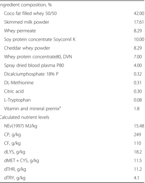
cal-culated nutrient composition of some important
nutrients is shown, expressed on DM basis. When ac-count for the dilution factor, it is possible to compare this milk formula with sow milk. For instance, it is clear
Table 1Composition of the milk replacer used for piglets from 3 d of age until weaning at d 19
Ingredient composition, %
Coco fat filled whey 50/50 42.00
Skimmed milk powder 17.61
Whey permeate 8.29
Soy protein concentrate Soycomil K 10.00
Cheddar whey powder 8.29
Whey protein concentrate80, DVN 7.00
Spray dried blood plasma P80 4.00
Dicalciumphosphate 18% P 0.32
DL-Methionine 0.31
Citric acid 0.30
L-Tryptophan 0.08
Vitamin and mineral premixa 1.8
Calculated nutrient levels
NEv(1997) MJ/kg 15.48
CP, g/kg 249
CF, g/kg 110
dLYS, g/kg 18.2
dMET + CYS, g/kg 11.5
dTHR, g/kg 11.2
dTRY, g/kg 4.1
a
The mineral and vitamin premix supplied as the following (per kg diet):
Vitamin A, 30, 000 IU; Vitamin D3, 5000 IU; Vitamin E; 75 mg; Fe2+, 120 mg; Zn2+,
that the crude protein level in the formula (49.8 g/kg milk) is low but comparable with what can be expected in sow milk (51–64 g/kg milk) ([34–36]). The crude fat content was calculated at 22 g/kg milk, which is consid-erably lower than the crude fat content of sow milk (5.3–6.5 g/kg milk) [34–36]. In contrast, the total amount of lactose in the liquid milk replacer was 80 g/kg milk. Values for sow milk range from 48 to 59 g/kg milk, and thus are lower than what was offered to the formula fed piglets. From this, it might be clear that the milk for-mula was not completely comparable to the nutrient composition of porcine milk. However, this tailor made milk formula was meant as a compromise of many exist-ing, commercially available milk replacer formulas, and as a copy of sow milk as such. A maximum of 4 NBW-LBW gender matched littermate pairs were co-housed. The starting ambient temperature was 32 °C and linearly decreased to 28 °C towards d19 post-natal. Heating and ventilation was automatically controlled in function of the temperature settings. Six pairs of sow-reared LBW and NBW piglets were sampled on d0, d3, d8 and d19 of age, and 6 pairs of artificially reared LBW and NBW pig-lets were sampled on d5, d8 and d19 of age. Pigpig-lets sam-pled on d0 were removed from the sow between 12 and 24 h after parturition. At d3, intramuscular iron injec-tions were given (Iron(III) Dextran, 200 mg/piglet, Uniferon). The selected animals did not receive any anti-biotic treatment prior or during the experiment.
Pigs were killed by exsanguination by severing the ca-rotid arteries and jugular veins following induction of terminal anesthesia by intramuscular injection of keta-mine (15 mg/kg BW) combined with xylazine (2 mg/kg BW). All piglets were weighed prior to euthanasia.
Blood was collected in EDTA and heparinized tubes containing supplemental bathophenanthroline disulfo-nate sodium salt. Erythrocytes were isolated by centri-fuging (3000 × g, 15 min) 0.5 mL of unclotted, heparinized blood. After removing the plasma, eryth-rocytes were lysed by adding 100 μL of a 70% meta-phosphoric acid solution, 600 μL milli Q and intense vortexing. These extracts were then centrifuged (3000 × g, 15 min), and 0.5 mL of the remaining acid extract was transferred to a vial containing 50 μL of a γ -glu-glu internal standard solution. After opening the abdo-men, the liver was isolated and samples of the left lat-eral lobe were dissected for acid and phosphate buffered aqueous extraction, as described for the small intestinal mucosa. Subsequently, the small intestine (SI), defined as the part of the gastrointestinal tract between the pylorus and the ileocecal valve, was dis-sected and its length was measured. A 10 cm segment of proximal and distal SI (5 and 75% of total SI length, respectively) was taken for Ussing chamber measure-ments. In addition, 20 cm segments at 5 and 75% of
the total SI length were emptied and carefully flushed with saline. The tissue of these 20 cm segments was placed on an ice-cold surface and the mucosa was re-trieved by gently scraping the mucosal surface with a glass slide. Aliquots of the mucosa were either used instantaneously for acid and phosphate buffered aque-ous extracts or transferred to plastic 2 mL screw-capped tubes, snap-frozen in liquid nitrogen and stored at -80 °C pending redox state analysis. Further-more, 5 cm segments at 5 or 75% of the total SI length were taken, flushed with saline, snap-frozen in liquid nitrogen and stored at -80 °C pending protein expres-sion analysis. Finally, a 5 cm segment at 5 or 75% of the total SI length was flushed with saline, divided in smaller pieces of max 1.5 cm in length and fixated for 2 h in 4% freshly prepared paraformaldehyde (in 0.01 mol/L phosphate-buffered saline) (volume tissue/ volume fixative: 1/5) and routinely processed for paraffin-embedding [37].
Small intestinal histo-morphological measurements In brief, 4 μm sections of paraffin-embedded samples were mounted on slides and stained with hematoxylin-eosin. Villus height, mid-villus width, and crypt depth were measured at 10× magnification using an Olympus BX61 microscope and image analysis software (analySIS Pro, Olympus Belgium, Aartselaar, Belgium) in 1–3 well-oriented villi and associated crypts in at least 12–15 sec-tions per tissue sample, to yield 30 measurements per small intestinal region.
measured using an Infinite M200 Pro spectrophotometer with X-Fluor software at 450 nm at 25 °C (Tecan Group Ltd., Männedorf, Switzerland). Values of protein expres-sion were determined per gram of total protein in a sam-ple, measured using a PierceTM BCA Protein Assay Kit (ThermoFisher Scientific, Belgium), and expressed as fmol/mg.
Ex vivo measurement of small intestinal permeability Intestinal mucosal permeability was assessed ex vivo by measuring the translocation of macromolecular markers using the Ussing chamber technique. The segments were first rinsed with saline. The mucosal layer was stripped from the seromuscular layer and pinned onto 1.07 cm2 sliders that were mounted into modified Ussing cham-bers (Dipl.-Ing. Muβler Scientific Instruments, Aachen, Germany). All tissues were mounted within 10 min fol-lowing euthanasia. Tissues were immersed in 6.5 mL Ringer solution (115 mmol/L NaCl, 5 mmol/L KCl, 25 mmol/L NaHCO3, 2.4 mmol/L Na2HPO4, 0.4 mmol/L NaH2PO4, 1.25 mmol/L CaCl2, 1 mmol/L MgSO4) with 6 mmol/L of mannitol or glucose in the luminal and se-rosal side, respectively. The system was water-jacketed to 37 °C and oxygenated with 95% O2and 5% CO2. After an equilibration period of 20 min, 4 kDa fluorescein isothiocyanate-dextran (FD-4, Sigma-Aldrich, Bornem, Belgium) and 40 kDa horseradish peroxidase (HRP, type IV, Sigma-Aldrich, Bornem, Belgium) were added to the mucosal side to a final concentration of 0.8 mg/mL of FD-4 and 0.FD-4 mg/mL of HRP. Samples of the buffer solution were taken from the serosal chamber at 20, 40, 60 and 100 min after adding markers. Meanwhile, the same vol-ume of buffer was taken from the mucosal side to keep the volume balance across sides. Fluorescence intensity of FD-4 in the medium was measured at excitation wave-length of 485 nm and emission wavewave-length of 538 nm using a fluorescence plate reader (Thermo Scientific, Marietta, OH, USA). HRP was measured according to the method described previously [38]. In short, HRP activity was measured by adding a ‘start solution’ (50 mL of 0.2 mol/L NaH2PO4, 1 mL of 0.2 mol/L Na2HPO4, 20.4μL of 30% H2O2, 1.7 mL of 1% dianisidine peroxide substrate, made up to 204 mL with water) to the HRP samples and left for 10 min, after which the reaction was terminated by addition of 120μL of 4% sodium azide and the absorbance was read at 460 nm. The relation between peroxidase concentration and absorbance is linear in the concentration range 10–100 pmol/L. The apparent per-meation coefficient (Papp) was calculated as:
Papp cmð =sÞ: ðdc=dtÞ V=c0=A;
Whereby dc/dt is the change of serosal concentration in the 20- to 100-min period (cm/s); V is the volume of
the chamber, c0is the initial marker concentration in the mucosal reservoirs and A the area of the exposed intes-tine in the chambers (cm2).
Mucosal, liver and blood homogenate extracts and biochemical assays
An acid extract was prepared from 1 g of homogenized (Braun homogenizer at 900 rpm) intestinal mucosa or liver that was placed in 10 mL ice-cold perchloric acid (PCA) 10% solution and centrifuged at 15,000 × g for 15 min at 4 °C. The resulting acid extract (0.5 mL) was transferred to tubes containing 50μLγ-glu-glu internal standard solution. Samples were snap frozen in liquid nitrogen and stored at -80 °C until analysis of GSH and glutathione disulfide (GSSG). The biuret reaction was applied to determine the total protein content. Mucosal GSH and GSSG were measured using a modified high performance liquid chromatography (HPLC) method [39, 40]. The derivation procedure included the reac-tion of 100 mmol/L iodoacetic acid solureac-tion with thiols to form S-carboxymethyl derivatives followed by chromophore derivation of primary amines with dini-trofluorobenzene (DNFB, 1% (v/v) in ethanol). GSH and GSSG were separated through EC250/4.6 Nucleosil
120-7 NH2 aminopropyl column (Machery-Nagel,
measure MDA concentration in EDTA plasma, liver and mucosa extracts [42].
Statistical analysis
Linear mixed models were fitted to assess the influence of birth weight category (NBW/LBW), feeding (artifi-cially reared/sow-reared) and days postnatal (as a cat-egorical variable) on the quantitative outcome variables. To model the dependence between observations within the same litter, random intercept terms for litter were added to the model. Depending on the research ques-tion, separate analyses were carried out in subgroups (e.g. only in sow-reared piglets) or time points were analyzed separately. In the subgroup analyses where no random effect terms was needed, a multiple linear re-gression model was fitted. Post hoc tests to compare mean values between the different time points (days postnatal) were carried out using Tukey’s honestly sig-nificant difference. Models were fitted using the Mixed Model procedure of the JMP Pro11 software (SAS Insti-tute, Cary, NC, USA). Significance for the fixed effects was tested using an F-test with Kenward-Roger correc-tion. Data are expressed as means and their standard er-rors (S.E.), andP <0.05 was considered significant.
Results
Body weight of piglets
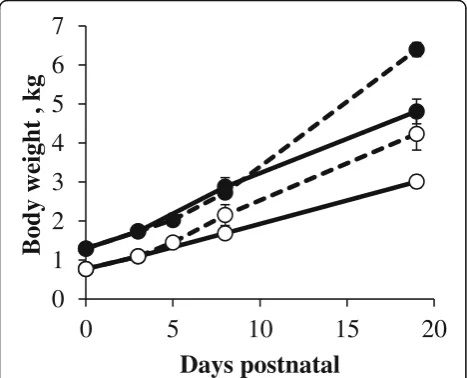
Average body weight of LBW and NBW piglets sampled at birth (d0) was 0.77 ± 0.07 and 1.29 ± 0.08 kg, respect-ively (Fig. 1). The body weight of both birth weight cat-egories did not significantly change during the first 3 days. Afterwards, in both sow- and artificially reared piglets the body weight gradually increased (P< 0.001). At each point in time, LBW sow-reared piglets showed lower body
weights compared to their NBW sow-reared littermates (P< 0.01). A similar observation was seen in artificially reared piglets at d5 (P< 0.001) and d19 (P< 0.05). Body weights of LBW and NBW artificially reared piglets were significantly higher, compared to respectively LBW and NBW sow-reared piglets at d19 (P< 0.05).
Histo-morphological measurements in the proximal and distal small intestine
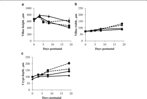
The histo-morphological parameters were stable be-tween d0 and d3 (Fig. 2). After d3, villus height signifi-cantly decreased (P< 0.05; Fig. 2a), whereas villus width (P< 0.05; Fig. 2b) and crypt depth significantly increased (P< 0.05; Fig. 2c). Furthermore, the transfer of piglets to a nursery caused villus atrophy in the proximal and dis-tal (P< 0.05) SI from d3 to d5. Villi were significantly wider (at d19: P< 0.001) and crypts deeper (at d8 and d19: P< 0.01) in artificially reared piglets compared to sow-reared piglets. In the distal SI of NBW sow-reared piglets villus heights were consistently higher compared to those of their LBW littermates (P< 0.05). In contrast, villus heights in the distal SI of NBW artificially reared piglets were on average 193 μm lower at d19 compared to their LBW littermates (P< 0.05).
PCNA and caspase-3 protein expression in the proximal and distal small intestine
PCNA protein expression remained at the same level dur-ing the suckldur-ing period in the proximal and distal SI of sow-reared piglets (Fig. 3a). Transferring the piglets at d3 to a nursery significantly reduced PCNA protein expres-sion in the proximal SI at d5 (P< 0.05) as compared to d3, whereas in the distal SI its level remained unaffected. PCNA protein expression in the proximal and distal SI of artificially reared piglets was significantly lower compared to sow-reared piglets at d8 (P< 0.05) and at d19 in the proximal SI (P< 0.001) (Fig. 3a).
Caspase-3 protein expression significantly decreased in the proximal SI of sow-reared (P< 0.05) and artifi-cially reared piglets (P< 0.001) piglets from d3 to d19 (Fig. 3b). In contrast, in the distal SI of sow- and artifi-cially reared piglets, no age-related differences were observed. Caspase-3 protein expression was signifi-cantly lower in the proximal (P< 0.05) and distal (P< 0.05) SI of artificially reared piglets compared to sow-reared piglets at d19.
Occludin and claudin-3 protein expression in the proximal and distal small intestine
Occludin expression in the proximal SI of sow-reared piglets decreased from d3 to d19 (P< 0.001), whereas in the distal SI its level remained unchanged (Fig. 4a). Occludin expression dropped significantly in the prox-imal (P< 0.05) but not in the distal SI of artificially
0
1
2
3
4
5
6
7
0
5
10
15
20
Body w
eight
, kg
Days postnatal
reared piglets, from d3 to d5, but returned to its initial levels afterwards. The expression of occludin in the SI was comparable for sow- and artificially reared piglets.
Claudin-3 expression in the proximal and in the distal SI of artificially reared piglets dropped significantly from d3 to d5 (P< 0.01; Fig. 4b). Claudin-3 expression increased from d5 to d19 and was significantly higher in the SI of artificially reared piglets compared to sow-reared piglets at d19 (P<
0.01). Claudin-3 expression in the distal SI of LBW sow-reared piglets was on average 33% higher compared to NBW sow-reared piglets at d3, d8 and d19 (P< 0.01).
Ex vivo permeability in the proximal and distal small intestine
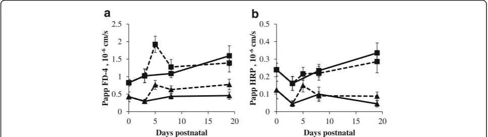
FD-4 and HRP permeability in the proximal and distal SI remained stable in sow-reared piglets (Fig. 5a and b). FD-4
0 200 400 600 800 1000
0 5 10 15 20
V
illus height , µm
Days postnatal
0 50 100 150 200 250
0 5 10 15 20
V
illus width , µm
Days postnatal
0 50 100 150 200 250
0 5 10 15 20
Crypt depth , µm
Days postnatal
a
b
c
Fig. 2Villus height (μm) (a), villus width (μm) (b) and crypt depth (μm) (c) in proximal (square) and distal (triangle) SI of sow- (full line) and artificially reared (dashed line) piglets expressed asμm. Values are means ± SE (n= 12 as LBW and NBW piglets were pooled together)
0 2 4 6 8 10 12 14 16
0 5 10 15 20
P
CNA
,
fm
ol/m
g
Days postnatal
0 2 4 6 8 10 12 14 16
0 5 10 15 20
Caspase-3 , fm
ol/m
g
Days postnatal
a
b
permeability in the proximal SI significantly increased in 5-day-old NBW artificially reared piglets when transferred to a nursery at d3 (P< 0.05; Fig. 5a). Given the observation that the Papp of FD-4 was already high at 3 days of age in the proximal SI of LBW piglets, no changes could be ob-served after the start of artificial rearing. In the distal SI, both FD-4 (P< 0.05; Fig. 5a) and HRP (P< 0.01; Fig. 5b) permeabilities were significantly increased in both 5-day-old LBW and NBW artificially reared piglets, compared to d3. LBW sow-reared piglets consistently showed signifi-cantly higher FD-4 (proximal: on average 0.5 × 10⁻⁶cm/s higher; distal: on average 0.2 × 10⁻⁶cm/s higher) and HRP (proximal: on average 0.09 × 10⁻⁶cm/s higher; distal: on average 0.06 × 10⁻⁶cm/s higher) permeability when com-pared to their NBW littermates (P< 0.05). No such differ-ences were observed in artificially reared piglets.
Mucosal redox state represented by GSH concentration, GSSG concentration, GSSG/GSH ratio, GSH-Px activity and MDA concentration
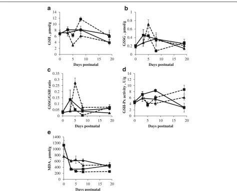
GSH concentration was constant from birth until d8. In the distal SI mucosa GSH concentrations remained stable SI, whereas in the proximal SI it decreased significantly
after d8 (P< 0.01) of sow-reared piglets (Fig. 6a). The concentration of GSSG and the GSSG/GSH ratio did not show any age-related changes in sow-reared pig-lets (P> 0.05; Fig. 6b and c). The activity of GSH-Px in the proximal and distal SI mucosae of sow-reared piglets in-creased from birth until d3 (P< 0.05). After d8, GSH-Px activity decreased significantly (P< 0.01), reaching a simi-lar activity at d19 as observed at birth (Fig. 6d).
In the proximal intestine of sow-reared pigs, the concentration of MDA abruptly dropped after birth (P< 0.001; Fig. 6e) and remained stable from d3 until d19, whereas in the distal intestine the decrease was more spread out in time (P< 0.01). Transferring piglets to a nursery decreased the concentration of GSH, whereas the GSSG concentration and GSSG/GSH ratio were increased in the SI of artificially reared piglets at d5. In the proximal SI of artificially reared piglets GSH concentration peaked, while GSSG concentration and GSSG/GSH ratio showed a minimum at d8 (P< 0.05). However by d19, these redox parameters returned to the values noted at birth (P <0.05). GSH-Px activity showed a minimum at d5 (P< 0.05), but recovered from d8 to d19 (P< 0.01) in the SI of artificially reared piglets.
,
Fig. 4Relative protein expression of occludin (fmol/mg) (a) and claudin-3 (fmol/mg) (b) in the proximal (square) and distal (triangle) SI of sow-(full line) and artificially reared (dashed line) piglets. Values are means ± SE (n= 12 as LBW and NBW piglets were pooled together)
0 0.5 1 1.5 2 2.5
0 5 10 15 20
P
a
pp F
D
-4
, 10
-6
cm
/s
Days postnatal
0 0.1 0.2 0.3 0.4 0.5
0 5 10 15 20
P
a
pp HRP
, 10
-6
cm
/s
Days postnatal
a
b
In the proximal SI, GSH concentration was significantly higher (P< 0.05), while GSSG concentration (P< 0.01) and consequently the GSSG/GSH ratio (P= 0.001) was signifi-cantly lower in artificially reared piglets compared to sow-reared piglets at d8. GSH-Px activity was significantly lower at d8 (P< 0.001) and significantly higher at d19 (P< 0.001) in both regions of the SI of artificially reared piglets compared to sow-reared piglets. GSH-Px activity in the SI of LBW sow-reared piglets was significantly higher than their NBW littermates (proximal: on average 1.38 U/g higher; distal: on average 0.64 U/g higher) (P< 0.001). A similar observation for both birth weight categories was made in the distal SI of artificially reared piglets (on average 1.24 higher,P< 0.01).
At d8, MDA concentration in the proximal SI of sow-reared piglets was significantly higher compared to artifi-cially reared piglets (P< 0.01).
Liver redox state represented by GSH concentration, GSSG concentration, GSSG/GSH ratio, GSH-Px activity and MDA concentration
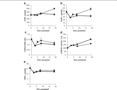
Liver GSH concentration, GSSG/GSH ratio and GSH-Px activity in sow-reared piglets did not significantly change (Fig. 7a, c and d). GSSG concentration decreased signifi-cantly from d0 to d3 (P< 0.05; Fig. 7b) in sow-reared piglets and returned to concentrations seen at birth afterwards (Fig. 7b). MDA concentration showed a sig-nificant increase after d3 in both LBW and NBW piglets (P< 0.05; Fig. 7e).
When piglets were introduced to a milk replacer, GSH (P< 0.05) and GSSG (P< 0.01) concentrations significantly decreased whereas GSSG/GSH ratio (P< 0.05) and MDA concentration (P< 0.001) significantly increased from d3 to d5. Meanwhile, GSH-Px activity remained unchanged. All redox parameters in the liver were stable from d5 to
0 2 4 6 8 10 12 14
0 5 10 15 20
G
S
H , µ
m
ol/g
Days postnatal
0 0.2 0.4 0.6 0.8 1
0 5 10 15 20
G
SSG
,
µ
m
ol/g
Days postnatal
0 0.05 0.1 0.15 0.2 0.25 0.3 0.35
0 5 10 15 20
G
SSG
/G
SH ratio
Days postnatal
0 2 4 6 8 10 12 14
0 5 10 15 20
G
S
H-P
x activity
,
U/g
Days postnatal
0 200 400 600 800 1000 1200 1400
0 5 10 15 20
MDA
, µ
m
ol/g
Days postnatal
a
b
c
d
e
d8 in artificially reared piglets, except GSSG/GSH ratio that showed a significant decrease (P< 0.01). Towards d19, GSH concentration (P< 0.01) and GSH-Px activity (P< 0.05) significantly increased while MDA concentra-tion (P< 0.01) significantly decreased in artificially reared piglets.
At d8, a lower GSH concentration and a higher MDA were observed in artificially reared piglets compared to sow-reared piglets (P< 0.001). At d19, higher GSH-Px activities were observed in artificially reared piglets com-pared to sow-reared piglets (P< 0.01).
Systemic redox state represented by GSH concentration, GSSG concentration, and GSSG/GSH ratio, GSH-Px activity and MDA concentration
The GSH-Px activity gradually, significantly increased dur-ing the investigated time frame in both sow- and artifi-cially reared piglets (P< 0.01; Fig. 8d). GSH concentration
in erythrocytes significantly increased in artificially reared piglets (P< 0.01) from d5 to d19, but remained constant in sow-reared piglets from d0 to d19 (Fig. 8a). Plasma MDA concentration decreased 6.77 μmol/g during the first 3 days of life in LBW piglets (P< 0.01; Fig. 8e). Plasma GSH-Px activity was significantly lower in artificially reared piglets compared to sow-reared piglets at d8 and d19 (P< 0.05). At d19, GSH-Px activity was still lower when LBW piglets were artificially reared (P< 0.05). Erythrocyte GSSG concentrations (P< 0.01; Fig. 8b) and GSSG/GSH ratio (P< 0.05; Fig. 8c) were significantly increased at d5 when artificially reared piglets were trans-ferred to a nursery at d3 (Fig. 8b and c). The concentra-tion of GSH (P< 0.01) and GSSG (P< 0.05) significantly increased in artificially reared piglets from d5 to d19. At d19, GSH concentration and GSSG concentration were significantly higher in artificially reared when compared to sow-reared piglets (P< 0.05). GSH concentration was
0 20 40 60 80 100 120 140
0 5 10 15 20
G
S
H , µ
m
ol/g
Days postnatal
0 2 4 6 8 10 12 14
0 5 10 15 20
G
SSG
,
µ
m
ol/g
Days postnatal
0 0.02 0.04 0.06 0.08 0.1 0.12 0.14 0.16
0 5 10 15 20
G
S
SG
/G
SH ratio
Days postnatal
0 5 10 15 20 25 30 35 40
0 5 10 15 20
G
S
H-P
x
activity , U/g
Days postnatal
0 0.2 0.4 0.6 0.8 1 1.2 1.4
0 5 10 15 20
MDA
, µ
m
ol/g
Days postnatal
a
b
c
d
e
significantly lower in LBW sow-reared piglets when com-pared to their NBW littermates. (P< 0.05).
Discussion
Given the need for an alternative rearing strategy that lowers the challenges that LBW and supernumerary pig-lets face during the suckling period, we aimed to investi-gate the responses to full artificial rearing. Our data demonstrated that artificial rearing beneficially affected piglet performance, notwithstanding impairing effects on small intestinal architecture, permeability and redox state in both LBW and NBW piglets.
Artificial rearing influences piglet performance
This study documents the implications of full artificial rearing of LBW and supernumerary piglets. Under standard rearing conditions these pigs are at high risk of succumbing due to insufficient nutrient intake, increased
disease susceptibility, and physiological deficits (e.g. lower energy reserves) [1]. Our study demonstrated that transitioning piglets to a nursery with ad libitum access to a milk replacer led to significantly higher body weights of LBW and NBW piglets compared to sow-reared piglets at d19. The experiment was terminated on the same day as when the conventionally reared piglets were weaned on the farm with a 3-week batch system. For this specific farm, the average age at weaning is 19.6 d. Furthermore, milk production of the sow strongly decreases towards the end of the suckling phase. Around d 18–19, sow milk starts to be very limit-ing and becomes hard to compare to the ad libitum ac-cess for the artificially reared piglets [43]. Using a weigh-suckle-weigh technique, De Vos et al. [26] showed that piglets with ad libitum access to milk replacer have a higher relative energy intake compared to sow-reared piglets [28]. Our findings confirm this and other
0 100 200 300 400 500 600
0 5 10 15 20
G
S
H , µ
m
ol/g
Days postnatal
0 10 20 30 40 50 60 70
0 5 10 15 20
G
SSG
,
µ
m
ol/g
Days postnatal
0 0.05 0.1 0.15 0.2
0 5 10 15 20
GS
S
G
/GS
H
ratio
Days postnatal
0 50 100 150 200 250 300
0 5 10 15 20
G
S
H-P
x
activity , U/g
Days postnatal
0 5 10 15 20
0 5 10 15 20
MDA
, µ
m
ol/g
Days postnatal
b
a
c
d
e
previous research where LBW piglets receiving an en-ergy rich diet—comparable with our LBW piglets fed a milk replacer ad libitum—presented a comparable body weight gain as NBW piglets receiving a lower energy intake—comparable with our NBW piglets fed by the sow [44]. In addition, the milk replacer used in our study contained spray-dried plasma which could have contrib-uted further to the higher weight gain in the artificially reared group. Ermer et al. [45] showed that spray-dried porcine plasma increased feed intake. Thus next to ad libitum access to feed, diet composition cannot be neglected.
Artificial rearing influences small intestinal architecture Small intestinal morphology is one of the major indica-tors reflecting gut health in pigs [46]. However, caution should be taken when evaluating morphology alone as a measure of gut health. For example, Enterotoxigenic
Escherichia coli, the major causal agent of neonatal diar-rhea, may occur without histological changes in the in-testine [47]. Notwithstanding, stereological analysis of small intestinal morphology will provide the most accur-ate estimation of the intestinal absorptive surface area [48], a proxy of the surface can be calculated using villus height and villus width [47]. In our study, the mucosal surface area of the proximal small intestine at d19 was markedly larger in artificially reared piglets than sow-reared piglets. In this regard, feeding a milk replacer shows promise as the increased mucosal surface suggests a higher ability to absorb nutrients. Moreover, previous research showed an increased activity of maltase and su-crase when piglets are fed a milk replacer [49–51]. Thus, artificial rearing seems to improve the digestive capacity at the level of the small intestine.
The deepening of the crypts could be a response to promote mucus secretion rather than lead to enterocyte maturation and proliferation and thus an increase in PCNA expression. Previous research showed that breast-fed infants showed a delay in the mucin degradation when compared to artificially reared infants [52]. Phillips [53] showed that crypt goblet cells have the ability to restitute the mucus layer and showed a decrease in the percentage of villus epithelial volume occupied by mucin secretory granules.
Transferring piglets to a nursery at d3 exposed them to stressful effects caused by psychological, environ-mental or nutritional factors similar to those encoun-tered during the conventional weaning process [54]. However, it is difficult to unravel the separate contri-butions of these factors. Previous studies showed sig-nificant villus atrophy at d4 and deeper crypts at d7 in piglets that were separated from the sow and still fed sow’s milk compared to unweaned piglets [55, 56]. Similarly, our study showed transient villus atrophy in
piglets transferred to a nursery at d3. This villus height reduction is analogue to the intestinal morpho-logical changes as a result of inadequate food intake immediately after conventional weaning [56–59]. In contrast to conventional weaning, villus length is rap-idly restored. This could be related to the inclusion of spray-dried plasma in the milk replacer since this is known to increase villus height [8, 60–63].
Artificial rearing affects small intestinal tight junction protein expression
We hypothesized that artificial rearing influences small intestinal physiology. The intestinal epithelium plays a critical role in the transport of nutrients and macromol-ecules. At the same time, it has to provide an effective barrier to harmful macromolecules and microorganisms [64]. Epithelial cells constitute a dynamic barrier where large molecules can be transported by transcytosis and this can be measured by HRP [65]. Tight junctions (TJs) are essential components of the physical intercellular barrier and their presence and functionality changes under different physiological and pathological conditions [66, 67]. Well-formed TJs are characterized by low sol-ute permeability which can be determined by measuring FD-4 permeability [65]. The family of junctional adhe-sion molecules, the claudin and occludin families, are structural transmembrane TJ components that have the potential to mediate cell–cell adhesion [66, 68]. Within TJs, claudins are the main determinants of the selective pore properties [68, 69], while the role of occludin in barrier functioning is more diverse [70, 71]. In mice, the expression of claudin-3 is promoted during the first 3 weeks of life concomitant with the establishment of the intestinal microbiota [72, 73]. Our study showed that claudin-3 expression rose particularly in artificially reared piglets. Possibly the different microbiota finger-print [74] and the absence of milk born IgA’s [34] can be held responsible. Claudin-3 is known to be a“tightening” claudin [75]. This could explain why permeability is seemingly unaffected by the observed increase in claudin-3. However, at the start of artificial rearing, claudin-3 as well as occludin protein levels transiently dropped. This drop is reflected in a concomitantly in-creased permeability for FD-4 and HRP. Previous studies showed lower abundances of occludin mRNA and pro-tein, claudin-3 propro-tein, and increased lactulose perme-ability after weaning [50, 76]. Thus artificial rearing induced a similar response as seen after conventional weaning.
Artificial rearing resulted in a redox imbalance
suckling [21]. Vergauwen et al. [12] and others showed a redistribution of TJ proteins during times of imposing reactive species and could relate these responses to a compromised permeability [12, 77]. The current study demonstrated that transfer of piglets to a nursery re-sulted in oxidative stress.
Glutathione (GSH) is an important regulator of the redox status within intestinal epithelial cells [13]. The liver is the major site of GSH biosynthesis and exports GSH via the bile to the proximal SI [78]. Thus GSH ori-ginating from the liver supports mucosal GSH by decreasing lipid peroxidation and maintaining the GSSG/GSH redox homeostasis in the proximal intestine [11, 79, 80]. In our study, artificial rearing resulted in the liver in a decreased GSH content, an increased GSSG/ GSH ratio and MDA concentration. After this first phase, the redox parameters returned to their initial values. GSH-Px activity in the liver seemed unaffected during the transition period, while GSH-Px activity tran-siently dropped in the proximal and distal SI when piglets were introduced to a nursery. Previously, conven-tional weaning caused a drop in GSH-Px activity but GSH-Px activity increased afterwards as part of a feed-back mechanism [31]. Our study shows that artificial rearing increases oxidative stress in the SI and as a result the GSH-Px activity increased and was higher than the activity seen in sow-reared piglets. Furthermore, LBW piglets showed a higher GSH-Px activity com-pared to their NBW littermates. It is clear that the antioxidant capacity of the liver helps protecting the proximal intestine by secreting GSH into the lumen of proximal SI [78, 79]. This could explain the in-creased mucosal GSH concentration of the proximal SI and the concomitant massive decrease of the liver GSH concentration. On the other hand, a remarkable drop of the GSH concentration in the distal SI was observed from d3 until d5. Consequently, this re-sulted in a high concentration of MDA and GSSG, resulting in a higher GSSG/GSH ratio in the distal SI. Furthermore, the GSSG/GSH ratio increased systemic-ally upon transfer to a nursery. Degroote et al. [31] already showed that conventional weaning increased the GSSG/GSH ratio.
Our research presents a window of opportunity for antioxidant supplementation to protect piglets from redox imbalance due to artificial rearing. NBW artifi-cially reared piglets were more susceptible to redox im-balance and loss of intestinal integrity upon transferal to a nursery (from d3 to d5). Taken together, these findings favor the transfer of both LBW and NBW piglets to a nursery as a solution for LBW and supernumerary pig-lets. Further research is necessary to elucidate how these artificially reared LBW and NBW piglets will respond to the introduction to solid food.
Conclusions
In conclusion, we demonstrated that artificial rearing in-fluences morphological and functional parameters in the small intestine, liver and blood in a way similar to what is seen after conventional weaning. Nevertheless, growth performance of artificially reared piglets was positively influenced. In addition, artificially reared piglets rapidly recovered from redox imbalances and restored intestinal permeability within a couple of days. Further research is needed to explore the possibility to supplement LBW and NBW piglets with antioxidants prior to initiating artificial rearing. Thus, artificial rearing is a valuable alternative to raise LBW or supernumerary piglets.
Abbreviations
ELISA:Enzyme linked immunosorbent assay; FD-4: Fluorescein isothiocyanate dextran 4 kDa; GSH: Glutathione; GSH-Px: Glutathione peroxidase; GSSG: Oxidized glutathione; HPLC: High performance liquid
chromatography; HRP: Horseradish peroxidase; LBW: Low birth weight; MDA: Malondialdehyde; NBW: Normal birth weight; SI: Small intestine; TJs: Tight junctions
Acknowledgments
We thank K. Huybrechts, G. Vrolix, K. Jennes, M. De Reys, S. Coolsaet, A. Ovyn and T. Van der Eecken for their technical assistance.
Funding
This work was supported by a grant from the government agency for Innovation by Science and Technology (IWT-LO 100856).
Availability of data and materials
The datasets supporting the conclusions of this article are included within the article.
Authors’contributions
Author contributions: CVG, JM and SDM concept and design of research; HV, JDG, WW, CVG and JM performed experiments; HV and JDG analyzed the data; HV and EF design and implementation of the statistical model; HV, JDG, CVG and JM interpreted the results of the experiments; HV and CVG prepared the figures; HV and CVG drafted manuscript; HV, JDG, CVG, JM, SDS, WW, CC, SVC and SP edited and revised the manuscript; CVG and JM approved the final version of the manuscript. All authors read and approved the final manuscript.
Competing interests
The authors declare that they have no competing interests.
Consent for publication Not applicable.
Ethics approval
All husbandry and experimental procedures were approved by the Ethical Committee for Animal Testing (ECD) of the University of Antwerp (Belgium) (EC number: 2015-02) in accordance with the European Commission directive for the humane care and use of animals in research (2010/63/EU).
Author details
1Laboratory of Applied Veterinary Morphology, Department of Veterinary
Received: 5 September 2016 Accepted: 16 March 2017
References
1. Lay Jr DC, Matteri RL, Carrol JA, Fangman TJ, Safranski TJ. Preweaning survival in swine. J Anim Sci. 2002;80(E. Suppl. 1):E74–86.
2. Tuchscherer M, Puppe B, Tuchscherer A, Tiemann U. Early identification of neonates at risk: traits of newborn piglets with respect to survival. Theriogenology. 2000;54(3):371–88. doi:10.1016/s0093-691x(00)00355-1. 3. Milligan BN, Dewey CE, de Grau AF. Neonatal-piglet weight variation and its
relation to pre-weaning mortality and weight gain on commercial farms. Prev Vet Med. 2002;56(2):119–27.
4. Friel JK, Diehl-Jones B, Cockell KA, Chiu A, Rabanni R, Davies SS, et al. Evidence of oxidative stress in relation to feeding type during early life in premature infants. Pediatr Res. 2011;69(2):160–4. doi:10.1203/PDR. 0b013e3182042a07.
5. Ahola T, Levonen AL, Fellman V, Lapatto R. Thiol metabolism in preterm infants during the first week of life. Scand J Clin Lab Invest. 2004;64(7):649–58. doi:10.1080/00365510410002959.
6. Rook D, Te Braake FW, Schierbeek H, Longini M, Buonocore G, Van Goudoever JB. Glutathione synthesis rates in early postnatal life. Pediatr Res. 2010;67(4):407–11. doi:10.1203/PDR.0b013e3181d22cf6. 7. Yin J, Ren W, Liu G, Duan J, Yang G, Wu L, et al. Birth oxidative stress and
the development of an antioxidant system in newborn piglets. Free Radic Res. 2013;47(12):1027–35. doi:10.3109/10715762.2013.848277.
8. Tran H, Bundy JW, Li YS, Carney-Hinkle EE, Miller PS, Burkey TE. Effects of spray-dried porcine plasma on growth performance, immune response, total antioxidant capacity, and gut morphology of nursery pigs. J Anim Sci. 2014;92(10):4494–504. doi:10.2527/jas.2014-7620.
9. Michiels J, De Vos M, Missotten J, Ovyn A, De Smet S, Van Ginneken C. Maturation of digestive function is retarded and plasma antioxidant capacity lowered in fully weaned low birth weight piglets. Br J Nutr. 2013;109(1):65–75. doi:10.1017/s0007114512000670.
10. Krueger R, Derno M, Goers S, Metzler-Zebeli BU, Nuernberg G, Martens K, et al. Higher body fatness in intrauterine growth retarded juvenile pigs is associated with lower fat and higher carbohydrate oxidation during ad libitum and restricted feeding. Eur J Nutr. 2014;53(2):583–97. doi:10.1007/ s00394-013-0567-x.
11. Aw TY. Intestinal glutathione: determinant of mucosal peroxide transport, metabolism, and oxidative susceptibility. Toxicol Appl Pharmacol. 2005;204(3):320–8. doi:10.1016/j.taap.2004.11.016.
12. Vergauwen H, Tambuyzer B, Jennes K, Degroote J, Wang W, De Smet S, et al. Trolox and ascorbic acid reduce direct and indirect oxidative stress in the IPEC-J2 cells, an in vitro model for the porcine gastrointestinal tract. PLoS One. 2015;10(3):e0120485. doi:10.1371/journal.pone.0120485.
13. Kelly FJ. Glutathione content of the small intestine: regulation and function. Br J Nutr. 1993;69(2):589–96.
14. Carrasco-Pozo C, Morales P, Gotteland M. Polyphenols Protect the Epithelial Barrier Function of Caco-2 Cells Exposed to Indomethacin through the Modulation of Occludin and Zonula Occludens-1 Expression. Journal of agricultural and food chemistry. 2013. doi:10.1021/jf400150p.
15. Bhattacharyya A, Chattopadhyay R, Mitra S, Crowe SE. Oxidative stress: an essential factor in the pathogenesis of gastrointestinal mucosal diseases. Physiol Rev. 2014;94(2):329–54. doi:10.1152/physrev.00040.2012. 16. Wang J, Chen L, Li D, Yin Y, Wang X, Li P, et al. Intrauterine growth
restriction affects the proteomes of the small intestine, liver, and skeletal muscle in newborn pigs. J Nutr. 2008;138(1):60–6.
17. Wang X, Wu W, Lin G, Li D, Wu G, Wang J. Temporal proteomic analysis reveals continuous impairment of intestinal development in neonatal piglets with intrauterine growth restriction. J Proteome Res. 2010;9(2):924–35. doi:10.1021/pr900747d.
18. Mickiewicz M, Zabielski R, Grenier B, Le Normand L, Savary G, Holst JJ, et al. Structural and functional development of small intestine in intrauterine growth retarded porcine offspring born to gilts fed diets with differing protein ratios throughout pregnancy. J Physiol Pharmacol. 2012;63(3):225–39.
19. Ferenc K, Pietrzak P, Godlewski MM, Piwowarski J, Kilianczyk R, Guilloteau P, et al. Intrauterine growth retarded piglet as a model for humans–studies on the perinatal development of the gut structure and function. Reprod Biol. 2014;14(1):51–60. doi:10.1016/j.repbio.2014.01.005.
20. D’Inca R, Gras-Le Guen C, Che L, Sangild PT, Le Huerou-Luron I. Intrauterine growth restriction delays feeding-induced gut adaptation in term newborn pigs. Neonatology. 2011;99(3):208–16. doi:10.1159/000314919.
21. Wang W, Degroote J, Van Ginneken C, Van Poucke M, Vergauwen H, Dam TM et al. Intrauterine growth restriction in neonatal piglets affects small intestinal mucosal permeability and mRNA expression of redox-sensitive genes. FASEB journal : official publication of the Federation of American Societies for Experimental Biology. 2015. doi:10.1096/fj.15-274779. 22. Huygelen V, De Vos M, Willemen S, Tambuyzer B, Casteleyn C, Knapen D, et al. Increased intestinal barrier function in the small intestine of formula-fed neonatal piglets. J Anim Sci. 2012;90 Suppl 4:315–7.
doi:10.2527/jas.53731.
23. Huygelen V, De Vos M, Willemen S, Fransen E, Casteleyn C, Van Cruchten S, et al. Age-related differences in mucosal barrier function and morphology of the small intestine in low and normal birth weight piglets. J Anim Sci. 2014;92(8):3398–406. doi:10.2527/jas.2014-7742.
24. Willemen S, Che L, De Vos M, Huygelen V, Tambuyzer B, Casteleyn C, et al. Perinatal growth restriction is not related to higher intestinal distribution and increased serum levels of 5-hydroxytryptamin in piglets. J Anim Sci. 2012;90 Suppl 4:305–7. doi:10.2527/jas.53730.
25. Ferrari CV, Sbardella PE, Bernardi ML, Coutinho ML, Vaz Jr IS, Wentz I, et al. Effect of birth weight and colostrum intake on mortality and performance of piglets after cross-fostering in sows of different parities. Prev Vet Med. 2014;114(3-4):259–66. doi:10.1016/j.prevetmed.2014.02.013.
26. De Vos M, Che L, Huygelen V, Willemen S, Michiels J, Van Cruchten S, et al. Nutritional interventions to prevent and rear low-birthweight piglets. J Anim Physiol Anim Nutr. 2014;98(4):609–19. doi:10.1111/jpn.12133. 27. Donovan TS, Dritz SS. Effect of split nursing on variation in pig growth from
birth to weaning. J Am Vet Med Assoc. 2000;217(1):79–81.
28. De Vos M, Huygelen V, Willemen S, Fransen E, Casteleyn C, Van Cruchten S, et al. Artificial rearing of piglets: Effects on small intestinal morphology and digestion capacity. Livest Sci. 2014;159:165–73. doi:10.1016/j.livsci. 2013.11.012.
29. Wedig J, Christian MS, Hoberman A, Diener RM, Thomas-Wedig R. A study to develop methodology for feeding 24-hour-old neonatal swine for 3 weeks. Int J Toxicol. 2002;21(5):361–70. doi:10.1080/10915810290096577. 30. Fiorotto ML, Reeds PJ, Cunningham JJ, Pond WG. A semiautomatic
device for feeding liquid milk-replacer diets to infant pigs. J Anim Sci. 1993;71(1):78–85.
31. Degroote J, Michiels J, Claeys E, Ovyn A, De Smet S. Changes in the pig small intestinal mucosal glutathione kinetics after weaning. J Anim Sci. 2012;90 Suppl 4:359–61. doi:10.2527/jas.53809.
32. Zhu LH, Zhao KL, Chen XL, Xu JX. Impact of weaning and an antioxidant blend on intestinal barrier function and antioxidant status in pigs. J Anim Sci. 2012;90(8):2581–9. doi:10.2527/jas.2012-4444.
33. Sauerwein H, Schmitz S, Hiss S. The acute phase protein haptoglobin and its relation to oxidative status in piglets undergoing weaning-induced stress. Redox Report. 2005;10(6):295–302. doi:10.1179/135100005x83725. 34. Klobasa F, Werhahn E, Butler JE. Composition of sow milk during lactation.
J Anim Sci. 1987;64(5):1458–66.
35. Csapo J, Martin TG, Csapo-Kiss ZS, Hazas Z. Protein, fats, vitamin and mineral concentration in porcine colostrum and milk from parturition to 60 days. Int Dairy J. 1996;6(8-9):881–902.
36. Ontsouka CE, Bruckmaier RM, Blum JW. Fractionized milk composition during removal of colostrum and mature milk. J Dairy Sci. 2003;86(6):2005–11. doi:10.3168/jds.S0022-0302(03)73789-8.
37. Buesa RJ, Peshkov MV. How much formalin is enough to fix tissues? Ann Diagn Pathol. 2012;16(3):202–9. doi:10.1016/j.anndiagpath.2011.12.003. 38. McKie AT, Zammit PS, Naftalin RJ. Comparison of cattle and sheep colonic
permeabilities to horseradish peroxidase and hamster scrapie prion protein in vitro. Gut. 1999;45(6):879–88.
39. Yoshida T. Determination of reduced and oxidized glutathione in erythrocytes by high-performance liquid chromatography with ultraviolet absorbance detection. J Chromatogr B Biomed Appl. 1996;678(2):157–64. 40. Reed DJ, Babson JR, Beatty PW, Brodie AE, Ellis WW, Potter DW.
High-performance liquid chromatography analysis of nanomole levels of glutathione, glutathione disulfide, and related thiols and disulfides. Anal Biochem. 1980;106(1):55–62.
42. Grotto D, Santa Maria LD, Boeira S, Valentini J, Charao MF, Moro AM, et al. Rapid quantification of malondialdehyde in plasma by high performance liquid chromatography-visible detection. J Pharm Biomed Anal. 2007;43(2):619–24. doi:10.1016/j.jpba.2006.07.030.
43. Aguinaga MA, Gomez-Carballar F, Nieto R, Aguilera JF. Production and composition of Iberian sow’s milk and use of milk nutrients by the suckling Iberian piglet. Animal. 2011;5(9):1390–7. doi:10.1017/s1751731111000474. 44. Han F, Hu L, Xuan Y, Ding X, Luo Y, Bai S, et al. Effects of high nutrient
intake on the growth performance, intestinal morphology and immune function of neonatal intra-uterine growth-retarded pigs. Br J Nutr. 2013;110(10):1819–27. doi:10.1017/s0007114513001232.
45. Ermer PM, Miller PS, Lewis AJ. Diet preference and meal patterns of weanling pigs offered diets containing either spray-dried porcine plasma or dried skim milk. J Anim Sci. 1994;72(6):1548–54.
46. Caspary WF. Physiology and pathophysiology of intestinal absorption. Am J Clin Nutr. 1992;55(1 Suppl):299s–308s.
47. Egberts HJ, de Groot EC, van Dijk JE, Vellenga L, Mouwen JM. Tight junctional structure and permeability of porcine jejunum after enterotoxic Escherichia coli infection. Res Vet Sci. 1993;55(1):10–4.
48. Van Ginneken C, Van Meir F, Weyns A. Stereologic characteristics of pig small intestine during normal development. Dig Dis Sci. 2002;47(4):868–78. 49. Cera KR, Mahan DC, Reinhart GA. Effect of weaning, week postweaning and
diet composition on pancreatic and small intestinal luminal lipase response in young swine. Journal of animal science. 1990;68(2). doi:10.2527/1990. 682384x.
50. Wang J, Zeng L, Tan B, Li G, Huang B, Xiong X, et al. Developmental changes in intercellular junctions and Kv channels in the intestine of piglets during the suckling and post-weaning periods. J Anim Sci Biotechnol. 2016;7:4. doi:10.1186/s40104-016-0063-2.
51. Huygelen V, De Vos M, Prims S, Vergauwen H, Fransen E, Casteleyn C, et al. Birth weight has no influence on the morphology, digestive capacity and motility of the small intestine in suckling pigs. Livest Sci. 2015;182:129–36. http://dx.doi.org/doi:10.1016/J.LIVSCI.2015.11.003.
52. Midtvedt AC, Carlstedt-Duke B, Midtvedt T. Establishment of a mucin-degrading intestinal microflora during the first two years of human life. J Pediatr Gastroenterol Nutr. 1994;18(3):321–6.
53. Phillips TE. Both crypt and villus intestinal goblet cells secrete mucin in response to cholinergic stimulation. Am J Physiol. 1992;262(2 Pt 1):G327–31. 54. Funderburke DW, Seerley RW. The effects of postweaning stressors on pig
weight change, blood, liver and digestive tract characteristics. J Anim Sci. 1990;68(1):155–62.
55. van Beers-Schreurs HM, Nabuurs MJ, Vellenga L, Kalsbeek-van der Valk HJ, Wensing T, Breukink HJ. Weaning and the weanling diet influence the villous height and crypt depth in the small intestine of pigs and alter the concentrations of short-chain fatty acids in the large intestine and blood. J Nutr. 1998;128(6):947–53.
56. Kelly D, Smyth JA, McCracken KJ. Digestive development of the early-weaned pig. 1. Effect of continuous nutrient supply on the development of the digestive tract and on changes in digestive enzyme activity during the first week post-weaning. Br J Nutr. 1991;65(2):169–80.
57. Kelly D, Smyth JA, McCracken KJ. Digestive development of the early-weaned pig. 2. Effect of level of food intake on digestive enzyme activity during the immediate post-weaning period. Br J Nutr. 1991;65(2):181–8. 58. Spreeuwenberg MA, Verdonk JM, Gaskins HR, Verstegen MW. Small intestine
epithelial barrier function is compromised in pigs with low feed intake at weaning. J Nutr. 2001;131(5):1520–7.
59. Hedemann MS, Hojsgaard S, Jensen BB. Small intestinal morphology and activity of intestinal peptidases in piglets around weaning. J Anim Physiol Anim Nutr. 2003;87(1-2):32–41.
60. Pierce JL, Cromwell GL, Lindemann MD, Russell LE, Weaver EM. Effects of spray-dried animal plasma and immunoglobulins on performance of early weaned pigs. J Anim Sci. 2005;83(12):2876–85.
61. Touchette KJ, Allee GL, Matteri RL, Dyer CJ, Carroll JA. Effect of spray-dried plasma and Escherichia coli on intestinal morphology and the hypothalamic–pituitary–adrenal (HPA) axis of the weaned pig. J Anim Sci. 1999;77(Supplement 1):56.
62. Jiang R, Chang X, Stoll B, Fan MZ, Arthington J, Weaver E, et al. Dietary plasma protein reduces small intestinal growth and lamina propria cell density in early weaned pigs. J Nutr. 2000;130(1):21–6.
63. van Dijk AJ, Niewold TA, Margry RJ, van den Hoven SG, Nabuurs MJ, Stockhofe-Zurwieden N, et al. Small intestinal morphology in weaned
piglets fed a diet containing spray-dried porcine plasma. Res Vet Sci. 2001;71(1):17–22. doi:10.1053/rvsc.2001.0478.
64. Ramanan D, Cadwell K. Intrinsic Defense Mechanisms of the Intestinal Epithelium. Cell host & microbe. 2016. doi:10.1016/j.chom.2016.03.003. 65. Boudry G. The Ussing chamber technique to evaluate alternatives to in-feed
antibiotics for young pigs. Animal Res. 2005;54:219–30.
66. Schneeberger EE, Lynch RD. The tight junction: a multifunctional complex. Am J Physiol Cell Physiol. 2004;286(6):C1213–28. doi:10.1152/ajpcell.00558.2003. 67. Lu Z, Ding L, Lu Q, Chen Y-H. Claudins in intestines: distribution and functional
significance in health and diseases. Tissue Barriers. 2013;1(3):e24978. doi:10.4161/tisb.24978.
68. Anderson JM, Van Itallie CM, Fanning AS. Setting up a selective barrier at the apical junction complex. Curr Opin Cell Biol. 2004;16(2):140–5. doi:10.1016/j.ceb. 2004.01.005.
69. Furuse M, Tsukita S. Claudins in occluding junctions of humans and flies. Trends Cell Biol. 2006;16(4):181–8. doi:10.1016/j.tcb.2006.02.006. 70. Yu AS, McCarthy KM, Francis SA, McCormack JM, Lai J, Rogers RA, et al.
Knockdown of occludin expression leads to diverse phenotypic alterations in epithelial cells. Am J Physiol Cell Physiol. 2005;288(6):C1231–41. doi:10.1152/ajpcell.00581.2004.
71. Raleigh DR, Boe DM, Yu D, Weber CR, Marchiando AM, Bradford EM, et al. Occludin S408 phosphorylation regulates tight junction protein interactions and barrier function. J Cell Biol. 2011;193(3):565–82. doi:10.1083/jcb. 201010065.
72. Patel RM, Myers LS, Kurundkar AR, Maheshwari A, Nusrat A, Lin PW. Probiotic bacteria induce maturation of intestinal claudin 3 expression and barrier function. Am J Pathol. 2012;180(2):626–35. doi:10.1016/j. ajpath.2011.10.025.
73. Holmes JL, Van Itallie CM, Rasmussen JE, Anderson JM. Claudin profiling in the mouse during postnatal intestinal development and along the gastrointestinal tract reveals complex expression patterns. Gene Expression Patterns. 2006;6(6):581–8. doi:10.1016/j.modgep.2005.12.001.
74. Mackie RI, Sghir A, Gaskins HR. Developmental microbial ecology of the neonatal gastrointestinal tract. Am J Clin Nutr. 1999;69(5):1035s–45s. 75. Milatz S, Krug SM, Rosenthal R, Gunzel D, Muller D, Schulzke JD, et al. Claudin-3
acts as a sealing component of the tight junction for ions of either charge and uncharged solutes. Biochim Biophys Acta. 2010;1798(11):2048–57. doi:10.1016/j. bbamem.2010.07.014.
76. Wang H, Zhang C, Wu G, Sun Y, Wang B, He B, et al. Glutamine enhances tight junction protein expression and modulates corticotropin-releasing factor signaling in the jejunum of weanling piglets. J Nutr. 2015;145(1):25–31. doi:10.3945/jn.114.202515.
77. Fukui A, Naito Y, Handa O, Kugai M, Tsuji T, Yoriki H et al. Acetyl salicylic acid induces damage to intestinal epithelial cells by oxidation-related modifications of ZO-1. American journal of physiology Gastrointestinal and liver physiology. 2012. doi:10.1152/ajpgi.00236.2012.
78. Lee TK, Li L, Ballatori N. Hepatic glutathione and glutathione S-conjugate transport mechanisms. Yale J Biol Med. 1997;70(4):287–300.
79. Aw TY. Biliary glutathione promotes the mucosal metabolism of luminal peroxidized lipids by rat small intestine in vivo. J Clin Investig. 1994;94(3):1218–25.
80. Ballatori N, Truong AT. Relation between biliary glutathione excretion and bile acid-independent bile flow. Am J Physiol. 1989;256(1 Pt 1):G22–30.
• We accept pre-submission inquiries
• Our selector tool helps you to find the most relevant journal
• We provide round the clock customer support
• Convenient online submission
• Thorough peer review
• Inclusion in PubMed and all major indexing services
• Maximum visibility for your research
Submit your manuscript at www.biomedcentral.com/submit