(UDC: 616.833.2-085 ; 602.9)
Finite element modeling of axonal elongation and use of stem cells
M. Obradovic1,*, S. Novak2, H. Jorg Meisel3, A. Dinnyes4, N. Filipovic1,5
1Faculty of Engineering, University of Kragujevac, Sestre Janjica 6, 34000 Kragujevac, Serbia
2J. Stefan Institute, Jamova cesta 39, 1000 Ljubljana, Slovenia.
3Universität Leipzig (ULEI), Translational Centre for Regenerative Medicine Leipzig (TRM),
Goethestraße 6, 04109Leipzig. [email protected]
4Biotalentum Ltd (BIOT), Aulich Lajos str. 26, 2100 Gödöllő, Hungary.
5
Bioengineering Research and Development Center, Prvoslava Stojanovica 6, 34000 Kragujevac, Serbia.
*Corresponding author
Abstract
The basic function of an axon is to conduct electrical impulses away from the cell body, using special molecular structures. Axons move through their environment via the growth cone, which is placed on the top of the axon, and where the mass is added. When an axon is damaged, there is no information flow. Using stem cells we can accomplish axon repair. Besides stem cells growth, mechanical tension leaves impact on the axonal elongation improvement. We modeled axonal elongation using finite element method. If we apply force at the growth cone with mass adding, the axon will be considered as a material with viscoelastic properties. To achieve a nonlinear elongation along the axon and to include a viscoelastic material in our finite element model, we made a function of Young‘s modulus along the axon. The results show that the axon has a nonlinear elongation as a viscoelastic material. Mass adding is considered as a change of material concentration (diffusion equation). The current model with axon as a viscoelastic material and the calculation of axonal elongation using the diffusion equation is a good approach to an appropriate model of axon. We also numerically analyzed the behavior of stem cells inside the scaffold mixed with hydrogel and collagen fibers in order to simulate nerve repair which goes to the spinal cord. Our goal is to create a model which will give more information about the processes of axon healing and growing, with a good match in comparison with the experimental results.
Keywords: Neuron, Finite element method, stem cells, growth cone
1. Introduction
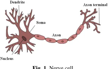
and an axon, beside length, is that the axon transmits signals, while dendrites receive signals from axons of other nerve cells, creating on that way a neural network. Therefore, the basic function of an axon is to conduct electrical impulses away from the neuron‘s cell body. Fig. 1 shows the structure of a nerve cell.
Fig. 1. Nerve cell.
An axon makes contact with other cells at junctions called synapses, where the membrane of the axon closely adjoins the membrane of the target cells, and special molecular structures serve to transmit electrical or electrochemical signals across the gap (http://en.wikipedia.org/wiki/Axon). Axon dysfunction causes neurological disorders which can affect both the peripheral and central neurons. When an axon is damaged there is no conduction of information from one part of the body to another. The axon needs to be repaired in order to provide the information flow.
Nerve cells are rarely regenerative cells. Repair of axon is possible to accomplish when axon has small cut, a few millimeters long. It can be done by extension and elongation, applying external force or adding material. When the rupture in connection is larger, about 1cm and more, it is hard to extend and to connect the axon. Currently, researches are trying to regenerate and repair a long rupture of the axon based on the stem cells application. Stem cells can produce bioactive molecules, stimulating axon regeneration, and some of them can differentiate into Schwann cells, which are the principal glia of the peripheral nervous system. The most important role of Schwann cells is to provide the conduction of nerve impulses along the axon.
The pluripotent stem cells can reduce and modulate the reaction of the immune system following injury.
The peripheral nerve repair can be enhanced by Schwann cells transplantation. Clinical application is limited by donor site morbidity, and it is difficult to get a sufficient number of cells quickly (Kingham P. J. et al., 2007).
Mesenchymal stem cells (MSC) can differentiate into Schwann cells, so they are an attractive cell source for the regeneration of nerve tissue. They are able to self-renew, have a high growth rate and are multi-potent differential stem cells, but they are collected from the bone marrow, which is a highly invasive and painful procedure. Beside them, adipose tissue has been identified as possessing a population of multi-potent stem cells.
Adipose-derived stem cells (ADSC) have the phenotypic and gene expression profiles similar to MSC. Unlike mesenchymal stem cells, they can be easily isolated by conventional liposuction procedures.
The ability of ADSC to promote neurite outgrowth was determined by examining their interaction with NG108-15 cells – a motor neuron-like cell line.
It was found experimentally (Kingham P. J. et al., 2007) that rat ADSC treated with a mixture of glial growth factors expressed GFAP, S100 and p75 proteins and enhanced neurite outgrowth in vitro, suggesting on that way the transition to a Schwann cell phenotype.
The conclusion is that adipose tissue possesses a lot of regenerative stem cells, which can be used to differentiate into Schwann cells, in the certain circumstances, and may be of benefit for the treatment of peripheral nerve injuries.
Damage of motoneurons of the spinal cord causes immobility, and break in connection is usually large, a few centimeters long. Human stem cells and Schwann-cells placed in chitosan tube, filled with hydrogel and collagen fibers, should be a good combination for a sufficient support and guidance for the motoneurons of the spinal cord in order to repair and accomplish nerve function after injury. This is a new investigation in the field of axon repair of motoneurons.
The paper is organized as following. First, we present methods for axon elongation with and without applied mechanical tension. Stem cells for repair of the axon that goes to the spinal cord are described as well. In the results section we give some numerical examples of axon elongation and comparison with experimental data. Only numerical results for the stem cells behavior inside the scaffold are described. At the end, some concluding remarks are given.
2. Materials and Methods
Axons move through their environment via the growth cone, which is placed on the top of the axon, and where the mass is added. Through the cytoplasm of the axon, the axoplasm, mitochondria, lipids, proteins and other organelles are moving to and from the cell body of a neuron. That is a cellular process called axoplasmic or axonal transport, which is responsible for the growth of neurites (axon and dendrites). Besides that, mechanical tension leaves impact on axonal elongation improvement.
2.1. Elongation of axon with applied mechanical tension
We create a model of an axon and calculate axonal elongation using finite element method. The axon is modeled as a material with viscoelastic properties (O‘Toole M. et al., 2007). To accomplish a nonlinear elongation through the axon and to include a viscoelastic material in our finite element model, we made a function of Young‘s modulus along the axon, according to Equation 1:
0
cosh( ( ) / )
( , ( )) ,
cosh( / )
L t G
E x L t G gA
F x G
(1) 0
9.6 constant of friction, interaction between the axon and substrate
3611.11 axson's axial viscosity constant force at growth cone
current position at axon ( ) maximal length of axon
g m s g g m s F x L t
cross-sectional area of axon
Applied force at the growth cone was 200 µdyn, the initial length of the axon was 200 µm, and radius 1µm. In this case we considered that axonal thinning is negligible (G=const).
Obtained results have shown that the axon has a nonlinear elongation.
2.2. Elongation of axon without applied mechanical tension
Elongation of the axon can be understood as a function of the production, transport, assembly and disassembly of tubulins into microtubules. Tubulins are proteins that make up microtubules (Fig. 2).
Fig. 2. The biology of neurite outgrowth.
The axon elongates at the growth tip as a result of a change in the tubulin concentration along the axon. The governing equation of neurite elongation is (McLean D. et al., 2004):
2 2
c c c
a D gc
t x x
(2)
where D is the diffusion of tubulin, a is an active transport, g is a constant rate of tubulin degradation, and c is the concentration of tubulin along the length of axon.
In the beginning model x=0 represents the soma/neurite interface and x=l length of neurite. Numerically solution is complicated by the fact that spatial domain changes over time due to changes in the length (Graham B. P. and van OOyen A., 2006).
The length of neurite is a function of time (ODE):
| , (0) 0
dl
r cg sg l l
x l
dt
(3)
elongation rate
s retraction rate rg
g
Boundary condition includes a steady flux of tubulin into the domain at x=0, and a flux at x=l(t), which is proportional to the quantity of tubulin there.
0 0| 0
Here and represent a flux-source rate and a flux-sink rate, respectively, is
microtubule disassembly rate and is tubulin scale.
3. Results and Discussion
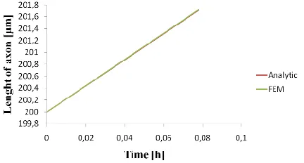
When an axon is described as a material with viscoelastic properties, a nonlinear elongation of the axon along is obtained. Elongation increases as the current position at axon, x, is further away from nerve cell body. The obtained results are compared with the experimental measurement from Matthew O‘Toole et al., 2007, where they trace docked mitochondria along the axon under same condition for applied force at the growth cone (Fig. 3).
Fig. 3. Elongation along the axon (simulation time 280s).
Mechanical tension leaves impact on axonal elongation, and can be usuful when there is a need to extend the axon for a few milimeters. Extension of the axon with applied force leads to the disorder of tubulin concentration and provokes the diffusion of tubulin toward the growth cone and axonal elongation and growing.
Elongation of the growth cone of the axon is almost constant under constant applied force (constant mechanical tension) as it has been shown in Fig. 4.
Fig.4. Elongation of growth cone after 280s.
A simple model, representing axonal growing and elongation in one direction as a change of the concentration of tubulin, without applied mechanical tension can be seen in Fig. 5. In this case we implemented equation for change of the concentration of tubulin in our PAK software,
0
l
l0
together with moving boundary conditions, due to changes of the spatial domain over time, and appropriate flux at the inlet and outlet boundaries, and obtained elongation. Fig. 5 shows just a possibility of modelling of this problem in our software.
Fig. 5. Elongation of axon as a change of tubulin concentration.
When it comes to stem cells there is an idea of developing an appropriate scaffold with stem cells inside of it, mixed with hydrogel and collagen fibers, (Fig. 6) in order to accomplish nerve repair which goes to the spinal cord (greater rupture in the neural network).
Fig. 6. Structure of chitosan tube filled with – a) hydrogel, b) collagen fibers.
Fig. 7. Chitosan tube with hydrogel and stem cells, differentiating into nerve cells.
Concentration inside the tube over the time and space in the tube is plotted in Fig. 8.
Fig. 8. Concentration of nerve cells inside the tube depending on their position in the tube and time.
Model of chitosantube with collagen fibers is presented in Fig. 9.
4. Conclusion
Some experimental results confirmed that using stem cells we can accomplish axon repair. Usually, they are placed in a certain scaffold, where they are cultured, and then placed on the growth cone to provoke axonal repair. Yet, this is not a standard procedure, and we need a model to simulate and predict all the possibilities of the elongation and axonal repair. For the moment, if we consider an axon as a viscoelastic material, it gives good results and comparison with experiments. Also, a calculation of axonal elongation using diffusion equation could be a good approach to an appropriate model of axon. We did some initial simulation for the stem cells behavior in the scaffold for axon repair which goes to the spinal cord. The goal is to create a model that can give us more information about the process of axon healing and growing, that is compatible with future experimental approaches in that area.
Acknowledgements The authors acknowledge the support of the Ministry of Science of Serbia, grants ON174028 and III41007.
Извод
Моделирање аксоналне елонгације методом коначних елемената и
примена матичних ћелија
M. Obradovic1,*, S. Novak2, H. Jorg Meisel3, A. Dinnyes4, N. Filipovic1,5
1Faculty of Engineering, University of Kragujevac, Sestre Janjica 6, 34000 Kragujevac, Serbia
2J. Stefan Institute, Jamova cesta 39, 1000 Ljubljana, Slovenia.
3Universität Leipzig (ULEI), Translational Centre for Regenerative Medicine Leipzig (TRM),
Goethestraße 6, 04109Leipzig. [email protected]
4Biotalentum Ltd (BIOT), Aulich Lajos str. 26, 2100 Gödöllő, Hungary.
5
Bioengineering Research and Development Center, Prvoslava Stojanovica 6, 34000 Kragujevac, Serbia.
*Corresponding author
Резиме
који је променљив дуж аксона. Резултати показују да аксон има нелинеарну елонгацију као вискоеластични материјал. Додавање масе се сматра променом концентрације материјала (једначина дифузије). Тренутни модел са аксоном који има вискоеластичне карактеристике и рачунање издуживања аксона помоћу једначине дифузије је добар прилаз одговарајућем моделу аксона. Такође смо нумерички анализирали понашање матичних ћелија унутар скафолда измешаних са хидрогелом и влакнима колагена, како би се симулирала обнова нерва који иде до кичменог стуба. Наш циљ је стварање модела који би дао довољно информација о процесу залечења и раста аксона, са добрим поклапањем у поређењу са експерименталним резултатима.
Кључне речи: Нервна ћелија, метод коначних елемената, матичне ћелије, конус раста
References
Graham B P and van OOyen A (2006). Mathematical Modeling and Numerical Simulation of the Morphological Developmentof Neurons, BMC Neuroscience, doi:10.1186/1471-2202-7-SI-S9.
Kingham P J, Kalbermatten D F, Mahay D, Armstrong S J, Wiberg M, Terenghi G (2007). Adipose-Derived Stem Cells Differentiate into a Schwann Cells Phenotype and Promote Neurite Outgrowth in Vitro, Experimental Neurology, 207, pp. 267-274.
O‘Toole M, Lamoureux P, Miller K E (2007). A Physical Model of Axonal Elongation: Force, Viscosity, and Adhesions Govern the Mode of Outgrowth, Biophysical Journal, 94, pp. 2610-2620.