*Corresponding author:Gaurav Gupta
PHARMACEUTICAL RESEARCH
ISSN: 2395-6429, Impact Factor: 4.656
Available Online at www.journalcmpr.com
Volume 4; Issue 11(A); November 2018; Page No. 3856-3861
DOI: http://dx.doi.org/10.24327/23956429.ijcmpr20180572
Research Article
EVALUATION OF RENAL FUNCTION BY PRE AND POST-OPERATIVE RADIOLOGICAL AND
BIOCHEMICAL INVESTIGATIONS IN PATIENTS OF OBSTRUCTIVE UROPATHY
DUE TO URINARY STONES
Gaurav Gupta
1*, Pawan Saini
2, Sangeeta Gupta
3, Ravi Batra
4and Pranav Sharma
51
Department of General Surgery, All India Institute of Medical Sciences,
Tatibandh, Raipur, Chhattisgarh (492099)
2
Department of General Surgery, Teerthankar Mahaveer Medical College and Research,
NH 24, Delhi road, Moradabad, Uttar Pradesh (244001)
3
Department of Physiology, All India Institute of Medical Sciences, Phulwarisharif, Patna, Bihar (801507)
4
Department of Urology, Maharishi Markandeshwar Institute of Medical Sciences & Research,
Mullana, Ambala, Haryana (133203)
5
Department of General Surgery, Dr Y S Parmar Government Medical College, Nahan,
Sirmour Himachal Pradesh (173001)
ARTICLE INFO ABSTRACT
Urolithiasis has affected the mankind for long years and these stones have caused progressive renal impairment. These patients usually present with characteristic loin pain, vomiting, sometimes fever, some patients may also be asymptomatic at times. Urinary calculi may cause various complications like obstruction, infection and renal failure. Obstructive uropathy refers to the structural impedance to the flow of urine anywhere along the urinary tract leading to hydronephrosis. In this study we evaluated the preoperative and postoperative radiological and biochemical changes of obstructive uropathy in 40 cases of renal or ureteric calculus with renal cortical thickness of minimum 4 mm. After 1 month and 3 months the relief in obstruction was studied by repeating the tests. It was found that urea, creatinine levels took about 3 months to get normalized. Ultrasonography & Intravenous pyelography at 3 months showed near complete normalization of hydronephrosis, however at one month few cases still had hydronephrosis.
Copyright © 2018 Gaurav Gupta et al. This is an open access article distributed under the Creative Commons Attribution License, which permits unrestricted use, distribution, and reproduction in any medium, provided the original work is properly cited.
INTRODUCTION
Urolithiasis has affected the mankind for long years and these stones have caused progressive renal impairment. These patients usually present with characteristic loin pain, vomiting and fever but may also be asymptomatic at times. Urinary stones can be classified on the basis of their size, location, X-ray characteristics, etiology, composition (mineralogy) and risk groups for recurrent stone formation. [1]
The standard evaluation of a patient includes taking a detailed history and General physical examination; the clinical diagnosis should be supported by appropriate investigations. Ultrasonography is used as the primary modality for diagnosis as it is safe, reproducible and remains an inexpensive method of renal stone detection. A KUB X-ray (kidney-ureter-bladder radiograph) can be helpful in differentiating between radiolucent and radio opaque stones and for comparison during follow up visits. Non-contrast enhanced computed tomography
(NCCT) is a standard method for diagnosing acute flank pain due to obstructive uropathy. Intravenous urography (IVU) can also identify the presence of the stone, its diameter and function [1-3]. A renal contrast study (CT Urography or Intravenous Urography) is indicated while planning treatment for a renal stone in addition to assess the function of the affected kidney.
Investigations routinely used to assess the renal functions are urea, creatinineand electrolytes. Whole blood examination and midstream urine microscopic culture and sensitivity are used to diagnose sepsis [4]. Although diagnosis of the underlying cause may be relatively straightforward, further information of renal function is essential for management as regards to decision of nephrectomy or conservation. Gamma-camera renography using I-hippuran or 99mTc-DTPA (Technetium labelled diethylene triamine pentaacetic acid) objectively analyses the accumulation, transit, and elimination of injected activity by each kidney to produce the well-known time-
Article History:
Received 6th August, 2018 Received in revised form 15th September, 2018
Accepted 12th October, 2018
Published online 28th November, 2018
Key words:
International Journal of Current Medical And Pharmaceutical Research
activity curves. The simple and non-invasive nature of the procedure has an added advantage and is used widely now day’s [5].
Stone analysis should be performed in all first
formers [6]. The majority of urinary stones that migrate to the ureter will pass spontaneously. The size of the stone and its location within the ureter are the major factors affectin probability of stone passage. Up to 70% of stones less than or equal to 6 mm in transverse diameter pass spontaneously. Recent randomized studies have shown that such incidence may increase to 90% with the addition of Tamsulosin 400 pg/day to the usual regimen of analgesia for lower ureteric stones. However the best analgesic and the drug of choice is nonsteroidal antiinflammatory drugs (NSAIDs) especially in cases of renal colic [7, 8].
There are absolute and relative indications for intervention in cases of renal or ureteric stones. The absolute indications are pyonephrosis, renal failure due to obstruction where as relative indications are ongoing or recurrent pain, stone larger than 6 mm which are unlikely to pass, however an obstructed infected kidney is a urological emergency. Urgent decompression of the system is necessary to prevent further complications in infected hydronephrosis secondary to a stone, single kidney with obstruction, and bilateral renal obstruction.
Currently, there are two options for urgent decompression of obstructed collecting systems:-
1. Placement of an indwelling ureteral catheter under anaesthesia;
2. Percutaneous placement of a nephrostomy catheter [9, 10].
The definitive treatment of the stone should be delayed until sepsis is resolved which includes endourology techniques like Per Cutaneous Nephro Litholopaxy (PCNL), Ureterorenoscopy (URS), Extracorporeal Shock Wave Lithotripsy (ESWL), Open and laparoscopic surgery [11-13].In our study we plan to evaluate the radiological and biochemical improvement in renal functions of patients post operatively who have undergone surgical interventionfor stones causing obstruction.
MATERIAL AND METHODS
This study comprised of 40 cases of obstructive uropathy due to renal & ureteric calculus admitted in the department of General Surgery at Maharishi Markandeshwar Institute of Medical Sciences and Research (MMIMSR),
during the period of July 2014 to June 2016.
Inclusion Criteria: All patients who were admitted in
Department of Surgery, MMIMSR with the diagnosis of obstructive uropathy due to stones with cortical thickness of minimum 4 mm and operated upon were included in the study.
Exclusion criteria: Age below 10 years and above 70 years,
severely moribund patient, patients with septicemia/ chronic renal failure/ known severe cardiac conditions, and p undergoing only decompression like Percutaneous nephrostomy (PCN) or Stenting without definitive procedure were excluded.
At the time of admission detailed history and examination of all the patients included in the study were done & findings noted down in the performa. All pre-operative investigations of the patients like complete blood count,
levels, urine microscopy and culture sensitivity
Current Medical And Pharmaceutical Research, Vol. 4, Issue, 11(A), pp. 3856-3861
invasive nature of the procedure has an added advantage and is used widely now a
Stone analysis should be performed in all first-time stone The majority of urinary stones that migrate to the ureter will pass spontaneously. The size of the stone and its location within the ureter are the major factors affecting the probability of stone passage. Up to 70% of stones less than or equal to 6 mm in transverse diameter pass spontaneously. Recent randomized studies have shown that such incidence may increase to 90% with the addition of Tamsulosin 400 al regimen of analgesia for lower ureteric stones. However the best analgesic and the drug of choice is nonsteroidal antiinflammatory drugs (NSAIDs) especially in
There are absolute and relative indications for intervention in cases of renal or ureteric stones. The absolute indications are pyonephrosis, renal failure due to obstruction where as relative indications are ongoing or recurrent pain, stone larger than 6 mm which are unlikely to pass, however an obstructed infected idney is a urological emergency. Urgent decompression of the system is necessary to prevent further complications in infected hydronephrosis secondary to a stone, single kidney with obstruction, and bilateral renal obstruction.
ptions for urgent decompression of
Placement of an indwelling ureteral catheter under
Percutaneous placement of a nephrostomy catheter [9,
The definitive treatment of the stone should be delayed until epsis is resolved which includes endourology techniques like Per Cutaneous Nephro Litholopaxy (PCNL), Ureterorenoscopy (URS), Extracorporeal Shock Wave Lithotripsy (ESWL), 13].In our study we plan to cal and biochemical improvement in renal functions of patients post operatively who have undergone surgical interventionfor stones causing obstruction.
This study comprised of 40 cases of obstructive uropathy due calculus admitted in the department of General Surgery at Maharishi Markandeshwar Institute of Medical Sciences and Research (MMIMSR), Mullana Ambala during the period of July 2014 to June 2016.
All patients who were admitted in Department of Surgery, MMIMSR with the diagnosis of with cortical thickness of minimum 4 mm and operated upon were included in the study.
Age below 10 years and above 70 years, nt, patients with septicemia/ chronic renal failure/ known severe cardiac conditions, and patients undergoing only decompression like Percutaneous nephrostomy (PCN) or Stenting without definitive procedure
history and examination of all the patients included in the study were done & findings operative investigations of the patients like complete blood count, urea creatinine urine microscopy and culture sensitivity reports were
noted down. Ultrasonography (USG) of abdomen, X and Intra Venous Pyelography/Urography (IVP/IVU)
were saved for comparing with the images postoperatively. CT scan of the abdomen and Renal scan were done in few cases. Definitive treatment in the form of hydrotherapy
expulsive therapy), endourology techniques like Percutaneous nephrolitholapaxy (PCNL), Ureterorenoscopy
and laparoscopic surgery, Extracorporeal Shock Wave Lithotripsy (ESWL) was planned depending
stone, location, surgeons judgment, patients preference and consent. Stone was sent for biochemical analysis and patient was discharged with supportive medications.
improvement was assessed by renal function tests and Ultrasonography done at 1, 2 and 3 month’s interval. IVP was performed after 3 months in all the cases during the follow and compared with the preoperative images.
Statistical Analysis: Complete data related to different
procedures was collected fed into the
16.0. Student’s T-test was used to assess the level of significance, P value less than 0.001 was taken as significant.
RESULTS
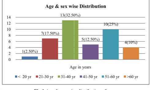
The incidence of urinary calculi was more in males 28(70%) as compared to females 12(30%).
wise distribution of cases, it is observed that urinary calculi is most commonly seen in age group of 31
followed by 51-60 years 10 (25%), 21 least number of cases in the age group of 41
Fig 1 Age & sex wise distribution of cases
Figure 2 shows the distribution of cases according to chief complaints, Majority of patients were admitted with pain 40(100%), followed by vomiting 15(37.5%), burning micturition10 (25%) and fever8 (20%).
Fig 2 Distribution of cases according to chief complaints 1(2.50%)
7(17.50%)
13(32.50%)
0 2 4 6 8 10 12 14
Age in years
Age & sex wise Distribution
< 20 yr 21-30 yr 31-40 yr
40(100%)
8(20%)
15(37.50%)
0 5 10 15 20 25 30 35 40 45
Distribution of cases according to chief complaints
3861, November, 2018
noted down. Ultrasonography (USG) of abdomen, X- ray KUB and Intra Venous Pyelography/Urography (IVP/IVU) images were saved for comparing with the images postoperatively. CT scan of the abdomen and Renal scan were done in few cases. treatment in the form of hydrotherapy (medical expulsive therapy), endourology techniques like Percutaneous nephrolitholapaxy (PCNL), Ureterorenoscopy (URS), Open and laparoscopic surgery, Extracorporeal Shock Wave was planned depending on the size of stone, location, surgeons judgment, patients preference and tone was sent for biochemical analysis and patient was discharged with supportive medications. Postoperative improvement was assessed by renal function tests and ography done at 1, 2 and 3 month’s interval. IVP was performed after 3 months in all the cases during the follow- up and compared with the preoperative images.
Complete data related to different procedures was collected fed into the computer SPSS version test was used to assess the level of significance, P value less than 0.001 was taken as significant.
The incidence of urinary calculi was more in males 28(70%) as compared to females 12(30%). Figure 1shows the age & sex wise distribution of cases, it is observed that urinary calculi is most commonly seen in age group of 31-40 years 13 (32.5%) 60 years 10 (25%), 21-30 years 07(17.5%) & least number of cases in the age group of 41-50 years 10(25%).
Age & sex wise distribution of cases
shows the distribution of cases according to chief complaints, Majority of patients were admitted with pain vomiting 15(37.5%), burning
8 (20%).
Distribution of cases according to chief complaints 13(32.50%)
5(12.50%) 10(25%)
4(10%)
Age in years
Age & sex wise Distribution
40 yr 41-50 yr 51-60 yr >60 yr
15(37.50%) 10(25%)
Distribution of cases according to chief complaints
Figure 3 a shows the pre-operative urea creatinine levels. Out of 40 patients 15(37.5%) patients had urea >40mg/dl, whereas 19(47.5%) patients had creatinine >1.4 mg/dl.
the post-operative urea creatinine levels after 1 month. Out of 40 patients 15(37.5%) patients had urea >40mg/dl, whereas 16(40%) patients had creatinine >1.4 mg/dl.
the post-operative urea creatinine levels after 3 months. 7(17.5%) patients had urea >40 mg/dl as compared to 15(37.5%) pre-operatively, similarly 11(27.5%) patients had creatinine >1.4 mg/dl as compared to 19(47.5%) pre operatively. 33(82.5%) patients had normal urea levels as compared to 25(62.5%) pre-operatively simil
patients had normal creatinine levels as compared to 21(52.5%) pre-operatively. P value for pre and post
urea level at 3 month was P-0.021 (not significant). P value for pre and post-operative creatinine level at 3 month was P (not significant).
Fig 3 a,b,c shows the preoperative urea creatinine levels, postoperatively at 1 month and 3 months
P value for pre and post-operative urea levels at (3 month). Urea preoperative: 36.09 +- 9.41. Urea postoperative: 34.66 + 9.78. P value: 0.021(not significant).
P value for pre and post-operative creatinine levels at (3 month).Creatinine preoperative: 1.12
+-postoperative: 1.11 +- 0.41. P value:0.589(not significant).
0 5 10 15 20 25
<40mg/dl >40mg/dl <1.4mg/dl >1.4mg/dl
UREA CREATININE
25(62.5%)
15(37.5%) 21(52.5%)
19(74.5%)
Distribution of cases showing pre operative urea/creatine levels
0 5 10 15 20 25
<40mg/dl >40mg/dl<1.4mg/dl>1.4mg/dl
UREA CREATININE
25(62.5%)
15(37.5%) 24(60%)
16(40%)
Distribution of cases showing post operatively urea/creatine levels after 1 months
operative urea creatinine levels. Out of 40 patients 15(37.5%) patients had urea >40mg/dl, whereas 19(47.5%) patients had creatinine >1.4 mg/dl. Figure 3b shows operative urea creatinine levels after 1 month. Out of 40 patients 15(37.5%) patients had urea >40mg/dl, whereas 16(40%) patients had creatinine >1.4 mg/dl. Figure 3c shows operative urea creatinine levels after 3 months. patients had urea >40 mg/dl as compared to operatively, similarly 11(27.5%) patients had creatinine >1.4 mg/dl as compared to 19(47.5%) pre-operatively. 33(82.5%) patients had normal urea levels as
operatively similarly 29(72.5%) patients had normal creatinine levels as compared to P value for pre and post-operative 0.021 (not significant). P value for operative creatinine level at 3 month was P-0.589
shows the preoperative urea creatinine levels, postoperatively at 1
operative urea levels at (3 month). 9.41. Urea postoperative: 34.66 +-
operative creatinine levels at (3 - .51. Creatinine not significant).
Fig 4 a,b,c shows the preoperative USG findings, postoperatively at 1
month and 3 months
Figure 4 a shows preoperative USG findings. 24(60%) patients had stones on right side in the renal pelvis or ureter, as compared to 11(27.5%) on the left side. 5(12.5%) patients had bilateral stones. 32(80%) patients had stones >10mm leading to hydronephrosis, whereas 4(10%) patients had stones <10mm causing hydronephrosis. Most of these were ureteric stones. USG diagnosed 33(82.5%) patients to be having hydronephrosis pre-operatively. 4(10%) cases had non obstructive calculus as per USG but these cases had hydronephrosis on IVP hence were included in the study. Figure 4b shows post-operative USG findings after 1 Month 3(7.5%) stones remained on the left side, 2(5%) stones on right side & 1(2.5%) patients had few stones left in both the kidneys. 2(5%) stones left were
<10mm, predominantly the cases with multiple renal calculi had stones left in the calyces. Postoperatively after 1 month 11(27.5%) patients had hydronephrosis and 21(52.5%) had normally functioning kidneys.
USG findings after 3 months, 4(10%) stones remained on left side, 3(7.5%) stones on right side & 1(2.5%) patients had few stones left on both sides. 2(5%) stones left were >10mm & 5(12.5%) were <10mm, predominantly the cases with multiple renal calculi had stones left in the calyces. Postoperatively after 3 months 4(10%) patients had hydronephrosis and 32(80%) had normally functioning kidneys.
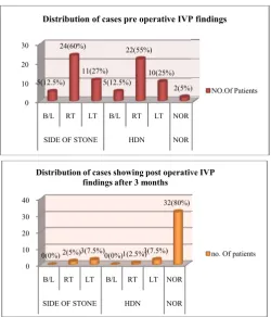
Figure 5a shows preoperative IVP findings. 24(60%) stones were on right side, 11(27.5%) stones on left side and 5
patients had bilateral stones. 22
hydronephrosis on right side, 10(25%) hydronephrosis on left
Distribution of cases showing pre operative
no of patients
Distribution of cases showing post operatively urea/creatine levels after 1 months
no. Of patients
preoperative USG findings, postoperatively at 1 month and 3 months
shows preoperative USG findings. 24(60%) patients had stones on right side in the renal pelvis or ureter, as compared to 11(27.5%) on the left side. 5(12.5%) patients had bilateral stones. 32(80%) patients had stones >10mm leading as 4(10%) patients had stones <10mm causing hydronephrosis. Most of these were ureteric stones. USG diagnosed 33(82.5%) patients to be having operatively. 4(10%) cases had non obstructive calculus as per USG but these cases had rosis on IVP hence were included in the study. operative USG findings after 1 Month 3(7.5%) stones remained on the left side, 2(5%) stones on right side & 1(2.5%) patients had few stones left in both the kidneys. 2(5%) stones left were >10mm & 4(10%) were <10mm, predominantly the cases with multiple renal calculi had stones left in the calyces. Postoperatively after 1 month 11(27.5%) patients had hydronephrosis and 21(52.5%) had normally functioning kidneys. Figure 4cshows postoperative USG findings after 3 months, 4(10%) stones remained on left side, 3(7.5%) stones on right side & 1(2.5%) patients had few stones left on both sides. 2(5%) stones left were >10mm & 5(12.5%) were <10mm, predominantly the cases with multiple d stones left in the calyces. Postoperatively after 3 months 4(10%) patients had hydronephrosis and 32(80%) had normally functioning kidneys.
International Journal of Current Medical And Pharmaceutical Research
side & 5(12.5%) had bilateral hydronephrosis. 2(5%) patients had IVP showing no hydronephrosis, however three patients had hydronephrosis on USG hence were included in the study Figure 5b shows Post-Operative IVP findings after 3 Months. 3(7.5%) patients had residual stones on left side, 2(5%) patients had residual stones on right side. 3(7.5%) patients had some hydronephrosis on left side & 1(2.5%) on right side. 32(80%) patients had hydronephrosis preoperatively as compared to 2(5%) postoperatively at 3 months.
Fig 5 a, b shows preoperative IVP findings and at 3 months postoperatively.
Figure 6 shows various surgical procedures performed. 22(55%) patients had right pyelolithotomy, 9(22.5%) patients had left pyelolithotomy, 3(7.5%) patients had right ureterolithotomy, 1(2.5%) patient had left ureterolithotomy. 3(7.5%) patients had right nephrolithotomy, 2(5%) patients had passed stones after hydrotherapy (medical expulsive therapy), 28(70%) patients had DJ stenting done along with other procedures.
Fig 6 shows various surgical procedures performed
To summarize out of 40 cases of obstructive u
patients were treated with hydrotherapy /Medical expulsive therapy. 22(55%) patients underwent right Pyelolithotomy, 9(22%) patients had left Pyelolithotomy, 1(2.5%) patient had left Ureterolithotomy. Out of them 28(70%) patients under
0 10 20 30
B/L RT LT B/L RT LT NOR
SIDE OF STONE HDN NOR
5(12.5%) 24(60%) 11(27%) 5(12.5%) 22(55%) 10(25%) 2(5%)
Distribution of cases pre operative IVP findings
0 10 20 30 40
B/L RT LT B/L RT LT NOR
SIDE OF STONE HDN NOR
0(0%) 2(5%)3(7.5%)0(0%)1(2.5%)3(7.5%)
32(80%)
Distribution of cases showing post operative IVP findings after 3 months
0 20 40 RT PL Y L T PYL DJ S RT NPL T L T URL RT/L T L A P H DT SURGERY 22(55%) 9(22.5%) 28(70%) 3(7.5%)1(2.5%)3(7.5%) 2(5%)
Distribution of cases showing various surgical procedures
Current Medical And Pharmaceutical Research, Vol. 4, Issue, 11(A), pp. 3856-3861
side & 5(12.5%) had bilateral hydronephrosis. 2(5%) patients had IVP showing no hydronephrosis, however three patients osis on USG hence were included in the study. Operative IVP findings after 3 Months. 3(7.5%) patients had residual stones on left side, 2(5%) patients had residual stones on right side. 3(7.5%) patients had side & 1(2.5%) on right side. 32(80%) patients had hydronephrosis preoperatively as compared to 2(5%) postoperatively at 3 months.
shows preoperative IVP findings and at 3 months postoperatively.
shows various surgical procedures performed. 22(55%) patients had right pyelolithotomy, 9(22.5%) patients had left pyelolithotomy, 3(7.5%) patients had right ureterolithotomy, 1(2.5%) patient had left ureterolithotomy. otomy, 2(5%) patients had passed stones after hydrotherapy (medical expulsive therapy), 28(70%) patients had DJ stenting done along with
s surgical procedures performed
To summarize out of 40 cases of obstructive uropathy 2(5%) patients were treated with hydrotherapy /Medical expulsive therapy. 22(55%) patients underwent right Pyelolithotomy, 9(22%) patients had left Pyelolithotomy, 1(2.5%) patient had left Ureterolithotomy. Out of them 28(70%) patients under
vent DJ stenting along with the surgery. 3(7.5%) patients had stone removal by laparoscopy. 3 patients underwent nephrectomy for non-functioning kidney for pyonephosis after Percutaneous Nephrostomy (PCN).
DISCUSSION
Urinary stones appear in 0.1-0.5% of world
relapse rate of about 80%. Most of kidney stones appear between age of 20 and 40 years.T
multiple with 40% of patients having them bilaterally [14 16].In our study we found 28(70%) males & 12(30%) females, majority of these were between 20
of 239 patients of renal and ureteric calculi with renal failure, there were 192 (80.5%) males and 47(19.5%) females. Majority of them belonged to 3rd and 4th decade [17].Stone formation is usually a result of urinary super saturation, any variation in urine saturation grade, urine pH and the concentration of crystallization inhibitors which can lead to urolithiasis [18, 19].Clinical manifestations of
are an episode of renal-ureteral colic
lumbar pain of sudden occurrence, may be accompanied by nausea and vomiting in some cases [20].
Urinary calculi may cause various complications like obstruction, infection and renal failure.
refers to the structural impedance to the flow of urine anywhere along the urinary tract leading to hydronephrosis, which is the dilation of the renal pelvis and calyces. Dilatation of the renal pelvis can occur even in the absence of urinary obstruction; therefore hydronephrosis and obstructive uropathy are not interchangeable or synonymous terms [21]. Unlike many other renal diseases, obstructive nephropathy if treated early is a potentially curable form of kidney disease [22].
In our study it was found that pain i.e.
the commonest complaint in 40(100%) patients, vomiting 15(37.5%) patients, burning micturition 10(25%) and fever 8(20%) patients. Drach (1992) has also stated that a urinary calculus “announces” its presence with an acute episode renal or ureteric colic. He further stated that uroliths create symptoms only when they become trapped in some segment of urinary tract. The other important symptoms were burning micturition (85%) and fever (77.5%), about 30% of patients had haematuria and around 11.25% of patients were admitted as acute retention of urine.[23].
Renal function test in the form of urea creatinine level is one of the most important predictor of renal change. We found that 14(35%) patients had pre-operative urea level >40
17(42.5%) patients had creatinine level >1.4mg/dl. levels were studied post operatively
the obstruction, it was seen that only 7(17.5%) patients had post-operative urea level >40mg/dl and only 11(27.5%) had post operative creatinine level >1.4mg/dl.M Hussain (2012) found in his study of 239 patients, blood urea from 70 550 mg and serum creatinine 3
admission. On post-operative evaluation, recover 183 patients, urine volume of more than 2 followed by 79 (33.2%) with urine output of 1
38 (15.8%) produced volume of less than 1 liter. On the other hand, 39.4% patients demonstrated massive natriuresis whil 46.0% showed normal urine sodium excretion, These findings were evaluated in two groups i.e., those who recovered and those who did not recover. It was found that in recoverable group 64.4% patients had urine output of 2
2(5%)
Distribution of cases pre operative IVP findings
NO.Of Patients
Distribution of cases showing post operative IVP
no. Of patients
H
DT
2(5%)
Distribution of cases showing various surgical
no. Of patients
3861, November, 2018
J stenting along with the surgery. 3(7.5%) patients had stone removal by laparoscopy. 3 patients underwent functioning kidney for pyonephosis after Percutaneous Nephrostomy (PCN).
0.5% of world population with Most of kidney stones appear between age of 20 and 40 years.T hey may be solitary or multiple with 40% of patients having them bilaterally [14-16].In our study we found 28(70%) males & 12(30%) females,
f these were between 20-40 years. In a similar study of 239 patients of renal and ureteric calculi with renal failure, there were 192 (80.5%) males and 47(19.5%) females. Majority of them belonged to 3rd and 4th decade [17].Stone ult of urinary super saturation, any variation in urine saturation grade, urine pH and the concentration of crystallization inhibitors which can lead to urolithiasis [18, 19].Clinical manifestations of urinary calculi ureteral colic or gross haematuria and
lumbar pain of sudden occurrence, may be accompanied by nausea and vomiting in some cases [20].
Urinary calculi may cause various complications like obstruction, infection and renal failure. Obstructive uropathy structural impedance to the flow of urine anywhere along the urinary tract leading to hydronephrosis, which is the dilation of the renal pelvis and calyces. Dilatation of the renal pelvis can occur even in the absence of urinary onephrosis and obstructive uropathy are not interchangeable or synonymous terms [21]. Unlike many other renal diseases, obstructive nephropathy if treated early is a potentially curable form of kidney disease [22].
In our study it was found that pain i.e. renal/ureteric colic was the commonest complaint in 40(100%) patients, vomiting 15(37.5%) patients, burning micturition 10(25%) and fever (1992) has also stated that a urinary calculus “announces” its presence with an acute episode of He further stated that uroliths create symptoms only when they become trapped in some segment of urinary tract. The other important symptoms were burning micturition (85%) and fever (77.5%), about 30% of patients and around 11.25% of patients were admitted as acute retention of urine.[23].
Renal function test in the form of urea creatinine level is one of the most important predictor of renal change. We found that operative urea level >40mg/dl and 17(42.5%) patients had creatinine level >1.4mg/dl. When these levels were studied post operatively at 3 month after relieving the obstruction, it was seen that only 7(17.5%) patients had 40mg/dl and only 11(27.5%) had post operative creatinine level >1.4mg/dl.M Hussain et al (2012) found in his study of 239 patients, blood urea from 70-550 mg and serum creatinine 3-35mg/100 ml at the time of
after relief of obstruction, while in non- recoverable group post-obstructive diuresis was noted in 4 (7.1%) only. Massive natriuresis was noted in 97(53.1%) in recoverable as compared to 12(21.4%) in non-recoverable group (P<0.001) [17].
Ultrasonography has become one of the most important tools for assessing urinary tract obstruction because it is rapid, low-cost, safe and sensitive. It is the diagnostic modality of choice in pregnancy [18]. The test is 98% sensitive for detecting hydronephrosis, but the specificity is 78% [24].Though operator-dependent, ultrasound detects the type and level of the lesion, shows hydronephrosis, can indicate pyonephrosis (echoes within the collecting system) and tells the thickness of the renal parenchyma as an indicator of the duration and severity of obstruction. Since up to 22% cases of hydronephrosis are not obstructive, there is room for error [25]. Dilatation without obstruction may be seen in vesicoureteral reflux, chronic massive diuresis, extra renal pelvis, calyceal diverticula, congenital megacalyces and ileal conduits. Obstruction without dilation may be seen in intrarenal crystals, nephrocalcinosis, staghorn calculi and retroperitoneal obstruction. Varma G et al (2009) concluded that both the plain X-ray KUB and USG should be performed in patients with suspected stone disease for identifying stone disease and also to exclude other pathology which may produce similar urinary symptoms [2]. When USG was done post operatively after 3 months, we found that hydronephrosis was present only in 4(10%) cases and 32(80%) cases had normal functioning kidney, 4(10%) cases had gross hydronephrosis and no improvement in the renal function was seen and were subjected to nephrectomy eventually.
Intravenous urogram (IVU) has been the gold standard for the detection of ureteral obstruction. The significant difference in IVU compared with Ultrasonography is that the IVU shows both increased anatomic detail and functional attributes of the urinary system. It tells about the exact site of obstruction, its cause, the degree of hydronephrosis, the status of renal parenchyma, guides the best treatment option, and also helps in deciding the best surgical approach for a particular patient. However, in view of nephrotoxicity of the contrast material, the usefulness of the test for patients already suspected of having obstructive nephropathy is questionable [25].Also poor renal function may render the test useless because of non-excretion of contrast. In general, a serum creatinine level of less than 2 mg% is needed for the test. Another dilemma with IVU is the significant time requirement, up to several hours, for performing serial delayed films. Still, IVU remains one of the most widely used imaging modality to guide the management of patients of obstructed kidney, because of its low cost and easy availability. A kidney which is non-visualized on IVU (absence of both nephrogram and pyelogram even on delayed films taken after 24-72 hours) should be assessed with a renal scan before labelling it as a non-functioning kidney (single kidney GFR which is not sufficient to take care of body wastes). A kidney may be non-visualized on IVU because of renal agenesis, post-nephrectomy, poor function with or without obstruction, or even in the presence of normal function during renal colic leading to renal artery spasm. When IVP was done after 3 month of treatment, hydronephrosis was found only in 1(2.5%) case on the right side and in 3(7.5%) cases on left side. 32(80%) cases had no hydronephrosis and 4(10%) cases were subjected to nephrectomy due to gross hydronephrosis. S Sourtzis et al (1999) in his study of 70 patients with
obstructive uropathy comparing IVP with CT scan and found that CT is better for identifying ureteral stones in patients with acute ureterolithiasis. Secondary CT signs of obstruction, including renal sinus fat blurring, were frequently present even when the stone was eliminated before imaging [1].NCCT (Non Contrast Computed Tomography) scan was done in three patients as the urea creatinine were more >2mg/dl in these patients making IVP not possible.
NCCT is an effective imaging tool for acute renal obstruction, with spiral scanners, images can be performed effectively without contrast media, take only 5 to 10 minutes to perform, and cost about the same as IVP. In terms of benefits, the CT equals the accuracy of the IVP in determining the presence of obstruction, but surpasses the IVP in detecting the specific cause of the obstruction[25].Various signs of obstruction on spiral CT include hydroureter, perinephric stranding, hydronephrosis, periureteral edema and renal swelling. However, NCCT does not indicate function of the kidneys.
MRI (Magnetic resonance imaging) gives no added advantage over CT scan. In addition it does not visualize the stone. However, it correctly identifies the point of obstruction and the non-calculus causes of obstruction. MRI excretory urography is a promising technique which affords equivalent functional and additional anatomical information to isotope renography. It is more accurate than Doppler ultrasound in the assessment of ureteric obstruction in pregnancy and is not associated with any risk of exposure to radiation, unlike IVU or Renal scan [26].
In the present study three cases which were non functioning on IVP were subjected to renal scan. Renal scan and diuretic renography is the most reliable technique to quantitatively assess the split and total renal function in the presence of hydronephrosis. It is a non-invasive study, can be done even in patients with deranged renal function, has no risk of contrast-induced nephrotoxicity and also has much less radiation exposure than IVP or CT scan. It can also be used to measure differential function and, therefore, is useful for treatment planning. Moreover, a functional obstruction can be differentiated from an anatomic cause. It can be used to follow up a patient after relief of obstruction. Also, the assessment of renal blood flow provides a sense of whether function may return upon relief of the obstruction. However, images from these scans lack the resolution to define the site of obstruction [27].
International Journal of Current Medical And Pharmaceutical Research, Vol. 4, Issue, 11(A), pp. 3856-3861, November, 2018
surgeons have also repaired both sides at the same sitting in the past but its routine application is questionable.
CONCLUSION
Urea, creatinine levels along with USG and IVP form the basis of detection and management of obstructive uropathy. It takes about three months for normalization of kidney function after definitive management to relieve the obstruction.
References
1. Sourtzis S, Thibeau J, Damry N, Raslan A, Vandendris M, Bellemans M. Radiologic investigation of renal colic: unenhanced helical CT compared with excretory urography. American Journal of Roentgenology 1999; 172 (6): 1491-94.
2. Varma G, Nair N, Salim A, FazilMarickar Y. Investigations for recognizing urinary stone. Urological Research 2009; 37(6): 349-52.
3. Heidenreich A, Desgrandschamps F, Terrier F. Modern approach of diagnosis and management of acute flank pain: review of all imaging modalities. European Urology 2002; 41 (4): 351-62.
4. Shine S. Urinary calculus: IVU vs. CT renal stone? A critically appraised topic. Abdominal Imaging 2008; 33(1): 41-43.
5. Buck A, Macleod M, Blacklock N. The advantages of 99mTc DTPA (Sn) in dynamic renal scintigraphy and measurement of renal function. British Journal of Urology 1980; 52 (3): 174-86.
6. Straub M, Strohmaier W, Berg W, Beck B, Hoppe B, Laube N, et al. Diagnosis and metaphylaxis of stone disease. Concensus concept of the National working committee on stone disease for the upcoming German Urolithiasis guideline. World Journal of Urology 2005; 23 (5): 309-23.
7. Ramos-Fernandez M, Serrano LA. Evaluation and management of renal colic in the emergency department. Bol Asoc Med P R. 2009 Jul-Sep; 101(3):29-32.
8. Engeler D, Schmid S, Schmid HP. The ideal analgesic treatment for acute renal colic-Theory and practice. Scandinavian Journal of Urology and Nephrology 2008; 42 (2): 137-42.
9. Ramsey S, Robertson A, Ablett MJ, Meddings RN, Hollins GW, Little B. Evidence-based drainage of infected hydronephrosis secondary to ureteric calculi. Journal of Endourology 2010; 24(2): 185-89.
10. Pearle MS, Pierce H, Miller GL, Summa JA, Mutz JM, Petty BA, et al. Optimal method of urgent decompression of the collecting system for obstruction and infection due to ureteral calculi. Journal of Urology 1998; 160 (4): 1260-64.
11. Honeck P, Wendt-Nordahl G, Krombach P, Bach T, Hacker A, Alken P, et al. Does open stone surgery still play a role in the treatment of urolithiasis? Data of a primary urolithiasis center. Journal of Endourology 2009; 23 (7): 1209-12.
12. Bichler K, Lahme S, Strohmaier W. Indications for open stone removal of urinary calculi. Urologia Internationalis 1997; 59 (2): 102-08.
13. Hruza M, Schulze M, Teber D, Gozen AS, Rassweiler JJ. Laparoscopic techniques for removal of renal and ureteral calculi. Journal of Endourology 2009; 23 (10): 1713-18.
14. Serie V et al. Metabolic and physico-chemical urolithiasis parameters in the first morning urine. Collegium Antropol. 2009; 33(2):85, 92-110.
15. Sharma RN, Goswami HL. Upper urinary calculi. International Journal of Surgery.1985; 47:1, 6-25. 16. Bartoletti R, Cai T, Mondaim N, Melone F, Travaglim
F, Carini M. Epidemiology and risk factors in urolithiasis. Urol Int.2007; 79(1): 3-7.
17. Manzoor Hussain, B. Ali, S. Ahmed, N. Zafar, S.A. Naqvi, S.A.H. Rizvi. Prediction of Renal Function Recovery in Obstructive Renal Failure Due to Stones. Sindh Institute of Urology and Transplantation (SlUT), Karachi. Pages with reference to book 2012; 55:159-61. 18. Micelyte S, Glinskis G, Cekauskas Z, Dasevicius V.
Hydronephrosis in pregnancy: Importance of urologic actions and their volume. Medicina (Kaunas) 2002; 38: Suppl 1:22-29.
19. Dretler SP. Ureteral stone disease: Options for management. UrolClin North Am 1990; 17:217-30. 20. Tiselius HG, Ackermann D, Alken P, Buck C, Conort P,
Gallucci M; Guidelines on urolithiasis. EurUrol 2001; 40(4): 362-71.
21. Walsh PC, RetnikAB, Vaughan ED, Wein AJ. Pathophysiology of urinary tract obstruction. In: Campbell's Urology. 7th Ed. Philadelphia: W.B. Saunders Company; 1998: 343-60.
22. Chevalier RL, Klahr S. Therapeutic approaches in obstructive uropathy. Semin Nephrol 1988; 18:652-58. 23. Drach GW. Urinary lithiasis; Etiology, diagnosis and
medical management. In Walsh P, Retik A, Stamey, Vaughan D. Eds. Campbell's Urology, 1992; 3(6):2085-156.
24. Rahman M, Smith MC. Chronic renal insufficiency: A diagnostic and therapeutic approach. Arch Intern Med 1998; 158:1743-52.
25. Smith RC, Rosenfield AT, Choe KA. Acute flank pain: Comparison of non-contrast-enhanced CT and intravenous urography. Radiology 1995; 194:789-94. 26. Zielonko J, Studniarek M, Markuszewski M. MR
urography of obstructive uropathy: Diagnostic value of the method in selected clinical groups. EurRadiol 2003; 13:802-09.
27. Finlayson B, Ackermann D. Overview of surgical treatment of urolithiasis with reference to lithotripsy. J Urol. 1989; 141: 778-79.
28. Gupta, M., Bolton, D.M. and Stoller, M.L. Improved renal function following agressive treatment of urolithiasis and concurrent mild to moderate renal insufficiency. J. Urol, 1994:152;l086.90
29. Bilic-Curcic Z: Urolithiasis and osteoporosis. Collegium Antropol. 2009; 33(2):189-92-106.
How to cite this article:
Gaurav Gupta et al (2018) 'Evaluation of Renal Function By Pre And Post-Operative Radiological And Biochemical Investigations In Patients of Obstructive Uropathy Due To Urinary Stones', International Journal of Current Medical and Pharmaceutical Research, 04(10), pp. 3856-3861.