Review
Drosophila
DNA-Binding Proteins in
Polycomb Repression
Maksim Erokhin, Pavel Georgiev and Darya Chetverina * ID
Department of the Control of Genetic Processes, Institute of Gene Biology, Russian Academy of Sciences, 34/5 Vavilov St., Moscow 119334, Russia; [email protected] (M.E.); [email protected] (P.G.)
* Correspondence: [email protected]
Received: 5 November 2017; Accepted: 10 January 2018; Published: 16 January 2018
Abstract: The formation of individual gene expression patterns in different cell types is required during differentiation and development of multicellular organisms. Polycomb group (PcG) proteins are key epigenetic regulators responsible for gene repression, and dysregulation of their activities leads to developmental abnormalities and diseases. PcG proteins were first identified inDrosophila, which still remains the most convenient system for studying PcG-dependent repression. In the Drosophilagenome, these proteins bind to DNA regions called Polycomb response elements (PREs). A major role in the recruitment of PcG proteins to PREs is played by DNA-binding factors, several of which have been characterized in detail. However, current knowledge is insufficient for comprehensively describing the mechanism of this process. In this review, we summarize and discuss the available data on the role of DNA-binding proteins in PcG recruitment to chromatin.
Keywords:DNA binding; Polycomb; Polycomb response elements; PRE; recruitment; epigenetics; chromatin; transcription regulation; repression; silencing
1. Introduction
Polycomb group (PcG) proteins are key epigenetic regulators of gene repression in multicellular organisms [1–5].
Polycomb group proteins were first identified inDrosophilaas regulators ofHoxgenes expression along the head-to-tail axis [6–9]. Mutations in the genes encoding PcG proteins lead to characteristic homeotic transformations resulting from derepression of Hox genes. A classic example is the development of extra sex combs on the second and third legs, with these structures being normally formed only on the first leg [10].
In addition to theHoxgenes, PcG proteins target numerousDrosophilagenes implicated in different cell processes [11–16]. These proteins are highly conserved and control multicellular development in plant and animal systems [17–21]. Most ofDrosophilaPolycomb proteins have paralogs in humans [21–23]. In mammals, PcG proteins have been shown to play a role in cancer [20,24–26], stem cell biology [17,20,27], X chromosome inactivation [28,29], imprinting [30], and in the situations of stress, ageing, regeneration and healing [31–37].
The Polycomb group proteins function in multisubunit complexes. The main complexes in Drosophilaare derived from the core subcomplexes named Polycomb repressive complex 1 (PRC1) and Polycomb repressive complex 2 (PRC2).
The PRC2 core subcomplex contains the Enhancer of zeste (E(z)), Extra sex combs (Esc), Suppressor of zeste 12 (Su(z)12), and Chromatin assembly factor 1 (Caf1) subunits [38,39]. The catalytic subunit is the E(z) protein that trimethylates histones 3 at lysine 27 (H3K27me3) [38–41]. The H3K27me3 modification is a specific hallmark required for PcG repression [42]. Inactivation of genes for E(z) [40,43–45], Esc [45,46], and Su(z)12 [47,48] leads to abrogation of H3K27me3 methylation, derepression ofHoxgenes, and emergence of homeotic phenotypes.
Epigenomes 2018,2, 1 2 of 24
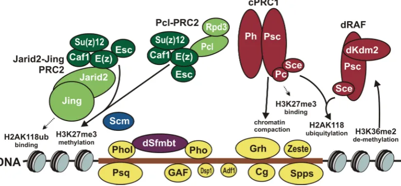
The PRC2 core subcomplex interacts with accessory protens, Polycomblike (Pcl) or Jarid2 (Jumonji, AT-rich interactive domain 2) and Jing, to form larger complexes: Pcl-PRC2 and Jarid2-Jing-PRC2, respectively (Figure1). The Pcl-PRC2 complex contains the Pcl protein required for high levels of H3K27me3 in vivo [48]. This complex can be additionally purified with the Reduced potassium dependency 3 (Rpd3) histone deacetylase [49], which removes the H3K27ac active chromatin hallmark [50]. The Jarid2-Jing-PRC2 complex contains the Jarid2 and Jing subunits [51]. This complex recognizes monoubiquitylation of histone H2A at lysine 118 (H2AK118ub), which is catalyzed by the PRC1 complexes [52,53]. Compared to the PRC2 core subcomplex, Jarid2–Jing–PRC2 methylates H3K27me3 in vitro more effectively, especially on H2AubK118 nucleosomes [54]. However, the role of this complex in vivo is questioned, since Jarid2 is absent on many regulatory elements of theHox genes [51] and repression can be maintained in the absence of H2AubK118 [55].
genes for E(z) [40,43–45], Esc [45,46], and Su(z)12 [47,48] leads to abrogation of H3K27me3 methylation, derepression of Hox genes, and emergence of homeotic phenotypes.
The PRC2 core subcomplex interacts with accessory protens, Polycomblike (Pcl) or Jarid2 (Jumonji, AT-rich interactive domain 2) and Jing, to form larger complexes: Pcl-PRC2 and Jarid2-Jing-PRC2, respectively (Figure 1). The Pcl-PRC2 complex contains the Pcl protein required for high levels of H3K27me3 in vivo [48]. This complex can be additionally purified with the Reduced potassium dependency 3 (Rpd3) histone deacetylase [49], which removes the H3K27ac active chromatin hallmark [50]. The Jarid2-Jing-PRC2 complex contains the Jarid2 and Jing subunits [51]. This complex recognizes monoubiquitylation of histone H2A at lysine 118 (H2AK118ub), which is catalyzed by the PRC1 complexes [52,53]. Compared to the PRC2 core subcomplex, Jarid2–Jing– PRC2 methylates H3K27me3 in vitro more effectively, especially on H2AubK118 nucleosomes [54]. However, the role of this complex in vivo is questioned, since Jarid2 is absent on many regulatory elements of the Hox genes [51] and repression can be maintained in the absence of H2AubK118 [55].
The PRC1 core subunits are Sex combs extra (Sce, also known as dRing) and Posterior sex combs (Psc). Interacting with accessory proteins, the PRC1 core forms at least two complexes: dRing Associated Factors (dRAF) and canonical PRC1 (Figure 1). The Sce subunit in both complexes has monoubiquitination activity and produces the H2AK118ub modification [53] recognized by Jarid2– Jing–PRC2. The dRAF complex contains the Lysine (K)-specific demethylase 2 (Kdm2) protein [52] and has higher ubiquitin ligase activity. The canonical PRC1 consists not only of the Sce–Psc pair, but also of Polycomb (Pc) and Polyhomeotic (Ph) proteins [56–58]. In addition, several non-core proteins, including the Sex comb on midleg (Scm) [57], can be purified as its minor components. The canonical PRC1 was discovered first, and its role in repression has been studied much better. This complex is able to repress transcription by compacting chromatin and inhibiting nucleosome remodeling [56,58–61]. Moreover, its Pc subunit interacts with H3K27me3 nucleosomes catalyzed by PRC2 [62,63].
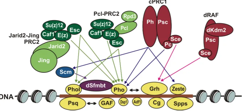
Figure 1. Functional activities of Drosophila Polycomb group (PcG) proteins. The Polycomb response element (PRE) shown as brown horizontal bar: DNA sequence depleted of nucleosomes (slanted ovals) on which PcG proteins are assembled. DNA-binding proteins: Pleiohomeotic (Pho), Pleiohomeotic-like (Phol), GAGA factor (GAF), Pipsqueak (Psq), Combgap (Cg), Sp1-like factor for pairing-sensitive silencing (Spps), Dorsal switch protein 1 (Dsp1), Grainy head (Grh), Zeste (Z) and Adh transcription factor 1 (Adf1). Pho and Phol interact with dSfmbt in mutually exclusive manner. Pho and dSfmbt form the Pho-repressive complex (PhoRC) complex. Other PcG complexes are made of Polycomb repressive complex 2 (PRC2) or of Polycomb repressive complex 1 (PRC1) core subunits. The PRC2 core subunits are Enhancer of zeste (E(z)), Extra sex combs (Esc), Suppressor of zeste 12 (Su(z)12) and Chromatin assembly factor 1 (Caf1) proteins. Together with Polycomblike (Pcl) and Reduced potassium dependency 3 (Rpd3) or Jarid2 (Jumonji, AT-rich interactive domain 2) and Figure 1.Functional activities ofDrosophilaPolycomb group (PcG) proteins. The Polycomb response element (PRE) shown as brown horizontal bar: DNA sequence depleted of nucleosomes (slanted ovals) on which PcG proteins are assembled. DNA-binding proteins: Pleiohomeotic (Pho), Pleiohomeotic-like (Phol), GAGA factor (GAF), Pipsqueak (Psq), Combgap (Cg), Sp1-like factor for pairing-sensitive silencing (Spps), Dorsal switch protein 1 (Dsp1), Grainy head (Grh), Zeste (Z) and Adh transcription factor 1 (Adf1). Pho and Phol interact with dSfmbt in mutually exclusive manner. Pho and dSfmbt form the Pho-repressive complex (PhoRC) complex. Other PcG complexes are made of Polycomb repressive complex 2 (PRC2) or of Polycomb repressive complex 1 (PRC1) core subunits. The PRC2 core subunits are Enhancer of zeste (E(z)), Extra sex combs (Esc), Suppressor of zeste 12 (Su(z)12) and Chromatin assembly factor 1 (Caf1) proteins. Together with Polycomblike (Pcl) and Reduced potassium dependency 3 (Rpd3) or Jarid2 (Jumonji, AT-rich interactive domain 2) and Jing PRC2 core forms Pcl-PRC2 and Jarid2-Jing-PRC2 complexes, respectively. The PRC1 core subunits are Sex combs extra (Sce, also known as dRing) and Posterior sex combs (Psc). Together with Lysine (K)-specific demethylase 2 (Kdm2) or Polycomb (Pc) and Polyhomeotic (Ph) proteins PRC1 core forms dRing Associated Factors (dRAF) and canonical PRC1 (cPRC1) complexes. The arrows pointing to and from the nucleosomes indicate the main activities of PcG complexes.
proteins, including the Sex comb on midleg (Scm) [57], can be purified as its minor components. The canonical PRC1 was discovered first, and its role in repression has been studied much better. This complex is able to repress transcription by compacting chromatin and inhibiting nucleosome remodeling [56,58–61]. Moreover, its Pc subunit interacts with H3K27me3 nucleosomes catalyzed by PRC2 [62,63].
InDrosophila, the PcG complexes are recruited to specialized DNA regulatory elements, named Polycomb response elements (PREs) [3,64–68], responsible for repression of target genes. PRE-like elements have been described in mouse and humans, suggesting similarity in the organization of the PcG system [69–73]. However, the mechanism of PcG protein recruitment to PRE DNA is as yet poorly understood.
The key role in the recruitment ofDrosophilaPcG proteins is usually assigned to DNA-binding factors. On the other hand, several models have been proposed, based mainly on the properties of the PcG system in mammals, that consider the role of long noncoding RNAs and histone modifications [3,5,64,66,74]. However, none of these models provides a detailed account of the mechanism of recruitment.
The role of DNA-binding proteins in the recruitment of PcG complexes to PREs is best studied inDrosophila, in which a number of DNA-binding factors have been characterized in detail [3,64–66]. However, none of the known DNA-binding proteins can associate with all predicted PREs, and their binding sites cannot independently provide for the recruitment of PcG complexes. Therefore, the cooperation of these proteins is necessary for the effective recruitment of PcG complexes to DNA. Moreover, recent data allow the possibility that more DNA-binding proteins implicated in PcG function will be soon discovered.
In this review, we summarize available information on theDrosophilaDNA-binding proteins implicated in PRE function and discuss possible mechanism of PcG protein recruitment to PREs.
2. Properties of Polycomb Response Elements
TheDrosophilaPREs were first characterized in the regulatory regions of the Bithorax (BX-C) [75–77] and the Antennapedia (ANT-C) [78] gene complexes. Further genome-wide studies identified more than a thousand peaks of PcG proteins, suggesting the existence of numerous PREs in the Drosophilagenome [13–16,79,80], which could be located either in close proximity or at a large distance (several tens of thousands of nucleotides) before or after transcription start site (TSS) [13,14].
Experiments with transgenes at different insertion sites have shown that PREs repress reporter gene expression and maintain repression during development [75–77,81]. Moreover, PREs in transgenes are sensitive to mutations in PcG genes, recruit the PRC1 and PRC2 complexes, and provide for the H3K27me3 modification, suggesting that they are independent units with repressor activity. However, PREs repress the reporter genes in only about 60% of independent transgenic lines, indicating that the site of transgene insertion is important for their activity [82,83].
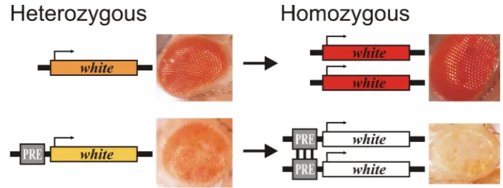
Figure 2. Pairing-sensitive silencing (PSS). The PSS assay is usually performed using the white reporter gene responsible for eye pigmentation. The eyes of homozygous flies without PRE become darker than in heterozygous flies; in the presence of PRE, the eyes of homozygous flies are lighter than in heterozygous flies.
The activity of PREs can change under certain conditions: instead of repression, they can provide for activation of reporter genes [86–90]. This is mediated by Trithorax group (TrxG) activator proteins, which also bind to PREs and can antagonize PcG activity. The TrxG is composed of heterogeneous proteins and includes components of different complexes: ATP-dependent chromatin remodelers, histone methyltransferases, Mediator, cohesin, etc. [1,4,5,91].
The PcG and TrxG group proteins can be present at PREs in both active and repressed states, and the direct competition between these two groups determines the resulting activity of PREs [11,14,80,92–94].
Both PRE-dependent repression and activation could be epigenetically inherited during mitosis and meiosis upon the disappearance of the initial stimulus from the system [75–77,86,95–98].
3. Role of PRE DNA-Binding Proteins in Repression
Polycomb response elements were first identified as extended DNA sequences several kilobase pairs long. Further studies have demonstrated, however, that the functional core of PREs resides in DNA fragments of several hundred nucleotides. PREs have typical properties of DNA regulatory elements actively involved in interactions with DNA-binding transcription proteins: they are hypersensitive to DNaseI and are characterized by low nucleosome occupancy.
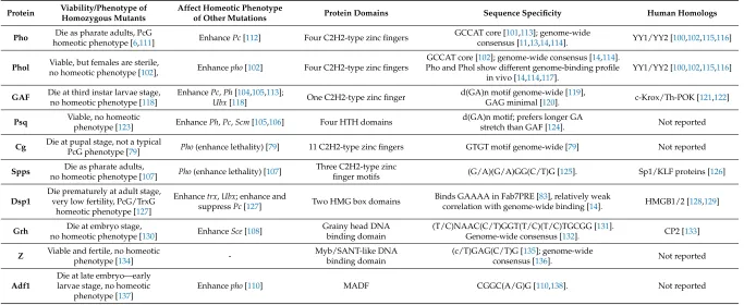
Core sequences of PREs are made up of binding sites for different DNA-binding proteins [65,66,99]. A number of DNA-binding factors important for PRE activity have been characterized (Table 1, Figure 1). They include Pleiohomeotic (Pho) [100,101] and its homologue Pleiohomeotic-like (Phol) [102], GAGA factor (GAF, also known as Trithorax-like (Trl)) [103,104], Pipsqueak (Psq) [105,106], Combgap (Cg) [79], Sp1-like factor for pairing-sensitive silencing (Spps)) [107], Dorsal switch protein 1 (Dsp1) [83], Grainy head (Grh, also known as NTF-1 (Neuronal transcription factor 1)) [108], Zeste (Z) [57,109] and Adh transcription factor 1 (Adf1) [110].
Table 1. PRE DNA-binding proteins.
Protein Viability/Phenotype of Homozygous Mutants Affect Homeotic Phenotype of Other Mutations Protein
Domains Sequence Specificity
Human Homologs
Pho
Die as pharate adults, PcG homeotic
phenotype [6,111]
Enhance Pc [112]
Four C2H2-type zinc
fingers
GCCAT core [101,113]; genome-wide consensus [11,13,14,114].
YY1/YY2 [100,102,115,116]
Phol
Viable, but females are sterile, no homeotic phenotype
[102],
Enhance pho [102]
Four C2H2-type zinc
fingers
GCCAT core [102]; genome-wide consensus [14,114]. Pho and Phol show different genome-binding profile in vivo
[14,114,117].
YY1/YY2 [100,102,115,116] Figure 2.Pairing-sensitive silencing (PSS). The PSS assay is usually performed using thewhitereporter
gene responsible for eye pigmentation. The eyes of homozygous flies without PRE become darker than in heterozygous flies; in the presence of PRE, the eyes of homozygous flies are lighter than in heterozygous flies.
The activity of PREs can change under certain conditions: instead of repression, they can provide for activation of reporter genes [86–90]. This is mediated by Trithorax group (TrxG) activator proteins, which also bind to PREs and can antagonize PcG activity. The TrxG is composed of heterogeneous proteins and includes components of different complexes: ATP-dependent chromatin remodelers, histone methyltransferases, Mediator, cohesin, etc. [1,4,5,91].
The PcG and TrxG group proteins can be present at PREs in both active and repressed states, and the direct competition between these two groups determines the resulting activity of PREs [11,14,80,92–94].
Both PRE-dependent repression and activation could be epigenetically inherited during mitosis and meiosis upon the disappearance of the initial stimulus from the system [75–77,86,95–98].
3. Role of PRE DNA-Binding Proteins in Repression
Polycomb response elements were first identified as extended DNA sequences several kilobase pairs long. Further studies have demonstrated, however, that the functional core of PREs resides in DNA fragments of several hundred nucleotides. PREs have typical properties of DNA regulatory elements actively involved in interactions with DNA-binding transcription proteins: they are hypersensitive to DNaseI and are characterized by low nucleosome occupancy.
Table 1.PRE DNA-binding proteins.
Protein Viability/Phenotype of Homozygous Mutants
Affect Homeotic Phenotype
of Other Mutations Protein Domains Sequence Specificity Human Homologs
Pho Die as pharate adults, PcG
homeotic phenotype [6,111] EnhancePc[112] Four C2H2-type zinc fingers
GCCAT core [101,113]; genome-wide
consensus [11,13,14,114]. YY1/YY2 [100,102,115,116]
Phol Viable, but females are sterile,
no homeotic phenotype [102], Enhancepho[102] Four C2H2-type zinc fingers
GCCAT core [102]; genome-wide consensus [14,114]. Pho and Phol show different genome-binding profile
in vivo [14,114,117].
YY1/YY2 [100,102,115,116]
GAF Die at third instar larvae stage,
no homeotic phenotype [118]
EnhancePc,Ph[104,105,113];
Ubx[118] One C2H2-type zinc finger
d(GA)n motif genome-wide [119],
GAG minimal [120]. c-Krox/Th-POK [121,122]
Psq Viable, no homeotic
phenotype [123] EnhancePh,Pc,Scm[105,106] Four HTH domains
d(GA)n motif; prefers longer GA
stretch than GAF [124]. Not reported
Cg Die at pupal stage, not a typicalPcG phenotype [79] Pho(enhance lethality) [79] 11 C2H2-type zinc fingers GTGT motif genome-wide [79] Not reported
Spps Die as pharate adults,
no homeotic phenotype [107] Pho(enhance lethality) [107]
Three C2H2-type zinc
finger motifs (G/A)(G/A)GG(C/T)G [125]. Sp1/KLF proteins [126]
Dsp1
Die prematurely at adult stage, very low fertility, PcG/TrxG
homeotic phenotype [127]
Enhancetrx,Ubx; enhance and
suppressPc[127] Two HMG box domains
Binds GAAAA in Fab7PRE [83], relatively weak
correlation with genome-wide binding [14]. HMGB1/2 [128,129]
Grh Die at embryo stage,
no homeotic phenotype [130] EnhanceSce[108]
Grainy head DNA binding domain
(T/C)NAAC(C/T)GGT(T/C)(T/C)TGCGG [131].
Genome-wide consensus [132]. CP2 [133]
Z Viable and fertile, no homeotic
phenotype [134]
-Myb/SANT-like DNA binding domain
(c/T)GAG(C/T)G [135]; genome-wide
consensus [136]. Not reported
Adf1
Die at late embryo—early larvae stage, no homeotic
phenotype [137]
The role of Pho protein in the PcG function is the most studied. Pho interacts with all functionally characterized in transgenes PREs and is suggested to play a central role in their silencing activity [66]. Pho binds to PREs together with dSfmbt protein as the Pho-repressive complex (PhoRC) complex (Figure1) [139]. The mutants of thephogene die as pharate adults with sex combs on the second and third legs demonstrating classical PcG phenotype [111] and are characterized by derepression ofHox genes [45,101,102].
The mutations in the genes encoding other PRE DNA-binding proteins do not result in PcG homeotic phenotypes [79,102,107,118,123,130,134,137]. One exception is the Dsp1 protein: thedsp1 mutants have a mixed PcG/TrxG phenotype, indicating that Dsp1 protein is also implicated in activation [127]. At the same time, it should be noted that the mutant alleles of genes for different PRE DNA-binding proteins are lethal at different stages of development and some of them die earlier than Pho and Dsp1 not allowing to compare their implication in PcG repression directly (Table1).
While mutations in genes for other PRE DNA-binding proteins do not themselves result in homeotic phenotypes, some of them enhance mutations in the PRC1 core genes orphogene resulting in stronger homeotic transformations. Thus, mutation of theTrl(gene encoding GAF) [104,105,113] andPsq[105,106] enhancePcmutation; mutation of thegrh[108] enhancesScemutation; the mutations of phol [102] and Adf1 [110] enhance pho mutation. While not enhancing homeotic phenotype, the mutations ofcg(Combgap) [79] andspps[107] enhance lethality of thephomutation, and flies die at earlier stage of development.
There are several standard tests with transgene reporter constructs for verifying the involvement of proteins in PRE-mediated repression and PSS. ThelacZ or whitegenes are commonly used as reporters in these test systems. ThelacZgene allows visual assessment of PRE-mediated repression of transcription in certain tissues at a selected stage of development. A mutation in the gene encoding a PcG protein leads to indiscriminate expression of the reporter [101,102,113]. Thewhitegene allows testing the role of protein in PRE-mediated repression in the eyes and is standardly used for PSS assessment [83,100,103,107,108,140]. Mutations in the genes encoding PcG proteins leads to darker eye pigmentation and the effect on PSS in homozygous lines is often more significant than that on the repression in heterozygotes.
Mutation of at least several genes encoding PRE DNA-binding proteins leads to partial derepression of lacZor whitereporter and decrease in PSS. In particular, they includepho[100–102,113,140–142], Trl(GAF) [103,113,140,143], spps[107],dsp1[83], and grh[108]. Mutation in thezestegene affects PSS [103], with Zeste being required for the maintenance ofUbxrepression in the embryo [109]. At the same time, mutation in zestehas also been shown to increase repression, acting opposite to PcG group factors [103,143]. This is clear evidence that the Zeste protein has a dual function in both gene repression and activation.
The participation in activation and TrxG function is characteristic not only of Dsp1 and Zeste but also of other PRE DNA-binding proteins, including GAF [109,118,144], Grh [109,130], Adf1 [145], Pho and Phol [141]. The Zeste, GAGA and Grh proteins together positively regulate the transcription of theUbxgene [109,144,146]. Moreover, the Pho and Phol proteins are required for the maintenance of both repressed and active states of theevePRE [141]. Currently this is an exceptional example for activating function of Pho/Phol and it remains to be tested whether it applies to other PREs.
However, in accordance with the role of PRE DNA-binding factors in activation, several proteins have been demonstrated to bind not only to PREs but also to the promoters of active genes [14,109,114]. Similar, PcG proteins can be involved in gene activation [147–150] demonstrating dual function of PcG proteins and close interplay between PcG and TrxG systems.
The highest colocalization is observed for the Pho and Combgap proteins: about 90% of PRC1 and PRC2 peaks overlap with these factors [11,14,79,114]. The second place belongs to GAF and Dsp1: both proteins colocalize with approximately 50% PRC1 peaks [14]. Then follow Zeste and Phol, which colocalize with approximately 25% and 21% of PRC1 peaks, respectively [14].
The degree of overlap between DNA-binding proteins is somewhat smaller: about 80% of Pho peaks colocalize with Combgap [79]; 50% of Pho peaks colocalize with GAF, and 10% with Zeste [13].
The genome-wide colocalization for Psq, Spps, Grh and Adf1 with PRC1 and PRC2 has not been reported. However, there are data on colocalization of Psq-Psc [106], Psc-Spps [107], Combgap-Spps [79] and Psq-GAF [151] on polytene chromosomes.
Despite different distribution of PRE DNA-binding proteins in the genome, at least some of them functionally interact with each other, indicating their mutual role in repression. In addition to the aforementioned interactions withpho, it has been shown that mutation of thegrhenhances the effect ofphomutation on PSS in transgenes [108] and that mutation of thecgenhances lethality ofspps mutation [79]. It is likely that other combinations of mutations can enhance each other, although this possibility has not been tested.
It is noteworthy that many of PRE DNA-binding proteins appear to have functions other than PcG repression. Extensive colocalization between these proteins is more likely to occur in case of PcG/TrxG linked function, while at other genomic regions they can act separately, forming connections with other DNA-binding proteins and protein complexes.
4. Role of Protein Binding Sites in Repression
PREs consist of binding sites for different DNA-binding proteins that are densely grouped together. The binding sites are short and fairly degenerate sequences with a core of about 3–8 bp (Table1). The sites for a particular protein in a PRE may differ in number and tend to form clusters. Such sites in PREs occur in different combinations, apparently in random order and at different distances from each other [82,152,153], suggesting that each DNA-binding factor can perform non-equivalent functions in different PREs.
It turned out that different DNA-binding proteins often have the same DNA-binding consensus site. The best-studied example is that of Pho and Phol, two proteins that are closely homologous within their DNA-binding region and short linker fragment [102] (Table1). The same binding site is recognized by the GAF and Psq protein pair, despite that they have different types of DNA-binding domains [124] (Table1). The binding site of the Zeste protein is also recognized by the Fs(1)h (female sterile (1) homeotic) protein, which has been characterized as a TrxG factor [154] and, at the same time, purifies with the PRC1 complex when tagged with the Pc protein [155,156]. The recruitment of Fs(1)h to PREs and its implication in PcG function are yet to be verified. Another example is the Sp1-like proteins and Krüppel-like factors (Sp1/KLF) family that shares the site with the Spps co-member [107,125].
The above examples may be not exceptional, and it is possible that the list of proteins interacting with these sites or with binding sites for other PRE DNA-binding proteins will be replenished. The affinity of different proteins for the same site should be especially taken into account when analyzing the role of the sites in repression.
The mutations of sites for Pho/Phol [100,101,113,140–142], GAF/Psq [95,105,113,140,141,143], Combgap (GTGT sites) [157], Spps/(Sp1/KLF) [95,125], Dsp1 (G(A)n) [83], Zeste [82] affect repression and/or PSS in transgenes. Similar functional tests of the sites for Grh and Adf1 have not been reported.
(e.g., Fs(1)h) binds to these sites instead of Zeste. However, it may well be that mutation of the test site can lead to inability of neighbor proteins to bind DNA affecting overall complex composition.
On the other hand, mutation of sites not obligatory leads to derepression. For example, the removal of Pho sites fromenPRE does not always result in the complete loss of PcG-mediated silencing [125]. This may be explained by the influence of PcG proteins bound to genome near transgene insertion site.
Therefore, the effects of site mutation and protein disruption are not the same and nonequivalence of the results obtained by mutation of the site and of the protein should always be taken into account.
5. Role of DNA-Binding Proteins and Their Binding Sites in Recruitment of Polycomb Group Complexes
Several attempts to construct synthetic PREs have been made. However, three [95] or four sites for Pho protein [113] or two Grh sites [108] failed to make up a functional PRE. Similar results were obtained with several combination of sites: three copies of a fragment containing Pho, Spps, GAF sites [95] or a synthetic fragment containing three Pho, two GAF, and three Zeste sites (Syn-Fab) [83] proved to be not enough to provide for repression and PSS.
As shown by a protein-binding test, the Pho protein is not recruited to a Syn-Fab fragment containing three sites for this protein [83]. It can be speculated that other nonfunctional combinations of sites also fail to recruit DNA-binding proteins, suggesting that cooperative interactions with other proteins are required.
It has been shown that the recruitment of Pho to the Syn-Fab fragment can be enhanced by G(A)n sequence (suggested Dsp1 site), with consequent association of Pho and Dsp1 with DNA that is also accompanied by the recruitment of E(z) and Ph proteins. Functionally, this results in repression in 47% of the lines, but is not sufficient for PSS [83]. Thus, additional factors are required for PSS activity.
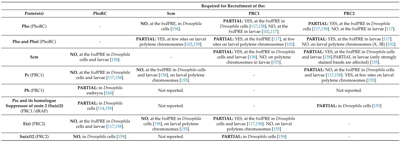
Table 2.Interdependence between the recruitment of different PcG complexes.
Required for Recruitment of the:
Protein(s) PhoRC Scm PRC1 PRC2
Pho(PhoRC) - NO, at thebxdcells [PRE, in158].Drosophila
PARTIAL:YES, at thebxdPRE in
Drosophilacells [117,158]; NO, at the
bxdPRE in larvae [102,117].
PARTIAL:YES, at thebxdPRE inDrosophila
cells [117,158]. NO, at thebxdPRE in larvae [117].
Pho and Phol(PhoRC) - PARTIAL:polytene chromosomes [YES, at few sites on larval102,159]. sites on larval polytene chromosomes [PARTIAL:YES, at thebxdPRE [117], at few102]. NO, on larval polytene chromosomes (X, 3R) [PARTIAL:YES, at thebxdPRE in larvae [117102].].
Scm NO, at thebxdPRE inDrosophila
cells and larvae [158].
-PARTIAL:YES, at thebxdPRE inDrosophila
cells and larvae [158]. NO, on polytene chromosomes in larvae [155].
PARTIAL:YES, at thebxdPRE inDrosophilacells and larvae [158].PARTIAL in larvae (only strongly
stained bands are affected) [155].
Pc(PRC1) NOcells and larvae [, at thebxdPRE in117Drosophila,158].
NO, at thebxdPRE inDrosophilacells and larvae [158], on larval polytene
chromosomes [155].
-PARTIAL:NO, at thebxdPRE inDrosophilacells and larvae [117,158]. YES, at few sites on larval
polytene chromosomes [155]. Ph(PRC1) PARTIAL:embryos [inDrosophila160] Not reported. - Not reported. Psc and its homologue
Suppressor of zeste 2 (Su(z)2) (PRC1/dRAF)
PARTIAL:inDrosophila
cells [114,159] Not reported. - PARTIAL:inDrosophilacells [159]
E(z)(PRC2) NOcells and larvae [, at thebxdPRE in117Drosophila,158].
NO, at thebxdPRE inDrosophila
cells [158], on larval polytene chromosomes [155].
PARTIAL:YES, at thebxdPRE inDrosophila
cells and larvae [117,158]. NO, on larval polytene chromosomes [155]
-Although studied in less detail thanPho/Phol, thedsp1mutation has been shown to affect the binding of Ph to transgene PRE on polytene chromosomes [83]. Likewise, mutation in thecggene reduces the binding of Ph, Psc and affects H3K27me3 in a small subset of genome PREs [79].
Mutations of binding sites for at least several PRE DNA-binding factors have a negative effect on the recruitment of PcG proteins. It has been shown that Pho site mutation reduces the recruitment of Pho, dSfmbt, Scm, Ph, E(z), and H3K27me3 proteins tobxdPRE [139,161] and affects the recruitment of Ph protein toFab7PRE [83] in transgenes. The absence of G(A)n prevents Dsp1 binding and reduces the recruitment of Ph, Pho, and E(z) proteins toFab7PRE [83]. Mutations of GAF sites affect the binding of Pc toFab7PRE andbxdPRE [83,143] and, slightly, Ph recruitment toFab7PRE [83]. As indicated above, the recruitment of not only the test factor but also of additional proteins may be affected. In particular, this can also explain why, in contrast to the mutation ofpho/pholgenes [102], mutation of Pho sites affects the recruitment of Ph protein [139,161]. Thus, a more detailed analysis is required to test the effect of site mutation on the ability of different proteins to bind DNA.
6. Multiple Interactions between Proteins on PRE
The important property of PcG proteins is their ability to establish multiple direct contacts with each other (Figure3). Contacts at three levels can be distinguished: between DNA-binding proteins, between DNA-binding proteins and subunits of PcG complexes, and between different PcG complexes.
Epigenomes 2018, 2, 1 9 of 22
of Pho, dSfmbt, Scm, Ph, E(z), and H3K27me3 proteins to bxdPRE [139,161] and affects the recruitment of Ph protein to Fab7PRE [83] in transgenes. The absence of G(A)n prevents Dsp1 binding and reduces the recruitment of Ph, Pho, and E(z) proteins to Fab7PRE [83]. Mutations of GAF sites affect the binding of Pc to Fab7PRE and bxdPRE [83,143] and, slightly, Ph recruitment to
Fab7PRE [83]. As indicated above, the recruitment of not only the test factor but also of additional proteins may be affected. In particular, this can also explain why, in contrast to the mutation of
pho/phol genes [102], mutation of Pho sites affects the recruitment of Ph protein [139,161]. Thus, a more detailed analysis is required to test the effect of site mutation on the ability of different proteins to bind DNA.
6. Multiple Interactions between Proteins on PRE
The important property of PcG proteins is their ability to establish multiple direct contacts with each other (Figure 3). Contacts at three levels can be distinguished: between DNA-binding proteins, between DNA-binding proteins and subunits of PcG complexes, and between different PcG complexes.
Figure 3. Direct protein-protein interactions between PcG proteins.Arrows show the characterized direct interactions between different PcG subunits and PRE DNA-binding proteins.
6.1. Contacts between DNA-Binding Proteins
This type of contacts has not been studied sufficiently, but at least two interacting pairs have been identified: Grh interacts with Pho [108], and GAF interacts with Psq [151,162]. It can be predicted that some other protein pairs will be found to interact, taking into account the existence of functional contacts between proteins, including genetic interaction and co-assistant function (see below). GAF co-purifies with Adf1 and Zeste, which makes them potential candidates as direct interactors [163]. The interactions between DNA-binding proteins appear to play a crucial role in the recruitment of PcG complexes, since combinations of proteins may be required for their effective binding to DNA.
6.2. Contacts between DNA-Binding Proteins and Subunits of PcG Complexes
Most of PRE DNA-binding proteins were purified together with PRC1 or PRC2 under certain conditions. Thus, Psq [164], Combgap [79], Grh [156], Zeste [57] and Adf1 [110] were shown to be closely linked to PRC1, and Pho [156,161,165] and GAF [163,165] were purified with both PRC1 and PRC2. Purification of Phol, Spps, and Dsp1 with the PRC1/PRC2 complexes has not been reported. Likely some of the DNA-binding proteins can have specificity to a certain PcG complex (for
Figure 3.Direct protein-protein interactions between PcG proteins. Arrows show the characterized direct interactions between different PcG subunits and PRE DNA-binding proteins.
6.1. Contacts between DNA-Binding Proteins
This type of contacts has not been studied sufficiently, but at least two interacting pairs have been identified: Grh interacts with Pho [108], and GAF interacts with Psq [151,162]. It can be predicted that some other protein pairs will be found to interact, taking into account the existence of functional contacts between proteins, including genetic interaction and co-assistant function (see below). GAF co-purifies with Adf1 and Zeste, which makes them potential candidates as direct interactors [163]. The interactions between DNA-binding proteins appear to play a crucial role in the recruitment of PcG complexes, since combinations of proteins may be required for their effective binding to DNA.
6.2. Contacts between DNA-Binding Proteins and Subunits of PcG Complexes
PRC2. Purification of Phol, Spps, and Dsp1 with the PRC1/PRC2 complexes has not been reported. Likely some of the DNA-binding proteins can have specificity to a certain PcG complex (for example, Psq, Combgap, Grh, Zeste or Adf1 to PRC1) and, thus, take part only in its recruitment to DNA.
A number of direct interactions were characterized. Similar to Pho, its homologue Phol directly interacts with the dSfmbt protein. While the Pho and Phol interactions with dSfmbt are mutually exclusive [139], both proteins can co-occupy the same genomic targets [14,114]. The distribution of Pho, Phol, and dSfmbt proteins is well correlated with PREs [114], however, the overall genomic distributions of these proteins are very different. While Pho shows strongest binding to PREs, the strongest peaks for Phol reside at TSS of a subset of transcriptionally active loci, outside Polycomb target regions [14,114]. Moreover, only about 50% of dSfmbt and Pho colocalize in the genome [13]. Thus, Pho, Phol and dSfmbt are the main but not the omnipresent partners and could bind DNA independently of each other.
Both Pho and Phol are directly connected to the PcG core complexes. Pho is able to form direct contacts with E(z), Esc [117], Ph, and Pc [166]. Phol was shown to interact directly with Esc, but not with E(z) [117]; its probable interaction with Ph and Pc has not been verified. Moreover, dSfmbt was shown to establish direct contacts with the Scm protein [161,167], which is supposed to be a connector between different complexes [155,161]. The ability of PhoRC (Pho, dSfmbt) and Phol to establish the above contacts is in accordance with requirement of Pho/Phol for PRC1 and PRC2 recruitment at several PREs (Table2).
The Pho/Phol protein pair plays the main role in the recruitment of PRC1 and PRC2 complexes to only a few genomic loci of polytene chromosomes. It suggests that other proteins can also provide for PRC1 and PRC2 binding, and therefore other connections with PcG proteins should exist.
Two such pairs are known: Zeste can be purified together with Ph [57] and interacts directly with this factor [168]; Grh can be purified together with Pc [156] and establish direct contacts with Sce [133]. Testing of other proteins for direct interactions has not been reported, and therefore more contacts of this type are expected to be found.
6.3. Contacts between Different PcG Complexes
The available data suggest that the repression may require interactions between different complexes with the involvement of connector proteins. One of them is the Scm protein that can link together the PhoRC, PRC1, and PRC2 complexes.
The binding of Scm is independent of PRC1 and PRC2 [155,158] (Table2). At the same time, Scm knockdown negatively affects the binding of PRC1 and PRC2 complexes tobxdPRE [158] and abrogates the binding of PRC2, but not PRC1, to strongly stained bands on polytene chromosomes [155]. This suggests that Scm can assist in the recruitment of PRC2 and, locally, of PRC1 complexes to PREs. It has been shown that Scm directly interacts with dSfmbt [161,167] and Ph [169,170] and in this way can link together the PhoRC and PRC1 complexes [161]. The probable Scm partner from the PRC2 complex has not yet been identified.
7. Consequences of Multiple Contacts Formed by PRE DNA-Binding Proteins
The ability of DNA-binding proteins to form multiple contacts accounts for a number of important properties of these factors, including the ability to establish indirect contacts with PREs, the redundant functions of DNA-binding proteins, and assistance between proteins in the recruitment to chromatin.
7.1. Indirect Contacts with PRE
A fairly common property of DNA-binding proteins is the ability to be recruited to PRE DNA indirectly, through contact with other proteins.
It is assumed that the Combgap protein can also be recruited in the same way. This may explain the fact that Combgap was purified using a 17-bp DNA-sequence that contained no binding site for it [79].
7.2. Redundant Activity of DNA-Binding Proteins
The redundant activity of different proteins suggests that they are implicated in common regulatory pathway and, thus, should directly or indirectly interact with the same subunits of PcG complexes.
Redundant activity clearly manifests itself in genetic tests. Carriers of mutations in most of characterized PRE DNA-binding factors are usually more viable than those of mutations in PRC1 and PRC2 core proteins, suggesting that their functional activities are partially duplicated by other proteins.
The redundant function has been well demonstrated for the Pho/Phol proteins. In wild-type flies, Phol appears to play no significant role, because its removal has no visible effect on PcG function [102]. However, both proteins are responsible for the recruitment of PcG complexes in larvae [102,117], and Phol can partially compensate the absence of Pho, since its level at PREs becomes higher upon Pho knockdown [114]. Thus, Phol may have a backup function when Pho is damaged. It is probable that the same redundancy exists in cases of some other PRE DNA-binding proteins.
7.3. Assistance between Proteins
The Pho protein has primer function in the recruitment of PRC1 and PRC2 complexes at several PREs [117,158]. At the same time, there is evidence that the binding of Pho is facilitated by other proteins.
The Pho protein binding to chromatinized PREs in vitro was shown to depend on GAF and Zeste proteins [171]. This experiment was performed with a PRE fragment containing sites for Pho, GAF and Zeste proteins. The three proteins can efficiently bind to naked DNA without cooperation. When tested on the chromatinized PRE, however, only GAF alone was capable of efficient binding, while Pho and Zeste were not. At the same time, Pho was efficiently recruited in the presence of GAF. In turn, Pho stimulated GAF binding. It is assumed that in this case GAF acts as a classic chromatin remodeler and a pioneer factor, releasing DNA from the nucleosomes to allow Pho binding. If this is the case, then GAF should facilitate the recruitment of other DNA-binding proteins to PRE.
A noteworthy fact is that, although the Zeste protein is not recruited alone, it can provide for the binding of Pho. Thus, Pho and Zeste cooperate to facilitate the binding of each other to chromatinized PRE. A similar requirement for cofactor binding may exist in vivo.
A similar cooperative interaction between the Pho and Grh proteins was observed in vitro [108]: Pho facilitated Grh binding to Grh sites, while Grh facilitated Pho binding to Pho sites on naked DNA. It was initially suggested that dSfmbt is tethered to PREs by Pho or Phol, since mutation of Pho/Phol sites negatively affects the recruitment of dSfmbt to PREs [139]. Subsequent experiments showed, however, that dSfmbt facilitates Pho binding to PREs, with the level of Pho bound to these elements decreasing after dSfmbt knockdown [114].
Moreover, Pho binding to chromatinized PRE in vitro can be promoted by PRC1 core [172]. In accordance, in vivo while recruitment of Pho is not sensitive to Pc depletion [117,158], it becomes reduced in Ph mutant embryos [160] and in Psc/Su(z)2 double null cells [114,159], suggesting the same dependence on PRC1 and possibly dRAF (Table2).
8. Much More DNA-Binding Proteins Waits to Be Identified
Several sequences bound by an unknown protein were reported to be important for repression and PSS [82,95]. In other studies, several potentially important motifs were computationally predicted [14,110]. Two of these motifs resemble sites for the Klumpfuss (KLU) and Adult enhancer factor 1 (AEF1) transcription factors [110]. However, it may well be that they can also interact with as yet unidentified proteins.
Other proteins probably implicated in PRE DNA-binding function include the Sp1/KLF family members [125] and Fs(1)h [154], the binding sites for which are present in PREs.
A DNA-binding potential is also characteristic of the Jing protein, a component of the Jarid2–Jing–PRC2 complex [51], the vertebrate homologue of which binds to DNA [177].
In addition, several proteins with a DNA-binding potential were isolated together with PcG proteins [155,156,163]. For example, Enok and Br140 were tagged by Pc [156], and more than 20 proteins with C2H2-type zinc finger motifs were purified via GAF [163]. At least three GAF-associated proteins with C2H2-type zinc finger motifs were confirmed to be GAF direct partners: CG2199, Pzg/Z4, and MEP-1 [163]. MEP-1 and Pzg/Z4 are known to be associated with chromatin remodeling activity [178,179]. Although GAF is a multifunctional factor implicated in different nuclear functions, these proteins can be implicated in the PcG/TrxG system as well.
Two proteins, Scm and dSfmbt, are of special interest with regard to identification of their recruiters. Scm is recruited irrespective of other complexes [155,158], and none of its recruiters has been identified to date. The majority of (but not all) predicted PREs bound by dSfmbt are co-bound by Pho/Phol proteins [114]. Thus, dSfmbt should apparently be recruited by other proteins, at least at some sites.
The CG9932 protein carrying C2H2-type zinc finger motifs was isolated together with Scm [155]. During purification of the dSfmbt complex, no DNA-binding proteins except Pho were enriched by immunoprecipitation [180]. Apparently, PRE DNA-binding proteins form multiple but fairly weak interactions with PcG proteins and therefore are present at low concentration in the immunoprecipitate (IP). Consequently, PRE DNA-binding proteins are not purified as stable components of any PcG complex. We observed a similar association pattern of DNA-binding proteins when analyzing the GAF interactome: the direct partners of GAF were found among proteins whose peptide scores in IP were not the highest [163]. Apparently, such “background” proteins found in IPs may represent PRE DNA-binding factors and potential recruiters of PcG complexes to chromatin.
9. Model of Combinatorial Recruitment
How does the recruitment of PcG complexes to PRE DNA is implemented?
Firstly, PRC1, PRC2, and Scm do not form high-affinity contacts with PhoRC or other DNA-binding proteins. Secondly, none of known DNA-binding proteins is responsible for genome-wide recruitment of PcG complexes. Thirdly, depletion of the non-DNA-binding subunits from one of the PcG complexes does not globally affect the recruitment of subunits of other PcG complexes.
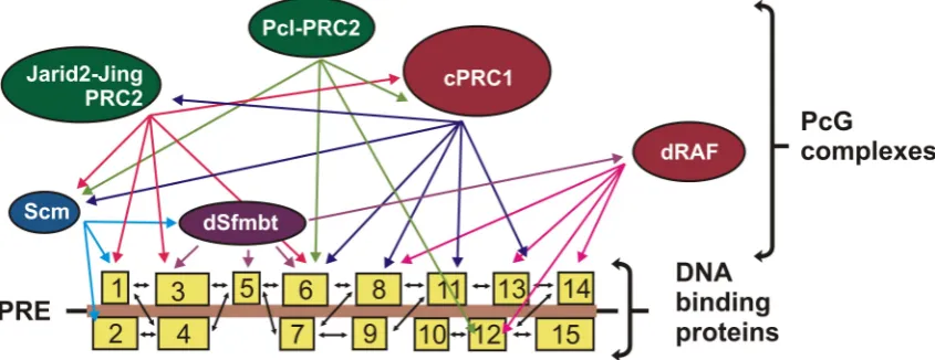
These properties can be explained by the fact that many DNA-binding proteins simultaneously take part in the recruitment of PcG complexes to chromatin (Figure4). The activity of many of these proteins is insured by functional clones that have a redundant activity and can interact with the same DNA-binding sites. DNA-binding proteins form multiple but fairly weak interactions with each other, thereby creating a kind of spider web for recruiting PcG complexes. In turn, PcG complexes bind to this platform due to multiple but weak interactions with DNA-binding proteins or mediator proteins.
What is the purpose for creating a combinatorial platform for the recruitment of PcG complexes? It is likely that the degree of affinity of DNA-binding proteins for individual PREs has a direct effect on the binding affinity of PcG complexes. In addition, specific combinations of DNA binding sites can regulate the recruitment of alternative or modified variants of PcG complexes. Apparently, some unknown combinations of DNA-binding proteins can play a decisive role in this process.
recognition of the PRE-binding platform occurs: whether DNA-binding proteins are recruited to PREs individually or preassembled in groups or complexes?
Epigenomes 2018, 2, 1 13 of 22
Figure 4.Model of combinatorial recruitment. Multiple DNA-binding proteins (boxes 1–15) interact with each other and with different PcG proteins, creating an efficient and specific platform for the recruitment of PcG complexes.
If most of the proteins are preassembled in groups or complexes, they may first recognize the DNA structure and then, at the second step, recognize specific DNA sites and form stronger contacts.
The same “preassembled or assembled” question fully applies to the recruitment of PRC1 and PRC2 complexes to chromatin. Do these complexes first recognize the platform created by DNA-binding proteins or bind together with them? Do they bind in preassembled state or are assembled on DNA? It has been shown that PRC2–Pcl is stable in the presence of ethidium bromide, indicating that DNA is not necessary for the formation of this complex [49]. If it is the case and other complexes are pre-assembled as well, efforts must be made to develop a combinatorial structural model for the recruitment of proteins to DNA.
10. Conclusions and Future Perspective
Despite the large amount of available information on the recruitment of PcG complexes to DNA via DNA-binding proteins, there still are many gaps that need to be filled.
Many presently unknown PRE-associated DNA-binding proteins are likely to be discovered in the nearest future. They will possibly include an important group of proteins with C2H2-type zinc fingers capable of specifically binding to various DNA motifs. In order to fully understand the process of PcG recruitment to chromatin, a more detailed study is needed of direct contacts between DNA-binding proteins and PcG complexes, as well as of other PcG proteins. A major breakthrough in understanding the recruitment process can be achieved using new genome editing technologies, such as Clustered regularly interspaced short palindromic repeat/CRISPR-associated 9 (CRISPR/Cas9) [181], which allow researchers to make desired deletions of genes and gene fragments coding for the domains participating in protein-protein interactions.
The PcG system is evolutionally very stable, and subunits of main PcG complexes have homologs in mammals exhibiting similar activities. It suggests that similar mechanisms can account for PcG targeting in Drosophila and mammals. At the same time, the vertebrate system is much more complex, and many Drosophila factors are represented in it by several paralogs. Therefore, the participation of a larger number of DNA-binding proteins in the recruitment is expected, which makes their identification more difficult.
Several Drosophila PRE DNA-binding proteins have direct homologs in mammals: Pho/Phol (YY1/YY2), GAF (c-Krox/Th-POK), Spps (Sp1/KLF), Dsp1 (HMGB1/2), Grh (CP2) (Table 1). Despite several connections, their participation in PcG recruitment requires further studies [64,66,133,182]. In addition, other DNA-binding proteins were linked to PcG repression in mammals, incl. AEBP2 (homolog of Jing) [177,183,184] that acts together with Jarid2 [54,185–188]; RUNX1 (homolog of Lozenge) [189]; REST (homolog of Charlatan) [69,70]; Snail (homolog of Snail) [69,190], etc.
Figure 4.Model of combinatorial recruitment. Multiple DNA-binding proteins (boxes 1–15) interact with each other and with different PcG proteins, creating an efficient and specific platform for the recruitment of PcG complexes.
If most of the proteins are preassembled in groups or complexes, they may first recognize the DNA structure and then, at the second step, recognize specific DNA sites and form stronger contacts.
The same “preassembled or assembled” question fully applies to the recruitment of PRC1 and PRC2 complexes to chromatin. Do these complexes first recognize the platform created by DNA-binding proteins or bind together with them? Do they bind in preassembled state or are assembled on DNA? It has been shown that PRC2–Pcl is stable in the presence of ethidium bromide, indicating that DNA is not necessary for the formation of this complex [49]. If it is the case and other complexes are pre-assembled as well, efforts must be made to develop a combinatorial structural model for the recruitment of proteins to DNA.
10. Conclusions and Future Perspective
Despite the large amount of available information on the recruitment of PcG complexes to DNA via DNA-binding proteins, there still are many gaps that need to be filled.
Many presently unknown PRE-associated DNA-binding proteins are likely to be discovered in the nearest future. They will possibly include an important group of proteins with C2H2-type zinc fingers capable of specifically binding to various DNA motifs. In order to fully understand the process of PcG recruitment to chromatin, a more detailed study is needed of direct contacts between DNA-binding proteins and PcG complexes, as well as of other PcG proteins. A major breakthrough in understanding the recruitment process can be achieved using new genome editing technologies, such as Clustered regularly interspaced short palindromic repeat/CRISPR-associated 9 (CRISPR/Cas9) [181], which allow researchers to make desired deletions of genes and gene fragments coding for the domains participating in protein-protein interactions.
The PcG system is evolutionally very stable, and subunits of main PcG complexes have homologs in mammals exhibiting similar activities. It suggests that similar mechanisms can account for PcG targeting inDrosophilaand mammals. At the same time, the vertebrate system is much more complex, and manyDrosophilafactors are represented in it by several paralogs. Therefore, the participation of a larger number of DNA-binding proteins in the recruitment is expected, which makes their identification more difficult.
several connections, their participation in PcG recruitment requires further studies [64,66,133,182]. In addition, other DNA-binding proteins were linked to PcG repression in mammals, incl. AEBP2 (homolog of Jing) [177,183,184] that acts together with Jarid2 [54,185–188]; RUNX1 (homolog of Lozenge) [189]; REST (homolog of Charlatan) [69,70]; Snail (homolog of Snail) [69,190], etc.
Several studies have confirmed the existence of PREs in the mammalian genomes [69–73]. Besides DNA-binding factors, the important role in PcG recruitment in mammals is assumed to belong to CpG islands or to combination of CpG islands with sites for DNA-binding proteins [69,70,191–193]. Other studies also suggest the large role of histone modifications and long non-coding RNAs in PcG recruitment in mammals [5,194–197]. The combinations of different features as well as the comprehensive search of PcG associated DNA-binding proteins could be possible strategies for identification of the mammalian PREs genome-wide. The role of histone modifications or long noncoding RNAs in PcG complexes recruitment inDrosophilais currently controversial and requires experimental verifications. Nevertheless, we believe that theDrosophilamodel will contribute to the understanding of general principles of PcG recruitment in different animals, including humans.
Acknowledgments:We are grateful to N.A. Gorgolyuk for his help in preparing the manuscript. This study was supported by Russian Science Foundation No. 14-24-00166.
Author Contributions:M.E., P.G. and D.C. wrote jointly this manuscript.
Conflicts of Interest:The authors declare no conflict of interest.
References
1. Chetverina, D.A.; Elizar’ev, P.V.; Lomaev, D.V.; Georgiev, P.G.; Erokhin, M.M. Control of the gene activity by polycomb and trithorax group proteins inDrosophila.Russ. J. Genet.2017,53, 157–177. [CrossRef]
2. Grossniklaus, U.; Paro, R. Transcriptional silencing by polycomb-group proteins. Cold Spring Harb. Perspect. Biol.2014,6, a019331. [CrossRef] [PubMed]
3. Kassis, J.A.; Kennison, J.A.; Tamkun, J.W. Polycomb and trithorax group genes inDrosophila.Genetics2017, 206, 1699–1725. [CrossRef] [PubMed]
4. Kingston, R.E.; Tamkun, J.W. Transcriptional regulation by trithorax-group proteins. Cold Spring Harb. Perspect. Biol.2014,6, a019349. [CrossRef] [PubMed]
5. Schuettengruber, B.; Bourbon, H.M.; Di Croce, L.; Cavalli, G. Genome regulation by polycomb and trithorax: 70 years and counting.Cell2017,171, 34–57. [CrossRef] [PubMed]
6. Breen, T.R.; Duncan, I.M. Maternal expression of genes that regulate the bithorax complex of Drosophila melanogaster.Dev. Biol.1986,118, 442–456. [CrossRef]
7. Jürgens, G. A group of genes controlling the spatial expression of the bithorax complex inDrosophila.Nature
1985,316, 153–155. [CrossRef]
8. Lewis, E.B. A gene complex controlling segmentation inDrosophila.Nature1978,276, 565–570. [CrossRef] [PubMed]
9. Struhl, G. A gene product required for correct initiation of segmental determination inDrosophila.Nature
1981,293, 36–41. [CrossRef] [PubMed]
10. Kennison, J.A.; Tamkun, J.W. Dosage-dependent modifiers of polycomb and antennapedia mutations in Drosophila.Proc. Natl. Acad. Sci. USA1988,85, 8136–8140. [CrossRef] [PubMed]
11. Kwong, C.; Adryan, B.; Bell, I.; Meadows, L.; Russell, S.; Manak, J.R.; White, R. Stability and dynamics of polycomb target sites inDrosophiladevelopment.PLoS Genet.2008,4, e1000178. [CrossRef] [PubMed] 12. Negre, N.; Hennetin, J.; Sun, L.V.; Lavrov, S.; Bellis, M.; White, K.P.; Cavalli, G. Chromosomal distribution of
PcG proteins duringDrosophiladevelopment.PLoS Biol.2006,4, e170. [CrossRef] [PubMed]
13. Oktaba, K.; Gutierrez, L.; Gagneur, J.; Girardot, C.; Sengupta, A.K.; Furlong, E.E.; Muller, J. Dynamic regulation by polycomb group protein complexes controls pattern formation and the cell cycle inDrosophila. Dev. Cell2008,15, 877–889. [CrossRef] [PubMed]
15. Schwartz, Y.B.; Kahn, T.G.; Nix, D.A.; Li, X.Y.; Bourgon, R.; Biggin, M.; Pirrotta, V. Genome-wide analysis of polycomb targets inDrosophila melanogaster.Nat. Genet.2006,38, 700–705. [CrossRef] [PubMed]
16. Tolhuis, B.; de Wit, E.; Muijrers, I.; Teunissen, H.; Talhout, W.; van Steensel, B.; van Lohuizen, M. Genome-wide profiling of PRC1 and PRC2 Polycomb chromatin binding in Drosophila melanogaster. Nat. Genet.2006,38, 694–699. [CrossRef] [PubMed]
17. Aloia, L.; Di Stefano, B.; Di Croce, L. Polycomb complexes in stem cells and embryonic development. Development2013,140, 2525–2534. [CrossRef] [PubMed]
18. Corley, M.; Kroll, K.L. The roles and regulation of polycomb complexes in neural development. Cell Tissue Res.2015,359, 65–85. [CrossRef] [PubMed]
19. Dressler, G.R.; Patel, S.R. Epigenetics in kidney development and renal disease. Transl. Res. 2015,165, 166–176. [CrossRef] [PubMed]
20. Laugesen, A.; Helin, K. Chromatin repressive complexes in stem cells, development, and cancer. Cell Stem Cell2014,14, 735–751. [CrossRef] [PubMed]
21. Mozgova, I.; Hennig, L. The polycomb group protein regulatory network.Annu. Rev. Plant Biol.2015,66, 269–296. [CrossRef] [PubMed]
22. Schwartz, Y.B.; Pirrotta, V. A new world of polycombs: Unexpected partnerships and emerging functions. Nat. Rev. Genet.2013,14, 853–864. [CrossRef] [PubMed]
23. Sowpati, D.T.; Ramamoorthy, S.; Mishra, R.K. Expansion of the polycomb system and evolution of complexity. Mech. Dev.2015,138 Pt 2, 97–112. [CrossRef] [PubMed]
24. Tan, J.Z.; Yan, Y.; Wang, X.X.; Jiang, Y.; Xu, H.E. EZH2: Biology, disease, and structure-based drug discovery. Acta Pharmacol. Sin.2014,35, 161–174. [CrossRef] [PubMed]
25. Tiffen, J.; Gallagher, S.J.; Hersey, P. Ezh2: An emerging role in melanoma biology and strategies for targeted therapy.Pigment Cell Melanoma Res.2015,28, 21–30. [CrossRef] [PubMed]
26. Pasini, D.; Di Croce, L. Emerging roles for polycomb proteins in cancer.Curr. Opin. Genet. Dev.2016,36, 50–58. [CrossRef] [PubMed]
27. Morey, L.; Santanach, A.; Di Croce, L. Pluripotency and epigenetic factors in mouse embryonic stem cell fate regulation.Mol. Cell. Biol.2015,35, 2716–2728. [CrossRef] [PubMed]
28. Brockdorff, N. Chromosome silencing mechanisms in X-chromosome inactivation: Unknown unknowns. Development2011,138, 5057–5065. [CrossRef] [PubMed]
29. Brockdorff, N.; Turner, B.M. Dosage compensation in mammals.Cold Spring Harb. Perspect. Biol.2015,7, a019406. [CrossRef] [PubMed]
30. Weaver, J.R.; Bartolomei, M.S. Chromatin regulators of genomic imprinting. Biochim. Biophys. Acta2014, 1839, 169–177. [CrossRef] [PubMed]
31. Kheradmand Kia, S.; Solaimani Kartalaei, P.; Farahbakhshian, E.; Pourfarzad, F.; von Lindern, M.; Verrijzer, C.P. EZH2-dependent chromatin looping controlsINK4aandINK4b, but notARF, during human progenitor cell differentiation and cellular senescence.Epigenet. Chromatin2009,2, 16. [CrossRef] [PubMed] 32. Wakeling, L.A.; Ions, L.J.; Escolme, S.M.; Cockell, S.J.; Su, T.; Dey, M.; Hampton, E.V.; Jenkins, G.; Wainwright, L.J.; McKay, J.A.; et al. SIRT1 affects DNA methylation of polycomb group protein target genes, a hotspot of the epigenetic shift observed in ageing.Hum. Genom.2015,9, 14. [CrossRef] [PubMed] 33. Marino, S.; Di Foggia, V. Polycomb group genes in the regeneration of the healthy and pathological skeletal
muscle.Neuropathol. Appl. Neurobiol.2015,42, 407–422. [CrossRef] [PubMed]
34. Hamada, Y.; Bando, T.; Nakamura, T.; Ishimaru, Y.; Mito, T.; Noji, S.; Tomioka, K.; Ohuchi, H. Leg regeneration is epigenetically regulated by histone H3K27 methylation in the cricketGryllus bimaculatus. Development
2015,142, 2916–2927. [CrossRef] [PubMed]
35. Cheedipudi, S.; Puri, D.; Saleh, A.; Gala, H.P.; Rumman, M.; Pillai, M.S.; Sreenivas, P.; Arora, R.; Sellathurai, J.; Schroder, H.D.; et al. A fine balance: Epigenetic control of cellular quiescence by the tumor suppressor PRDM2/RIZ at a bivalent domain in the cyclin a gene.Nucleic Acids Res.2015,43, 6236–6256. [CrossRef] [PubMed]
36. Chou, R.H.; Chiu, L.; Yu, Y.L.; Shyu, W.C. The potential roles of EZH2 in regenerative medicine.Cell Transpl.
2015,24, 313–317. [CrossRef] [PubMed]
38. Czermin, B.; Melfi, R.; McCabe, D.; Seitz, V.; Imhof, A.; Pirrotta, V.Drosophilaenhancer of Zeste/ESC complexes have a histone H3 methyltransferase activity that marks chromosomal polycomb sites.Cell2002, 111, 185–196. [CrossRef]
39. Muller, J.; Hart, C.M.; Francis, N.J.; Vargas, M.L.; Sengupta, A.; Wild, B.; Miller, E.L.; O’Connor, M.B.; Kingston, R.E.; Simon, J.A. Histone methyltransferase activity of aDrosophilapolycomb group repressor complex.Cell2002,111, 197–208. [CrossRef]
40. Cao, R.; Wang, L.; Wang, H.; Xia, L.; Erdjument-Bromage, H.; Tempst, P.; Jones, R.S.; Zhang, Y. Role of histone H3 lysine 27 methylation in polycomb-group silencing.Science2002,298, 1039–1043. [CrossRef] [PubMed] 41. Kuzmichev, A.; Nishioka, K.; Erdjument-Bromage, H.; Tempst, P.; Reinberg, D. Histone methyltransferase
activity associated with a human multiprotein complex containing the enhancer of Zeste protein.Genes Dev.
2002,16, 2893–2905. [CrossRef] [PubMed]
42. Pengelly, A.R.; Copur, O.; Jackle, H.; Herzig, A.; Muller, J. A histone mutant reproduces the phenotype caused by loss of histone-modifying factor polycomb.Science2013,339, 698–699. [CrossRef] [PubMed] 43. Ebert, A.; Schotta, G.; Lein, S.; Kubicek, S.; Krauss, V.; Jenuwein, T.; Reuter, G. Su(var) genes regulate the
balance between euchromatin and heterochromatin inDrosophila.Genes Dev.2004,18, 2973–2983. [CrossRef] [PubMed]
44. Jones, R.S.; Gelbart, W.M. Genetic analysis of the enhancer of zeste locus and its role in gene regulation in Drosophila melanogaster.Genetics1990,126, 185–199. [PubMed]
45. Simon, J.; Chiang, A.; Bender, W. Ten differentPolycombgroup genes are required for spatial control of the abdAandAbdBhomeotic products.Development1992,114, 493–505. [PubMed]
46. Ketel, C.S.; Andersen, E.F.; Vargas, M.L.; Suh, J.; Strome, S.; Simon, J.A. Subunit contributions to histone methyltransferase activities of fly and worm polycomb group complexes.Mol. Cell. Biol.2005,25, 6857–6868. [CrossRef] [PubMed]
47. Birve, A.; Sengupta, A.K.; Beuchle, D.; Larsson, J.; Kennison, J.A.; Rasmuson-Lestander, A.; Muller, J. Su(z)12, a novelDrosophilapolycomb group gene that is conserved in vertebrates and plants.Development2001,128, 3371–3379. [PubMed]
48. Nekrasov, M.; Klymenko, T.; Fraterman, S.; Papp, B.; Oktaba, K.; Kocher, T.; Cohen, A.; Stunnenberg, H.G.; Wilm, M.; Muller, J. Pcl-PRC2 is needed to generate high levels of H3-K27 trimethylation at Polycomb target genes.EMBO J.2007,26, 4078–4088. [CrossRef] [PubMed]
49. Tie, F.; Prasad-Sinha, J.; Birve, A.; Rasmuson-Lestander, A.; Harte, P.J. A 1-megadalton ESC/E(Z) complex fromDrosophilathat contains polycomblike and RPD3. Mol. Cell. Biol. 2003,23, 3352–3362. [CrossRef] [PubMed]
50. Tie, F.; Banerjee, R.; Stratton, C.A.; Prasad-Sinha, J.; Stepanik, V.; Zlobin, A.; Diaz, M.O.; Scacheri, P.C.; Harte, P.J. CBP-mediated acetylation of histone H3 lysine 27 antagonizesDrosophilaPolycomb silencing. Development2009,136, 3131–3141. [CrossRef] [PubMed]
51. Herz, H.M.; Mohan, M.; Garrett, A.S.; Miller, C.; Casto, D.; Zhang, Y.; Seidel, C.; Haug, J.S.; Florens, L.; Washburn, M.P.; et al. Polycomb repressive complex 2-dependent and -independent functions of Jarid2 in transcriptional regulation inDrosophila.Mol. Cell. Biol.2012,32, 1683–1693. [CrossRef] [PubMed]
52. Lagarou, A.; Mohd-Sarip, A.; Moshkin, Y.M.; Chalkley, G.E.; Bezstarosti, K.; Demmers, J.A.; Verrijzer, C.P. dKMD2 couples histone H2A ubiquitylation to histone H3 demethylation during polycomb group silencing. Genes Dev.2008,22, 2799–2810. [CrossRef] [PubMed]
53. Wang, H.; Wang, L.; Erdjument-Bromage, H.; Vidal, M.; Tempst, P.; Jones, R.S.; Zhang, Y. Role of histone H2A ubiquitination in polycomb silencing.Nature2004,431, 873–878. [CrossRef] [PubMed]
54. Kalb, R.; Latwiel, S.; Baymaz, H.I.; Jansen, P.W.; Muller, C.W.; Vermeulen, M.; Muller, J. Histone H2A monoubiquitination promotes histone H3 methylation in polycomb repression.Nat. Struct. Mol. Biol.2014, 21, 569–571. [CrossRef] [PubMed]
55. Pengelly, A.R.; Kalb, R.; Finkl, K.; Muller, J. Transcriptional repression by PRC1 in the absence of H2A monoubiquitylation.Genes Dev.2015,29, 1487–1492. [CrossRef] [PubMed]
56. Francis, N.J.; Saurin, A.J.; Shao, Z.; Kingston, R.E. Reconstitution of a functional core polycomb repressive complex.Mol. Cell2001,8, 545–556. [CrossRef]
58. Shao, Z.; Raible, F.; Mollaaghababa, R.; Guyon, J.R.; Wu, C.T.; Bender, W.; Kingston, R.E. Stabilization of chromatin structure by PRC1, a polycomb complex.Cell1999,98, 37–46. [CrossRef]
59. Francis, N.J.; Kingston, R.E.; Woodcock, C.L. Chromatin compaction by a polycomb group protein complex. Science2004,306, 1574–1577. [CrossRef] [PubMed]
60. King, I.F.; Emmons, R.B.; Francis, N.J.; Wild, B.; Muller, J.; Kingston, R.E.; Wu, C.T. Analysis of a polycomb group protein defines regions that link repressive activity on nucleosomal templates to in vivo function. Mol. Cell. Biol.2005,25, 6578–6591. [CrossRef] [PubMed]
61. King, I.F.; Francis, N.J.; Kingston, R.E. Native and recombinant polycomb group complexes establish a selective block to template accessibility to repress transcription in vitro.Mol. Cell. Biol.2002,22, 7919–7928. [CrossRef] [PubMed]
62. Fischle, W.; Wang, Y.; Jacobs, S.A.; Kim, Y.; Allis, C.D.; Khorasanizadeh, S. Molecular basis for the discrimination of repressive methyl-lysine marks in histone H3 by polycomb and HP1 chromodomains. Genes Dev.2003,17, 1870–1881. [CrossRef] [PubMed]
63. Min, J.; Zhang, Y.; Xu, R.M. Structural basis for specific binding of polycomb chromodomain to histone H3 methylated at Lys 27.Genes Dev.2003,17, 1823–1828. [CrossRef] [PubMed]
64. Bauer, M.; Trupke, J.; Ringrose, L. The quest for mammalian Polycomb response elements: Are we there yet? Chromosoma2016,125, 471–496. [CrossRef] [PubMed]
65. Di Croce, L.; Helin, K. Transcriptional regulation by polycomb group proteins.Nat. Struct. Mol. Biol.2013, 20, 1147–1155. [CrossRef] [PubMed]
66. Kassis, J.A.; Brown, J.L. Polycomb group response elements inDrosophilaand vertebrates.Adv. Genet.2013, 81, 83–118. [PubMed]
67. Steffen, P.A.; Ringrose, L. What are memories made of? How polycomb and trithorax proteins mediate epigenetic memory.Nat. Rev. Mol. Cell Biol.2014,15, 340–356. [CrossRef] [PubMed]
68. van Kruijsbergen, I.; Hontelez, S.; Veenstra, G.J. Recruiting polycomb to chromatin.Int. J. Biochem. Cell Biol.
2015,67, 177–187. [CrossRef] [PubMed]
69. Arnold, P.; Scholer, A.; Pachkov, M.; Balwierz, P.J.; Jorgensen, H.; Stadler, M.B.; van Nimwegen, E.; Schubeler, D. Modeling of epigenome dynamics identifies transcription factors that mediate polycomb targeting.Genome Res.2013,23, 60–73. [CrossRef] [PubMed]
70. Dietrich, N.; Lerdrup, M.; Landt, E.; Agrawal-Singh, S.; Bak, M.; Tommerup, N.; Rappsilber, J.; Sodersten, E.; Hansen, K. Rest-mediated recruitment of polycomb repressor complexes in mammalian cells.PLoS Genet.
2012,8, e1002494. [CrossRef] [PubMed]
71. Schorderet, P.; Lonfat, N.; Darbellay, F.; Tschopp, P.; Gitto, S.; Soshnikova, N.; Duboule, D. A genetic approach to the recruitment of PRC2 at theHoxDlocus.PLoS Genet.2013,9, e1003951. [CrossRef] [PubMed]
72. Sing, A.; Pannell, D.; Karaiskakis, A.; Sturgeon, K.; Djabali, M.; Ellis, J.; Lipshitz, H.D.; Cordes, S.P. A vertebrate polycomb response element governs segmentation of the posterior hindbrain. Cell2009,138, 885–897. [CrossRef] [PubMed]
73. Woo, C.J.; Kharchenko, P.V.; Daheron, L.; Park, P.J.; Kingston, R.E. A region of the humanHoxDcluster that confers polycomb-group responsiveness.Cell2010,140, 99–110. [CrossRef] [PubMed]
74. Aranda, S.; Mas, G.; Di Croce, L. Regulation of gene transcription by polycomb proteins.Sci. Adv.2015,1, e1500737. [CrossRef] [PubMed]
75. Chan, C.S.; Rastelli, L.; Pirrotta, V. A Polycomb response element in the Ubx gene that determines an epigenetically inherited state of repression.EMBO J.1994,13, 2553–2564. [PubMed]
76. Chiang, A.; O‘Connor, M.B.; Paro, R.; Simon, J.; Bender, W. Discrete polycomb-binding sites in each parasegmental domain of the bithorax complex.Development1995,121, 1681–1689. [PubMed]
77. Simon, J.; Chiang, A.; Bender, W.; Shimell, M.J.; O‘Connor, M. Elements of theDrosophilabithorax complex that mediate repression by polycomb group products.Dev. Biol.1993,158, 131–144. [CrossRef] [PubMed] 78. Gindhart, J.G., Jr.; Kaufman, T.C. Identification of polycomb and trithorax group responsive elements in the
regulatory region of theDrosophilahomeotic gene Sex combs reduced.Genetics1995,139, 797–814. [PubMed] 79. Ray, P.; De, S.; Mitra, A.; Bezstarosti, K.; Demmers, J.A.; Pfeifer, K.; Kassis, J.A. Combgap contributes to recruitment of Polycomb group proteins inDrosophila. Proc. Natl. Acad. Sci. USA2016,113, 3826–3831. [CrossRef] [PubMed]