Vascular Health and Risk Management
Dovepress
R e V i e w
open access to scientific and medical research
Open Access Full Text Article
The causes, consequences, and treatment
of left or right heart failure
Pablo Pazos-López Jesús Peteiro-Vázquez Ana Carcía-Campos Lourdes García-Bueno Juan Pablo Abugattas de Torres
Alfonso Castro-Beiras Department of Cardiology, Complejo hospitalario Universitario A Coruña, A Coruña, Spain
Correspondence: Pablo Pazos-López Juan Bautista Andrade 64 4°, C.P. 36005, Pontevedra, Spain
Tel +34 61 990 7556 Fax +34 98 117 8258
email [email protected]
Abstract: Chronic heart failure (HF) is a cardiovascular disease of cardinal importance because of several factors: a) an increasing occurrence due to the aging of the population, primary and secondary prevention of cardiovascular events, and modern advances in therapy, b) a bad prognosis: around 65% of patients are dead within 5 years of diagnosis, c) a high economic cost: HF accounts for 1% to 2% of total health care expenditure. This review focuses on the main causes, consequences in terms of morbidity, mortality and costs and treatment of HF.
Keywords: heart failure, cause, consequence, treatment
Definition, classification, and occurrence
of heart failure
Heart failure (HF) is a complex clinical syndrome caused by structural or functional cardiac disorders that impair the ability of one or both ventricles to fill with or eject blood.1 For the diagnosis of HF, symptoms (typically shortness of breath at rest or during exertion and/or fatigue), signs of fluid retention (such as pulmonary congestion and/or ankle swelling), and objective evidence of a decrease in myocardial performance at rest (normally demonstrated in an echocardiography study) are required.2 According to the time from ventricular dysfunction to clinical manifestations, left ventricular ejection fraction (LVEF), and the main site of congestion, HF can be divided into dif-ferent groups (Figure 1). Generally, it is a chronic condition with bouts of worsening symptoms that may require medical attention (decompensations). However, it may present acutely within just 24 hours in the form of pulmonary edema or even cardiogenic shock. Conventionally, HF was seen to result from the failure of the heart to pump enough blood into the circulation due to ventricular systolic dysfunction defined as LVEF , 40% to 50% (HF with depressed ejection fraction [HFDEF]).2 Nevertheless, patients with nondecreased LVEF can develop HF when higher filling pressures are needed to achieve a normal end-diastolic ventricular volume (HF with preserved ejec-tion fracejec-tion [HFPEF]). The occurrence of this condiejec-tion is more common in women, in the elderly, and in persons with longstanding high blood pressure (HBP) and is associated with a similar prognosis to HFDEF.3,4 Right and left HF refer to syndromes presenting predominantly with systemic or pulmonary congestion leading to jugular venous ingurgitation and ankle swelling or pulmonary edema, respectively.
Generally speaking, the prevalence of HF can be estimated at 1% to 2% in Western countries and the incidence approaches 5 to 10 per 1000 persons per year. Data on the occurrence of HF in the developing world are largely absent. This high prevalence is
Vascular Health and Risk Management downloaded from https://www.dovepress.com/ by 118.70.13.36 on 27-Aug-2020
For personal use only.
Number of times this article has been viewed
This article was published in the following Dove Press journal: Vascular Health and Risk Management
Dovepress Pazos-López et al
increasing because of the aging population, the success in primary and secondary prevention of coronary events, and the development of modern treatments. The mean age of patients with HF in industrialized societies is approximately 75 years.5
This review will focus on the main causes, consequences (in terms of morbidity, mortality, and economic costs), and treatment of chronic HF.
Causes of HF
Although many conditions may lead to HF, the predominant etiologies are myocardial ischemia and HBP (Table 1). The discrepancies in the frequency of causes reported in the medical literature can be explained by differences in the study population (from highly selected participants in clinical trials to relatively unselected subjects in population-based studies, respectively), definitions (eg, consensus on a cut-off value for LVEF to define HFPEF has not been reached), and time period (eg, the Framingham heart study originated in 1948). Furthermore, it has become clear that by using only noninvasive techniques, precise etiology cannot always be determined. For instance, in the Bromley HF study after nuclear testing and cardiac catheterization the percentage of HF with unknown cause declined from 42% to 10%, while the
percentage of patients with ischemic heart disease increased from 29% to 52%.6
Myocardial ischemia
Coronary artery disease (CAD) may be the initiating cause in ≈70% of cases of HF.7 Even in HF individuals clinically classified as nonischemic, evidence of ischemia has been found. Up to 25% may have significant atherosclerosis plaques in the coronary trees at autopsy,8 which points out the limited diagnostic accuracy of image techniques such as angiography. Besides, it has been reported that these patients may suffer ischemic events on the follow-up, a find-ing that suggests that CAD may not be just a ‘bystander’.9 Moreover, the high prevalence of a reduced flow reserve demonstrated in subjects with HF and nonsignificant steno-sis in the main coronary arteries incriminates microvascular impairment as a potential contributor to their myocardial dysfunction.10,11
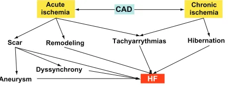
Mechanisms of HF in CAD (Figure 2)
1. Acute myocardial infarction (MI) frequently causes the death of the myocytes of one or more ventricular segments that become scarred, resulting in inadequate relaxation in diastole and impaired contraction in systole. Further decrease in ventricular performance may occur if an aneurysm is developed. MI can also predispose to HF by the dyssynchronous movement of the infarct area that can lessen the efficiency of pump function.
2. Whereas the initial myocardial scarring results in local dysfunction, remodeling in remote areas of the left ventricle (LV) may take place resulting in a distortion of the ventricular structure and geometry, which can contribute to an additional decrement of ventricular function.12 Besides, ventricular enlargement may promote dysfunction of the mitral apparatus with the consequent mitral regurgitation (MR), which can pre-dispose to HF.
Time from ventricular dysfunction to clinical manifestations
Acute HF Chronic HF
Ejection fraction
Main site of congestion
≥ 40%–50%: HFPEF < 40%–50%: HFDEF
Systemic circulation: Right HF
Pulmonary circulation: Left HF
Figure 1 Types of heart failure.
Abbreviations: HF, heart failure; HFDeF, heart failure with depressed ejection fraction; HFPeF, heart failure with preserved ejection faction.
Table 1 Main causes of heart failure
• Myocardial ischemia
• HBP
• Cardiomyopathies
• Valvular heart disease
• PHT
• Congenital heart disease
Abbreviations: HBP, high blood pressure; PHT, pulmonary hypertension. See text for details.
Acute
ischemia CAD ischemiaChronic
HF
Scar Remodeling
Dyssynchrony Aneurysm
Tachyarrythmias Hibernation
Figure 2 From coronary artery disease to heart failure.
Abbreviations: CAD, coronary artery disease; HF, heart failure.
Vascular Health and Risk Management downloaded from https://www.dovepress.com/ by 118.70.13.36 on 27-Aug-2020
Dovepress Causes, consequences, and treatment of heart failure
3. Several tachyarrhythmias such as nonsustained ventricu-lar tachycardia (NSVT) or atrial fibrillation/atrial flutter (AF), common in patients with CAD, can deteriorate cardiac function.
• Loss of atrial contraction in AF decreases ventricular filling and stroke volume.
• Systolic volume is diminished during NSVT due to atrioventricular dissociation and the consequent drop of preload.
• Diastolic time shortening, found in both arrhythmias, also contributes to the lessening of filling and cardiac output.
• When AF and NSVT are persistent they may lead to tachycardia-induced cardiomyopathy (tachycardiomyopathy).13
4. Maintained reduction in blood supply due to chronic severe coronary stenosis may result in low myocardial performance (hibernation).14
HBP
HBP boosts HF risk by two- to three-fold.15,16 Although the relative risk of developing HF in HBP is modest, its high prevalence renders it a cause in approximately one-third of cases.17 Besides, HBP is an independent risk factor for CAD.
effects of HBP in the heart
In hypertensive patients, the myocardium has to pump blood against a higher afterload posed by the elevated resistance of the peripheral vasculature.This condition leads to a compensatory increase in myocardial mass in order to maintain normal cardiac output. Left ventricular hypertrophy (LVH) in HBP is characterized not only by enlargement of the cardiac myocytes but also by an increase of interstitial and perivascular fibrosis.18 The progression from a structurally normal heart to LVH is not solely a consequence of enhanced afterload; many mechanisms are known to be involved, particularly the renin–angiotensin–aldosterone system (RAAS).19
Mechanisms of HF in HBP (Figure 3)
1. Hypertrophic ventricles are characterized by a higher myocardial stiffness and a decreased ability to relax and fill.20–23
2. HBP is associated with myocardial ischemia even in the absence of CAD. Three main mechanisms are implicated:24
• In HBP, the growth of the coronary bed does not keep pace with the increase in cardiac mass, proving a ‘set up’ for chronic ischemia.
• Coronary arteries travel across ventricular walls from the epicardium to the endocardium, perfusing the myocardium mainly during diastole due to a positive ratio between the intravessel and parietal pressures. Such a ratio is reduced in HBP as a consequence of the high ventricular chamber pressure transmitted to the walls, which promotes ischemia. This effect is more pronounced at the subendocardial level. • Coronary flow reserve is impaired due to structural
and functional changes in the arterioles (medial wall thickening and perivascular fibrosis boost vessel resis-tance, endothelial dysfunction impairs vasodilatation capacity).
These conditions may lead to HFPEF. Persistent work overload, hypoxia, and neurohumoral stimulation in long-standing uncontrolled HBP can promote myocyte apoptosis and eventually systolic dysfunction.25
Cardiomyopathies
According to the new classification recently published by the Working Group on Myocardial and Pericardial Diseases of the European Society of Cardiology, a cardiomyopathy is a
↑ RAAS ↑ VR
LVH HBP
↑ Myocardial
stiffness
HFPEF
HFDEF Maintained uncontrolled HBP
Myocytes apoptosis
Ischemia
Figure 3 From high blood pressure to heart failure.
Abbreviations: HBP, high blood pressure; HFDeF, heart failure with depressed ejection fraction; HFPeF, heart failure with preserved ejection fraction; LVH, left ventricular hypertrophy; RAAS, renin–angiotensin–aldosterone system; VR, vascular resistance.
Vascular Health and Risk Management downloaded from https://www.dovepress.com/ by 118.70.13.36 on 27-Aug-2020
Dovepress Pazos-López et al
myocardial disorder in which the heart muscle is structurally and functionally abnormal in the absence of CAD, HBP, valvular, or congenital heart disease severe enough to cause the observed myocardial abnormality. Cardiomyopathies are grouped into five specific phenotypes: dilated cardiomyopathy (DCM), hypertrophic cardiomyopathy (HCM), restrictive cardiomyopathy (RCM), arrhythmogenic right ventricular cardiomyopathy (ARVC), and other unclassified cardiomyo-pathies (isolated noncompaction of the left ventricle [INLV] and Takotsubo syndrome are included in this category). Each phenotype is subclassified into familial and nonfamilial forms taking into account the presence or absence in more than one member of the family, of either the same disorder or a phenotype that is (or could be) caused by the same genetic mutation. Nonfamilial cardiomyopathies are divided into idiopathic (when no identifiable cause is found) or acquired forms (in which ventricular dysfunction is a complication of the disorder rather than an intrinsic feature of the disease).26
DCM
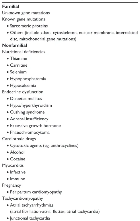
This entity is much more common by far than the other major types of cardiomyopathies. DCM is a heterogeneous disease characterized by left ventricular and sometimes atrial dilation (right ventricular enlargement and dysfunc-tion may be present but is not necessary for the diagnosis), with normal or reduced wall thickness eventually leading to varying degrees of impaired systolic function. The clinical picture at the time of diagnosis can vary widely from patient to patient; some have no symptoms, whereas others develop a progressive refractory HF. When the etiology is investigated with specialized techniques, at least 25% of patients have evidence of familial disease with predominantly autosomal dominant inheritance.27,28 Frequent nonfamilial acquired causes of DCM include cardiotoxic drugs such as anthracy-clines, alcohol, or cocaine, late stage of cardiac infectious and inflammatory diseases (myocarditis), and persistent tachyarrhythmias (tachycardiomyopathy) (Table 2).
HCM
The new classification has redefined HCM as the presence of an increased ventricular wall thickness or mass in the absence of loading conditions (HBP, valve disease) sufficient to cause the observed abnormality.26 Following this new definition, those diseases in which ventricular mass is increased due to interstitial infiltration or intracellular accumulation of meta-bolic substrates are included in this category (Table 3).
LVH in the absence of HBP and valve disease occurs in approximately 1 in 500 of the general population.
Many individuals have a familial disease with an autosomal dominant pattern of inheritance. Mutations identified in these patients affect genes that encode different proteins of the cardiac sarcomere. HCM caused by such mutations predominantly has an asymmetrical pattern of LVH, the interventricular septum segments being the most frequently affected, and myocyte disarray.29 LV volume is usually small and LVEF preserved. Symptoms are related to an impaired ventricular filling (HFPEF), and in some cases, to left outflow track dynamic obstruction. Progression to LV dilatation and systolic dysfunction is rare (from 2.5% up to 15% in some series).30
Although all types of LVH may be present in sarcomeric protein disease, concentric forms are more common in metabolic disorders (eg, Anderson–Fabry disease). Other differential features that may help in the diagnosis of these entities are the inheritance pattern (X-linked, autosomal recessive) and the presence of signs and symptoms derived from multisystemic affectation.26
Table 2 Causes of dilated cardiomyopathy Familial
Unknown gene mutations Known gene mutations
• Sarcomeric proteins
• Others (include z-ban, cytoskeleton, nuclear membrane, intercalated disc, mitochondrial gene mutations)
Nonfamilial Nutritional deficiencies
• Thiamine
• Carnitine
• Selenium
• Hypophosphatemia
• Hypocalcemia endocrine dysfunction
• Diabetes mellitus
• Hypo/hyperthyroidism
• Cushing syndrome
• Adrenal insufficiency
• excessive growth hormone
• Phaeochromocytoma Cardiotoxic drugs
• Cytotoxic agents (eg, anthracyclines)
• Alcohol
• Cocaine Myocarditis
• infective
• immune Pregnancy
• Peripartum cardiomyopathy Tachycardiomyopathy
• Atrial tachyarrhythmias
(atrial fibrillation-atrial flutter, atrial tachycardia)
• Junctional tachycardia
Vascular Health and Risk Management downloaded from https://www.dovepress.com/ by 118.70.13.36 on 27-Aug-2020
Dovepress Causes, consequences, and treatment of heart failure
Athletic training is associated with physiological LVH that can be misinterpreted as a pathological phenotype, but the degree of wall thickness is much less pronounced (,13 mm in septal segments in most cases) and diastolic function is normal.31
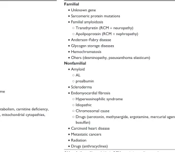
RCM
This cardiomyopathy is characterized by a severe diastolic dysfunction (restrictive physiology) in the presence of nor-mal or reduced diastolic volumes (of one or both ventricles), normal or reduced systolic volumes, and normal ventricular wall thickness. Although historically, systolic function has been said to be preserved in RCM, contractility is not completely normal in this entity.26 Although the exact prevalence of RCM is unknown it is probably the least prevalent type of cardiomyo-pathy. RCM may be idiopathic, familial, or result from various systemic disorders (Table 4). As can be seen from comparing Tables 3 and 4, several genetic mutations and infiltrative diseases may present with a restrictive or hypertrophic phenotype.
ARVC
Ventricular arrhythmias and sudden cardiac death (SCD) are the main manifestations of this rare cardiomyopathy Table 3 Causes of hypertrophic cardiomyopathy
Familial
• Unknown gene mutations
• Sarcomeric protein mutations
• Glycogen storage disease ○ Pompe
○ PRKAG2 ○ Forbes’ ○ Danon
• Lysosomal storage diseases ○ Anderson–Fabry ○ Hurler’s
• Syndromic HCM ○ Noonan’s syndrome ○ LeOPARD syndrome ○ Friedreich’s ataxia
○ Beckwith–wiedermann syndrome ○ Swyer’s syndrome
• Familial amyloid
• Others (disorders of fatty acid metabolism, carnitine deficiency, phosphorylase B kinase deficiency, mitochondrial cytopathies, phospholamban promoter)
Nonfamilial
• Obesity
• infants of diabetic mothers
• Athletic training
• Amyloid ○ AL ○ Prealbumin
Abbreviations: AL, amyloidosis; HCM, hypertrophic cardiomyopathy.
Table 4 Causes of restrictive cardiomyopathy Familial
• Unknown gene
• Sarcomeric protein mutations
• Familial amyloidosis
○ Transthyretin (RCM + neuropathy)
○ Apolipoprotein (RCM + nephropathy)
• Anderson–Fabry disease
• Glycogen storage diseases
• Hemochromatosis
• Ohers (desminopathy, pseuxanthoma elasticum) Nonfamilial
• Amyloid
○ AL
○ prealbumin
• Scleroderma
• Endomyocardial fibrosis
○ Hypereosinophilic syndrome
○ idiopathic
○ Chromosomal cause
○ Drugs (serotonin, methysergide, ergotamine, mercurial agents, busulfan)
• Carcinoid heart disease
• Metastatic cancers
• Radiation
• Drugs (anthracyclines)
Abbreviations: AL, amyloidosis; RCM, restrictive cardiomyopathy.
(1/1000–5000). Although uncommon, right ventricular or biventricular HF mimicking a DCM may be present.32
iNLV
INLV is an infrequent unclassified cardiomyopathy (0.014% of consecutive echocardiograms) assumed to occur due to an arrest in the compaction process of the LV during the normal development of the heart. This entity is morphologically charac-terized by the appearance of prominent trabeculations and deep intertrabecular recesses mainly in the apex and in the inferior and lateral mid segments, with an endsystolic ratio between the noncompacted subendocardial layer and the compacted subepicardial layer .2. The clinical presentation of INLV includes a high prevalence of HF, thromboembolic events and arrhythmias including ventricular tachycardia and AF.33 INLV is commonly familial, with at least 25% of asymptomatic relatives having a range of echocardiographic abnormalities.34 Causative mutations in several genes have been identified.26
Valvulopathies
A severe increase in ventricular afterload may lead to HF in severe aortic and pulmonary stenosis. In valve regurgitation, a persistent volume overload may cause ventricular enlarge-ment and functional impairenlarge-ment.
Vascular Health and Risk Management downloaded from https://www.dovepress.com/ by 118.70.13.36 on 27-Aug-2020
Dovepress Pazos-López et al
Other causes of HF
The elevated resistance of the pulmonary vasculature in pulmonary hypertension may promote right HF. A special case of this condition is observed in patients with chronic obstructive pulmonary disease (cor pulmonale).
Congenital heart diseases such as interventricular and interatrial communication or persistent arterial ductus can lead to HF due to maintained volume overload.
Consequences of HF
Reduction of functional capacity
Functional capacity in HF patients is limited by shortness of breath and fatigue on exertion. A basic pathophysiology of these symptoms can be summarized in two points (Figure 4):
1. When diastolic dysfunction is developed the failing heart requires a higher LV filling pressure to maintain output, particularly during exertion. The filling pressure of the LV can become high enough to cause stiff lungs or even transudation of fluid into the alveoli leading to breathlessness.
2. If systolic function is impaired, the failing heart may be unable to increase the stroke volume adequately in response to exercise. In turn, this leads to the inability to perfuse the exercising muscle effectively. The affected skeletal muscle signals the brain, and this sensation is interpreted as fatigue.
Although this view regards HF as a hemodynamic disorder, many studies have indicated that measurements of cardiac performance and symptoms produced by the disease are
poorly related. For instance, patients with a very low LVEF may be asymptomatic, whereas subjects with just a slightly depressed LVEF may have severe disability.35 The apparent discordance is not well understood but may be explained in part by many noncardiac factors that contribute considerably to exercise tolerance such as changes in peripheral vascular function, skeletal muscle physiology, pulmonary dynamics, or neurohormonal and reflex autonomic activity.36,37 The existence of such noncardiac factors may explain why the hemodynamic improvement produced by several drugs may not be instantly or necessarily translated into improvement in clinical status.
An approach used to quantify the degree of functional limitation imposed by HF was first developed by the New York Heart Association (NYHA). Severity ranges from essentially asymptomatic status – well-treated patients in whom symptoms have been relieved (NYHA I) – or slight limitation in physical activity (NYHA II), to symptoms while walking on the flat (NYHA III) or even breathless at rest and essentially housebound (NYHA IV). To get an objective evaluation of exercise performance, some form of exercise testing is necessary. Corridor walk tests, particularly the six-minute walk test, are commonly used due to their low cost and simplicity.38 However, to explore exercise limitation in greater detail, testing with metabolic gas exchange measurement is more useful. Although the functional class tends to deteriorate over time, most patients do not typically show an inexorable worsening of symptoms. Instead, their severity characteristically fluctuates. Medical therapy and diet can have either favorable or adverse effects on functional capacity even in the absence of significant changes in ventricular function. Some patients may show notable recovery associated with improvement in structural and functional abnormalities. When such improvement is associated with drug therapy, that therapy should be continued indefinitely.
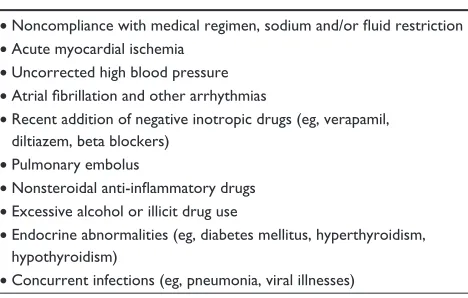
Hospitalizations
As HF progresses, decompensations are more frequent and, therefore, hospitalizations increasingly required. Admissions with HF may be triggered by a concomitant cardiovascular event such as a symptomatic tachyarrhythmia or unstable coronary syndrome, medical or dietary noncompliance, or a noncardiac condition such as infections or a newly diagnosed anemia (Table 5). Unfortunately, the precipitating event is not always apparent. HF hospitalizations account for a substantial portion of the overall costs of caring and may be associated with an astounding worsening in prognosis, particularly in the elderly population. In fact, 50% of patients are readmitted at six months and 25% to 35% are dead at
HF
Exercise
Inadequate ↑ stroke volume
Failure of muscle
perfusion Stiff/wet lungs
Fatigue Breathlessness
↑ Filling pressure
↓ Functional capacity
Figure 4 Physiopathology of symptoms in heart failure. Adapted fromClark with permission from BMJ Group Ltd.135 Abbreviation: HF, heart failure.
Vascular Health and Risk Management downloaded from https://www.dovepress.com/ by 118.70.13.36 on 27-Aug-2020
Dovepress Causes, consequences, and treatment of heart failure
twelve months.39–43 Indeed, many HF trials now incorporate the need for hospital admission as an important endpoint to evaluate new therapies.
A decline in hospitalization rates has been observed in Western countries during the past decade.44 This may come as a surprise in the face of the predicted increase in age-adjusted prevalence of HF. However, admissions for HF do not reflect the occurrence and prognosis of the disease in the commu-nity, as they relate only to the more severe stages that need in-hospital evaluation and treatment. A fall in hospitalization rates may well be due to improved treatment and manage-ment of patients (eg, developmanage-ment of new drugs and devices, HF clinics). In addition, a growing number of patients in the terminal stages are being cared for in a home-based setting by their general practitioner, rather than admitted to a hospital.
Mortality
The most comprehensive figures on the prognosis of the ‘average’ HF patients have been obtained in population-based research, in which incident cases were followed up carefully.45–49 Mortality rate is relatively high in the first few weeks after the occurrence of HF. However, following that period, the survival curve shows a much more gradual slope. According to data from different studies conducted in America and Europe, 30-day, 1-year, and 5-year mortality are around 10% to 20%, 30%, and 65% respectively. The mortality rates are higher when only patients hospitalized for HF are taken into account.50 In contrast, the risk of death in the placebo-treated arm of large randomized trials tends to be lower.51 To understand such discrepancies, we have to bear in mind that the severity of disease in these patients is different from the average patient. HF subjects admitted to hospital are often at advanced stages, whereas participants in medical trials tend to be healthier.
The vast majority of HF deaths are related to cardiovascular causes. Estimates vary from 50% to 90%, again, depending on the population analyzed. Patients with relatively mild HF (NYHA functional class I and II) are more susceptible to arrhythmias and SCD while those in NYHA functional class III and IV often die from end-stage ventricular dysfunction.5
Continuous advances in therapy are changing the prog-nosis in HF, and improving survival. For example, in the Framingham heart study, the 1- and 5-year mortality rates from HF in men declined from 30% and 70%, respectively, in the period 1950 to 1969 to 28% and 59% in the period 1990 to 1999. In women, 1-year mortality rates decreased from 28% to 24% and the 5-year mortality rates decreased from 57% to 45% during the same period.46 These results have been confirmed in other population-based studies.52
Prognostic determinants in HF can be arbitrarily catego-rized in: patient characteristics and comorbidity, laboratory measurements, functional parameters and ventricular func-tion, and interventions received (Table 6).53,54 Importantly, these prognostic determinants need not be causally related to the prognostic outcome. Age, for example, is an important prognostic marker in many diseases even after adjustment for other factors. Although age per se may not be causally implicated, it is associated with other, often immeasur-able, conditions that are etiologically involved. Apart from age, the NYHA classification has long been recognized as an important indicator of survival. The prognosis of HF obviously relates to the cause of HF; patients with HF caused by alcohol abuse may recover completely, while the 1-year mortality in an acute MI complicated by HF exceeds 50%. Comorbidities known to influence survival unfavorably include renal dysfunction, depression, and anemia. For the reasons outlined above, this does not imply that correction, if possible, of these factors improved survival (eg, treatment with darbepoetin alpha failed to reduce outcomes in HF patients with anemia55).
By using a combination of variables, prognostic scores have been developed. One of the most popular is the Seattle HF model (validated extensively in five cohorts with a total of 9942 HF patients).53 A web-based calculator is available at www.seattleheartfailuremodel.org.
economic costs
The economic burden of HF is significant. Approximately 1% to 2% of total health care expenditure is attributed to the diagnosis, treatment, and prevention of HF.56,57 A large share of this expenditure is related to the costs of long-term Table 5 Common factors that precipitate hospitalization for
heart failure
• Noncompliance with medical regimen, sodium and/or fluid restriction
• Acute myocardial ischemia
• Uncorrected high blood pressure
• Atrial fibrillation and other arrhythmias
• Recent addition of negative inotropic drugs (eg, verapamil, diltiazem, beta blockers)
• Pulmonary embolus
• Nonsteroidal anti-inflammatory drugs
• excessive alcohol or illicit drug use
• endocrine abnormalities (eg, diabetes mellitus, hyperthyroidism, hypothyroidism)
• Concurrent infections (eg, pneumonia, viral illnesses)
Vascular Health and Risk Management downloaded from https://www.dovepress.com/ by 118.70.13.36 on 27-Aug-2020
Dovepress Pazos-López et al
Table 6 Main prognostic factors in heart failure Patient characteristics
and comorbidity
Functional parameters and ventricular function indices
Laboratory measurements
Interventions received
Age NYHA class CT ratio ACeis/ARBs
Gender 6 min walk test BNP/NTproBNP BBs
Aetiology LVeF Hemoglobin Aldosterone antagonists
Diabetes Ventricular mass Creatinine HDZ-nitrates
Renal dysfunction QRS duration iCD
Anemia Sodium levels CRT
Depression LVAD
Heart transplantation
Adapted from Mosterd and Hoes with permission from BMJ Group Ltd.5
Abbreviations: ACeis, angiotensin converting enzyme inhibitors; ARBs, angiotensin receptor blockers; BNP, brain natriuretic peptide; CRT, cardiac resynchronization therapy; CT ratio, cardiothoracic index; HDZ, hydralazide; ICD, implantable cardioverterdefibrillator; LVAD, left ventricle assist device; LVEF, left ventricular ejection fraction; NT, N-terminal; NYHA, New York Heart Association.
complications and productivity losses. In order to manage these costs, health care providers increasingly have to focus on economically attractive interventions. Pharmacoeconomic analyses aid the systematic selection of cost-effective drug therapy in an era of increasing cost-containment.
Treatment
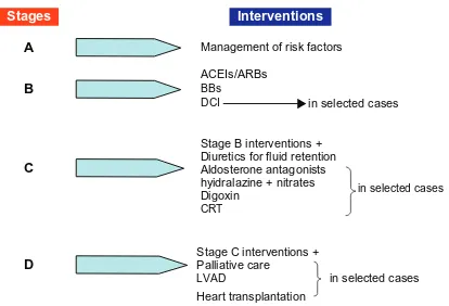
The American College of Cardiology (ACC)/American Heart Association (AHA) guidelines have identified four stages of HF.1 The first two stages are evidently not appropriately termed as HF. Stage A consists of patients with conditions that are associated with an increased risk for developing HF. Subjects with asymptomatic structural and/or functional LV disease constitute stage B. Symptomatic and terminal HF patients are included in stages C and D, respectively. Importantly, this classification provides a stepwise approach to HF care; in every stage, several interventions are indicated in order to improve clinical status and/or prognosis (Figure 4). Each of these will be discussed in detail below.
The goals of treatment are reduction in symptoms, a decrease in the rate of hospitalization, and the prevention of premature death. The cornerstone of treatment is pharma-cologic therapy. Lifestyle modification may also be needed. Surgery, implantable devices, or even heart transplantation may be required in selected cases.
Organization of care
Multidisciplinary intervention focused on educating patients, their families, and caregivers about HF, its treatment, in order to warrant adherence and optimize its effects,58 and the early recognition of and response to clinical worsening (eg, decrease in functional capacity or weight gain), with a guidance on flexible dosing of diuretics, has been shown to reduce the rate of hospital admissions and mortality.59 The Leonardo project, a care management initiative involving nurses, general practitioners, specialists, and patients conducted in a primary health care system, demonstrated pronounced effectiveness in increasing subjects’ knowledge about their disease, self-care, and ability to make changes in health behaviors.60
Remote device monitoring (eg, electrocardiogram, blood pressure, body weight) coupled with medical telephone support (telemonitoring) may help to detect early signs of cardiac decompensation, allowing optimization of therapy. A recent Cochrane review concluded that telemonitoring reduced the rate of death from any cause by 44% and the rate of HF-related hospitalizations by 21%.61 However, the quality of the methods used in the included studies was variable and many were small. The Tele-HF (Telemonitoring to Improve Heart Failure Outcomes) trial, a large study that randomized 1653 patients who had recently been hospitalized for HF to undergo either telemonitoring or usual care, found no benefits of this strategy.62 The ongoing TIMI-HF (Telemedical Interventional Monitoring In Heart Failure) trial will provide more data on the effects of this intervention.63
Stages Interventions
A
B
C
D
Management of risk factors
ACEIs/ARBs
BBs
DCI in selected cases
in selected cases
in selected cases Stage B interventions +
Stage C interventions + Diuretics for fluid retention Aldosterone antagonists hyidralazine + nitrates Digoxin
CRT
Palliative care
LVAD
Heart transplantation
Figure 5 American College of Cardiology (ACC)/American Heart Association (AHA) stages and therapeutic management of HF.
Abbreviations: BBs, beta-blockers; ACeis, angiotensin converting enzyme inhibitors; ARBs, angiotensin receptor blockers; CRT, cardiac resynchronization therapy; DCi, Diseases and Conditions index; iCD, implantable cardioverter-defibrillator; LVAD, left ventricular assist device.
Vascular Health and Risk Management downloaded from https://www.dovepress.com/ by 118.70.13.36 on 27-Aug-2020
Dovepress Causes, consequences, and treatment of heart failure
Tests such as standard blood analysis or echocardiograms should be scheduled only when needed (eg, to check ion levels and renal function in patients on diuretics, to reassess EF in cases of clinical deterioration or improvement). Despite the efficacy of brain natriuretic peptide (BNP) and N-terminal pro–B-type natriuretic peptide (NT-proBNP) measurements for the diagnosis of HF,64 their serial determinations to guide treatment is controversial.65
End-of-life palliative care should be available for indi-viduals with end-stage HF.66
Lifestyle and exercise
Restriction of sodium intake is routinely recommended although it is based on little evidence.1,2 Several stud-ies have demonstrated that exercise training improves quality of life as well as functional capacity, systolic– diastolic function and BNP and NT-proBNP expression.67 Nevertheless, it remains questionable whether this inter-vention reduces cardiac deaths and hospitalizations and whether disease severity predicts which patients are most likely to benefit.68,69
Pharmacological therapy
Although several therapeutic agents have proved to reduce morbidity and mortality in patients with HFDEF, no treat-ment has yet been shown to substantially improve clinical outcomes in patients with HFPEF. Therefore, we will focus our review on pharmacological therapy for HF patients with depressed systolic function.
The pathophysiological aspects of HF are essential for understanding drug actions and clinical trial designs. The development of HF is characterized by an initial cardiac injury that triggers a cascade of neurohormonal responses. Either an acute (MI) or a chronic (HBP) insult may alter the loading conditions of a normal ventricle, inducing stretch-ing of myocardial fibers or their loss. This condition evokes activation of the RAAS and the sympathetic nervous system. In the short term, these actions are beneficial and adaptive, maintaining organ perfusion. However, in the long run, this abnormal neurohormonal setting leads to myocyte hypertro-phy, apoptosis, and fibrotic proliferation, resulting in adverse remodeling and pump dysfunction. The consequences of these structural changes are a reduction in stroke volume, an increase in systemic vascular resistance, and the develop-ment of the signs and symptoms of congestion and hypop-erfusion. Neurohormonal stimulation, therefore, will be the main target of most pharmacological agents for HF. Others, such as diuretic or digoxin, will not actuate in the feedback
mechanisms of the disease but will be helpful in symptomatic release (Figure 6).70
Diuretics
Diuretics provide rapid relief for the signs and symptoms of congestion. Because no studies have yet demonstrated the long-term benefit of these drugs in terms of mortal-ity, they should be prescribed only to patients who have evidence of, or a prior history of, fluid retention. Due to their potency, loop diuretics have emerged as the preferred agents for use in most subjects; however, thiazide diuretics may be preferred in hypertensive HF with mild congestion because they present a more persistent effect on BP. The timing of administration can be altered for social conve-nience. Managing diuretic dosage is very important. The lowest dose needed to achieve an edema-free state (‘dry weight’) should be used (Table 7). Inappropriately low doses will result in fluid retention, which can increase the risk of treatment with beta-blockers, whereas inappropriately high doses will lead to volume contraction, which can lead to hypotension and renal insufficiency. Patients with mild HF respond favorably to low doses because absorption of the drug from the bowel and delivery to the renal tubules is fast. However, at advanced stages, bowel edema and hypoperfusion may delay absorption and delivery,71 and therefore, increasing doses may be required to achieve an appropriate effect. In some cases even high doses of diuretic are ineffective. The intake of large amounts of dietary sodium, treatment with agents that can block their effects,
Adverse ventricular remodeling
Patient at risk
Filling pressure Stroke volume
Digoxin
Triggering event (MI, HTA)
Worsening
heart function Initial hemodynamicresponse
Compensatory mechanisms
Hemodynamic effects Ivabradine
LV dysfunction Congestion Diuretics
ACEIs
ARBs
AId. ant.
BBs Activation of RAAS Activation of SNS
ADH HR
HDZ-nitrates Ventricular contraction Vascular resistance
Figure 6 Physiopathology of heart failure and sites of action of the main pharmacological agents.
Adapted from Ramani et al.70
Abbreviations: ACeis, angiotensin converting enzyme inhibitors; ADH, antidiuretic hormone; ald. ant., aldosterone antagonist; ARBs, angiotensin receptor blockers; BBs, beta-blockers; HDZ, hydralazide; HR, heart rate; HTA, heart transplant alone; LV, left ventricle; Mi, myocardial infarction; RAAS, renin-angiotensin-aldosterone system; SNS, sympathetic nervous system.
Vascular Health and Risk Management downloaded from https://www.dovepress.com/ by 118.70.13.36 on 27-Aug-2020
Dovepress Pazos-López et al
such as nonsteroidal anti-inflammatories,72 or significant impairment of renal function or perfusion73 may be the cause of such a situation. Generally, diuretic resistance can be overcome by parenteral administration (continuous infu-sion may be more helpful than bolus injection),74 the use of two or more diuretics in combination (eg, furosemide and metolazone),75 or the addition of drugs that increase renal blood flow (eg, positive inotropic agents). Diuretics can cause the depletion of important cations (potassium and magnesium), which may predispose patients to serious cardiac arrhythmias. Potassium deficits can be corrected by the short-term use of potassium supplements. Magnesium supplements may also be required. Concomitant administra-tion of angiotensin- converting enzyme inhibitors (ACEIs) alone or in combination with potassium-retaining agents (such as spironolactone) can prevent electrolyte depletion in most patients, and thus, when these drugs are prescribed
long-term, oral potassium supplementation frequently is not needed and may be deleterious.
ACeis
ACEIs are the most comprehensively studied agents in HF. These drugs not only interfere with the RAAS but also enhance the action of kinins and kinin-mediated prostaglandins, which may have beneficial effects in cardiac remodeling as seen in animal models.76–78 ACEIs have been evaluated in a large number of placebo-controlled trials involving patients with a reduced LVEF (,35% to 40%) and a wide range of severity of HF (from asymptomatic LV impairment to NYHA functional class IV). These studies demonstrated that ACEIs improve symptoms, mortality, and the combined risk of death or hospitalization.79 Although available data suggests that there are no differences among ACEIs in their effects, those used in clinical trials are recommended (Table 6). Treatment should be initiated at low doses followed by gradual increments attempting to reach target doses of clinical trials or the highest tolerated.1,2 The clinical response is generally delayed and may require several weeks, months, or longer to become apparent.80 However, even if symptoms do not improve, long-term treat-ment with an ACEI should be maintained to reduce the risk of death or hospitalization. The most common side effect of these agents is related to the suppression of angiotensin stimulation (hypotension and worsening of renal function) and to kinin enhancement (angioedema and cough).
• Symptomatic hypotension (eg, dizziness) often improves with time although the dose of diuretics and other hypotensive agents may need to be reduced.
• An increase in serum creatinine is expected after the initia-tion of an ACEI but is not considered clinically important unless rapid and substantial. Renal function is usually restored after a reduction in the dose of concomitantly administered diuretics, so these patients can generally be managed without withdrawing treatment with ACEIs.81
• Hyperkaliemia is generally seen in patients whose renal function deteriorates or who are taking oral potassium supplements or potassium-sparing diuretics.
• Cough is the most common reason for the withdrawal of long-term treatment with ACEIs.82 Its frequency is approximately 5% to 10% in white patients of European descent and rises to nearly 50% in Chinese patients.83
• Angioedema occurs in ,1% of patents taking an ACEI but is more frequent in black patients. Because its occur-rence may be life-threatening, the clinical suspicion of this reaction justifies subsequent avoidance of all ACEIs for the lifetime of the patient.82
Table 7 Diuretics, beta-blockers, angiotensin converting enzyme inhibitors, angiotensin receptor blockers, and aldosterone antagonists recommended in heart failure
Loop diureticsa Initial dose (mg) Usual daily dose (mg)
Furosemide 20–40 40–240
Bumetanide 0.5–1.0 1–5
Torasemide 5–10 10–20
Thiazidesb Initial dose (mg) Usual daily dose (mg)
Bendroflumethiazide 2.5 2.5–10 Hydrochlorothiazide 25 12.5–100
Metolazone 2.5 2.5–10
lndapamidec 2.5 2.5–5
ACEIs Initial dose (mg) Target dose (mg)
Captopril 6.25 ttd 50–100 ttd
enalapril 2.5 td 10–20 td
iisinopril 2.5–5.0 od 20–35 od
Ramipril 2.5 od 5 td
Trandolapril 0.5 od 4 od
ARBs Initial dose (mg) Target dose (mg)
Candesartan 4 to 8 od 32 od
Valsartan 20 to 40 td 160 td
Losartan 25 to 50 od 50 to 100 od BBs Initial dose (mg) Target dose (mg)
Carvedilol 3.125 td 25 td
Bisoprolol 1.25 od 10 od
Metoprolol 12.5 od 200 od
Nevibolol 1.25 od 10 od
Ald. ant. Initial dose (mg) Target dose (mg)
Spironolactone 25 od 25–50 od
eplerenone 25 od 50 od
Notes: aDose might need to be adjusted according to volume status/weight;
excessive dose may cause renal impairment and ototoxicity; bDo not use thiazides
if eGFR. 30 mL/min, except when prescribed synergistically with loop duretics;
cindapamide is non-thiazide sulfonamide.
Abbreviations: BBs, beta-blockers; ACeis, angiotensin-converting enzyme inhibitors; Ald. ant., aldosterone antagonists; ARBs, angiotensin receptor blockers; eGFR, estimated glomerular filtration rate; od, once daily; td, twice daily; ttd, three times daily.
Vascular Health and Risk Management downloaded from https://www.dovepress.com/ by 118.70.13.36 on 27-Aug-2020
Dovepress Causes, consequences, and treatment of heart failure
Angiotensin receptor blockers
Circulating levels of angiotensin II increase to pretreatment levels with long-term ACEI inhibition.84 Angiotensin receptor blockers (ARBs) bind competitively to the AT1 receptor, providing a downstream effect and thereby dampening this escape phenomenon.85 Besides, interference with the RAAS without inhibition of kininase would provide the benefits of ACEIs while minimizing their adverse reactions.86 However, as mentioned before, it is now known that some of the benefits of ACEIs may be related to the accumulation of kinins, whereas some of the their side effects are related to the suppression of angiotensin II formation. A large meta-analysis of 24 randomized trials showed the superiority of ARBs to placebo in patients with intolerance to ACEIs and their noninferiority in all-cause mortality or hospitalizations when compared directly with ACEIs.87 Taking into account this evidence, ARBs are used as an alternative to ACEIs when cough or angioedema is developed. Because combination therapy of ACEIs and ARBs results in a modest decrease in hospitalization (relative risk reduction [RRR]: 17%), an increase in side effects, and no benefits on mortality,88 its use is controversial.1,2 ARBs recommended in guidelines are listed in Table 7.
Beta blockers
The benefits of three beta-blockers (BBs), bisoprolol, metoprolol succinate (β1 receptor selective blockers), and carvedilol (which blocks α1, β1, and β2 receptors), have been assessed in several placebo-controlled trials enroll-ing patients with a reduced LVEF (,35% to 45%), mostly under ACEIs therapy, from initial to advance stages of HF.89–93 Long-term treatment with BBs has been shown to lessen symptoms, reduce mortality and the combined risk of death and hospital admission. In the SENIORS (Study of the Effects of Nebivolol Intervention on Outcomes and Rehospitalization in Seniors with heart failure) trial, nebivolol, another β1 receptor blocker, demonstrated efficacy in reducing the combined endpoint of death or cardiovascular hospitalization in elderly patients (.70 years) with HF.94 Although no randomized controlled trials of BBs in individu-als with asymptomatic LV dysfunction have been completed, their use (especially in those with CAD) is encouraged.2 Bearing in mind that most participants in BB trials were not on target doses on ACEIs and the fact that adding a BB produces a greater improvement in clinical status and mor-tality than increasing ACEIs,95,96 patients do not need to be on high doses of ACEIs before initiating therapy with BBs. Dose titration and time to clinical response considerations
for ACEIs also apply to BBs (Table 7). Because long-term treatment with BBs reduces the risk of worsening HF, dis-continuation of these drugs after an episode of decompensa-tion is not recommended.1,2 However, in unstable subjects it may be prudent to reduce its dose temporarily until the basal status is recovered.
The most common side effects of BBs include:
• Fatigue or weakness. In many cases it resolves sponta-neously within several weeks; however, in some cases, it may be severe enough to limit dose increase or even necessitate treatment withdrawal.
• Bradycardia. The slowing of heart rate (HR) is generally well tolerated. Nevertheless, when symptoms or an advanced heart block is developed BB dose should be reduced.
• Symptomatic hypotension may be managed by adminis-tering BBs and ACEIs at different times during the day. If this is ineffective, a temporary reduction in the dose of diuretics (in patients who are volume depleted) or ACEIs may be effective.
Aldosterone antagonists
Although short-term therapy with both ACEIs and ARBs can lower circulating levels of aldosterone, such suppres-sion may not be sustained during long-term treatment.97 This observation may be important because aldosterone promotes sodium retention, electrolyte imbalances, and endothelial dysfunction and may directly contribute to myocardial fibrosis.98 In the RALES (Randomized Spironolactone Evaluation Study) trial, a low dose of spironolactone, the most widely used aldosterone antagonist, added to ACE therapy in NYHA class III and IV patients, reduced mortality and HF hospitalizations. Functional class also improved.99 The EPHESUS (Epler-enone Post-Acute Myocardial Infarction Heart Failure Efficacy and Survival Study) trial investigated the newer aldosterone antagonist eplerenone in subjects with LVEF
# 40% and clinical evidence of HF or diabetes mellitus within 14 days after MI, and found a decrease in the risk of death.100 The recently published EMPHASIS-trial, whose participants had an LVEF # 35% and were in NYHA functional class II, showed that eplerenone sig-nificantly reduced mortality and hospital admissions.101 This promising result may extend the actual indication for aldosterone antagonists (NYHA functional class III–IV) in HF to early stages of the disease. The main side effects related to aldosterone antagonists are hyperkalemia and gynecomastia.
Vascular Health and Risk Management downloaded from https://www.dovepress.com/ by 118.70.13.36 on 27-Aug-2020
Dovepress Pazos-López et al
• Hyperkalemia is a concern, especially in subjects with underlying chronic kidney disease, so serum potassium levels must be closely monitored.
• Gynecomastia can occur during therapy with spironolac-tone but not in patients treated with eplerenone, and thus, switching from one agent to the other one may solve the problem.
The dosage of aldosterone antagonists is listed in Table 7.
Hydralazine and nitrates
Hydralazine produces arterial vasodilation and systemic vascular resistance reduction via modulation of intracellular calcium kinetics, and nitrates are transformed in smooth muscle cells into nitric oxide, which stimulates cyclic guanosine monophosphate production and subsequent arterial vasodilation. This combination increases survival in HF, but not as much as with ACEIs or ARBs.102,103 The A-Heft trial, conducted in African Americans with advanced HF who were receiving standard therapy (including BBs and ACEIs), found that the addition of a fixed-dose isosorbidedinitrate and hydral-azine enhanced survival and decreased hospitalizations.104 According to these data, combination therapy with hydralazine and nitrates may be an option for patients in which ACEIs or ARBs are contraindicated and also in African American HF subjects who remain symptomatic (NYHA functional class III–IV) despite optimal medical therapy.1,2
Digoxine
In addition to their mild inotropic effect, digitalis glycosides attenuate carotid sinus baroreceptors and have sympathoin-hibitory effects that produce a decrease in norepinephrine, renin, and possibly aldosterone levels.105,106 In the only randomized-controlled trial of digoxin therapy (DIG trial) a reduction in hospitalizations (RRR 28%) among individuals with HF and LVEF , 45% was found, although no overall mortality benefit was achieved.107 In daily practice, digoxin is used in addition to BBs to control HR in patients with atrial fibrillation as well as in those with persisting symptoms even with optimal medical treatment.1,2 Dosage should be adjusted to obtain serum levels between 0.6 and 1.2 ng/mL. Common side effects include bradyarrhythmias and tachyarrhythmias (digoxin-specific Fab antibody fragments may be required in some cases), gastrointestinal symptoms (eg, anorexia, nausea, and vomiting), and neurological complaints (eg, visual disturbances, disorientation, and confusion). Although digitalis toxicity is commonly associated with high serum digoxin levels (.2 ng/mL) it may occur with lower levels, especially if hypokalemia coexists.
Surgery
Coronary revascularization
A meta-analysis of 24 studies investigating late survival in patients with ischemic HF treated with coronary artery bypass grafting (CABG) or medical therapy showed a RRR of 79% in annual mortality in those with myocardial viability. Nevertheless, no incremental benefit with CABG was found in individuals without substantial viability.108 Revascularization is strongly supported in patients with angina, whereas there is no evidence to sustain it in individuals with multives-sel CAD but no ischemic symptoms. An ongoing randomized trial will clarify this matter.109
Mitral valve repair
Functional MR, characterized by annular dilatation and leaflet non-coaptation in the setting of anatomically normal papillary muscles, chordal structures, and valve leaflets, is frequent in patients with HF. Although severe functional MR is associated with poor prognosis, it has been demonstrated that valve repair improves only symptoms but does not reduce mortality. Besides, recurrence in the first year may reach up to 35% of cases. Therefore the indication for this surgical technique is controversial and must be individualized.110
Ischemic MR found in patients with previous MI is typically associated with leaflet tethering and displacement related to abnormal LV wall motion and geometry. Despite current guidelines recommending mitral valve repair in patients with moderate and severe ischemic MR who are considered for CABG surgery, long-term benefits are still unclear.111
Ventricular reconstruction
Multiple surgical techniques to reduce LV volume and thereby alleviate wall stress have been utilized. The recently published STICH (Surgical Treatment of Ischemic Heart Failure) trial randomized patients with ischemic LV dysfunction undergoing CABG to isolated CABG versus CABG plus surgical ventricular reconstruction. Although surgical reconstruction reduced LV volumes, no difference in mortality or hospital admissions was found.112 On the basis of these results, routine surgical LV reconstruction in addition to CABG is not recommended.
Devices
Implantable cardioverter-defibrillator
Implantable cardioverter-defibrillator (ICD) reduces the risk of SCD due to severe ventricular arrhythmias. Patients at higher risk are those with a previous MI and a severely
Vascular Health and Risk Management downloaded from https://www.dovepress.com/ by 118.70.13.36 on 27-Aug-2020
Dovepress Causes, consequences, and treatment of heart failure
reduced LVEF. The MADIT II (Multicentric Automatic Defibrillator Implantation II) trial, whose participants had a nonrecent MI and LVEF , 30%, found a significant survival benefit after ICD implantation.113 Similar results were showed SCD-HeFT (Sudden Cardiac Death in Heart Failure) trial which evaluated patients with nonischemic or ischemic LV systolic dysfunction (LVEF < 35%).114 According to current guidelines, ICD is indicated for secondary prevention in patients who survive an unprovoked episode of ventricular fibrillation or sustained ventricular tachycardia and for pri-mary prevention in patients with LV dysfunction (LVEF #
35%). In ischemic LV dysfunction, evaluation of LVEF and subsequent device implantation should be done three months after an elective revascularization and 40 days after a prior MI. NYHA functional class II or III, optimal medical therapy, and expected survival with good functional status for .1 year are required to implant an ICD in nonischemic LV systolic dysfunction. In cases of ischemic LV dysfunction this indica-tion is extended to NYHA funcindica-tional class I.115
Cardiac-resynchronization therapy (CRT)
Intraventricular conduction delays, identified by a QRS interval of .120 ms, occur in up to one-third of patients with severe systolic HF, and are associated with dyssynchronous contraction of the LV, which leads to impaired emptying and, in some patients, MR.116 Early studies showed that CRT has a positive effect on reverse remodeling (manifested by a decrease in LV volumes and an increase in LVEF), improves exercise capacity, and reduces symptoms.117 The CARE-HF (Cardiac Resynchronization in Heart Failure) trial was the first study to demonstrate a benefit in all-cause mortality with this treatment.118 A meta-analysis of 14 randomized trials of CRT confirmed a significant lessening in morbidity and mortality.119 In contrast, this therapy has not been successful in HF patients with narrow QRS.120 Although many echocardiographic parameters of dyssynchronous have been proposed to identify candidates for CRT, no one has shown efficacy for predicting responders,121 and therefore, to date, they are not routinely used in daily practice. The recently published MADIT-CRT trial, which involved patients with NYHA functional class I or II, LVEF of 30% or less, and wide QRS intervals ($130 ms), found that CRT added to an ICD improved ventricular function and reduced the risk of worsening HF in comparison with ICD alone. These effects were most marked in subjects with a QRS interval of $150 ms.122 Participants in the REVERSE (Resynchronization Reverses Remodeling in Systolic Left Ventricular Dysfunction) trial (NYHA function class I or II, LVEF # 40%, QRS duration $120 ms, and LV end-diastolic
diameter $55 mm) were randomly assigned to CRT activated versus CRT off. After 12 months, a positive effect on reverse remodeling was achieved in the CRT-activated group, but no difference in clinical outcomes was found.123 The European sample of REVERSE comprised 262 patients, followed up to 24 months. In this population, fewer patients assigned to CRT worsened clinically.124 Similarly, the time to first hospitalization for HF or to death from any cause was significantly delayed. The results from these two trials have recently changed the European Society of Cardiology recommendations for CRT (Table 8).125 The advantage of CRT plus ICD (CRT-D) over CRT alone in terms of survival has not been adequately addressed. However, because of the documented effectiveness of ICD therapy for prevention of SCD, the use of a CRT-D device is commonly preferred.
Left ventricular assist devices
Given the shortage of organ donors there has been a grow-ing interest in left ventricular assist devices (LVAD) as a bridge to transplantation or even as a definitive therapy for end-stage HF individuals. The REMATCH trial, conducted in patients with terminal HF ineligible for heart trans-plantation, showed that LVAD increased 1-year survival rates as compared with medical treatment (52% vs 25% respectively).126 Technological advances have led to the devel-opment of more effective and safer devices. In a recent study Table 8 Actual european recommendations for cardiac resynchronization therapy
Sinus rhythm
NYHA function class iii/iV
• LVeF # 35%
• QRS $ 120 ms
• Optimal medical therapy
• Class iV patients should be ambulatorya
NYHA function class ii
• LVeF # 35%
• QRS $ 150 ms
• Optimal medical therapy Atrial fibrillation NYHA function class lll/iV
• LVeF # 35%
• QRS $ 130 ms
• Optimal medical therapy
• Pacemaker dependency induced by AV nodal ablation or slow ventricular rate and frequent pacingb
Pacemaker indication
• LVeF # 35%c
Notes: aNo admissions for HF during the last month and a reasonable expectation
of survival .6 months; bFrequent pacing is defined as $95% pacemaker dependence;
cindication iiaC in NYHA iii-iV and iibC in NYHA ii.
Abbreviations: AV, atrioventricular; HF, heart failure; LVeF, left ventricular ejection fraction; NYHA, New York Heart Association.
Vascular Health and Risk Management downloaded from https://www.dovepress.com/ by 118.70.13.36 on 27-Aug-2020
Dovepress Pazos-López et al
comparing new continuous flow LVAD with old pulsatile volume-displacement LVAD in subjects with end-stage HF, the 2-year survival was significantly higher with the new device than with the older (46% vs 11%).127 Despite these promising results, current devices are expensive and may cause serious complications such as infection, bleeding, and malfunction. Therefore, candidates for this therapy must be carefully selected.
Heart transplantation
Heart transplantation is a last resort for patients with refractory HF. The development of immunosuppressive agents has dramatically reduced the rate of graft rejection and led to good survival rates (the estimated half-life heart transplantation exceeds 10 years) with a good quality of life.128 However, serious comorbidities must be ruled out and an ability to adhere to the intensive postoperative medical treatment and follow-up required should be confirmed before offering a patient this therapeutic option.
Novel therapies
Ivabradine acts on the If ion current, which is highly expressed in the sinoatrial node. If is a mixed Na+–K+ inward current activated by hyperpolarization and modulated by the autonomic nervous system. It is one of the most important ionic currents for regulating pacemaker activity in the sino-atrial node. Ivabradine selectively inhibits this current in a dose-dependent manner, slowing the HR. The drug has few other, if any, known cardiac effects. The SHIFT (Systolic Heart Failure Treatment with the IF Inhibitor Ivabradine) trial investigated the effects of ivabradine in HF patients with reduced LVEF (#35%) and HR . 70 beats per minute, showing a reduction in the rate of cardiovascular death and cardiovascular hospital admissions.129 These results confirm the importance of HR in HF and support the concept that its decrease contributes significantly to reduce outcomes.
Treatment of HFPeF
Identification of specific therapeutic agents has been disap-pointing. Effective drugs in HFDEF such as ACEIs, ARBs, BBs, and digoxin showed no benefits in HFPED.130–133 Therefore, guidelines focus on appropriate treatment of the underlying cause (basically BP and ischemia), rate/rhythm control if AF is developed, and control of pulmonary con-gestion with diuretic agents.1,2 Venodilators such as nitrates should be used with caution because decreases in preload may lead to an excessive reduction in LV filling, resulting in hypotension and syncope.
Treatment of isolated right HF
The most important aspect of managing right HF is tailoring therapy to its specific cause. In contrast to patients with chronic left HF, patients with right HF often have signifi-cantly abnormal afterload (eg, pulmonary hypertension) or valvular heart disease (eg, acquired or congenital pulmonary or tricuspid disease). It is therefore not surprising that the selected treatment should primarily target the etiology of the disease. Sodium and fluid restriction and judicious use of diuretics all help to optimize preload which, in the major-ity of patients, is achieved at normal right atrial pressure (,6 mmHg). Only small studies suggest a beneficial role for beta blockade or ACE inhibitors. Primary prevention of sudden death using defibrillators is recommended mainly in patients with ARVC and tetralogy of Fallot. In the setting of acute right HF, every effort should be made to avoid systemic hypotension, as this could lead to myocardial ischemia and further hypotension.134
Disclosure
The authors declare no conflicts of interest.
References
1. Hunt SA, Abraham WT, Chin MS, et al. 2009 Focused update incor-porated into the ACC/AHA 2005 Guidelines for the Diagnosis and Management of Heart Failure in Adults: a report of the American College of Cardiology Foundation/American Heart Association Task Force on Practice Guidelines: developed in collaboration with the Inter-national Society for Heart and Lung Transplantation. J Am CollCardiol. 2009;53(15):e1–e90.
2. Dickstein K, Cohen-Solal A, Filippatos G, et al. ESC Guidelines for the diagnosis and treatment of acute and chronic heart failure 2008: the Task Force for the Diagnosis and Treatment of Acute and Chronic Heart Failure 2008 of the European Society of Cardiology. Developed in collaboration with the Heart Failure Association of the ESC (HFA) and endorsed by the European Society of Intensive Care Medicine (ESICM). Eur Heart J. 2008;29(19):2388–2442.
3. Bhatia RS, Tu JV, Lee DS, et al. Outcome of heart failure with preserved ejection fraction in a population-based study. N Engl J Med. 2006; 355(3):260–269.
4. Owan TE, Hodge DO, Herges RM, Jacobsen SJ, Roger VL, Redfield MM. Trends in prevalence and outcome of heart failure with preserved ejection fraction. N Engl J Med. 2006;355(3): 251–259.
5. Mosterd A, Hoes AW. Clinical epidemiology of heart failure. Heart. 2007;93(9):1137–1146.
6. Fox KF, Cowie MR, Wood DA, et al. Coronary artery disease as the cause of incident heart failure in the population. Eur Heart J. 2001; 22(3):228–236.
7. Gheorghiade M, Bonow RO. Chronic heart failure in the United States: a manifestation of coronary artery disease. Circulation. 1998; 97(3):282–289.
8. Repetto A, Dal Bello B, Pasotti M, et al. Coronary atherosclerosis in end stage idiopathic dilated cardiomyopathy: an innocent bystander?
Eur Heart J. 2005;26(15):1519–1527.
9. Hedrich O, Jacob M, Hauptman PJ. Progression of coronary artery disease in non-ischemic dilated cardiomyopathy. Coron Artery Dis. 2004;15(5):291–297.
Vascular Health and Risk Management downloaded from https://www.dovepress.com/ by 118.70.13.36 on 27-Aug-2020