Original Article
Role of multi-parametric MRI of the prostate
for screening and staging: Experience with
over 1500 cases
Geoffrey Gaunay
a, Vinay Patel
a, Paras Shah
a, Daniel Moreira
b,
Simon J. Hall
a, Manish A. Vira
a, Michael Schwartz
a,
Jessica Kreshover
a, Eran Ben-Levi
a, Robert Villani
a,
Ardeshir Rastinehad
c, Lee Richstone
a,*
a
Department of Urology, The Smith Institute for Urology, Northwell Health, New Hyde Park, NY, USA
b
Department of Urology, University of Illinois Chicago, Chicago, IL, USA
cDepartment of Urology & Interventional Radiology, Mount Sinai Health System, New York City, NY,
USA
Received 30 August 2016; accepted 5 September 2016
Available online 22 November 2016
KEYWORDS Prostate cancer; Magnetic resonance imaging; Staging; Prostatectomy; Screening; Margins; Gleason score; Novel use
Abstract Objective:Contemporary prostate cancer (PCa) screening modalities such as pros-tate specific antigen (PSA) and digital rectal examination (DRE) are limited in their ability to predict the detection of clinically significant disease. Multi-parametric magnetic resonance im-aging (mpMRI) of the prostate has been explored as a stim-aging modality for PCa. Less is known regarding its utility as a primary screening modality. We examined our experience with mpMRI as both a screening and staging instrument.
Methods:mpMRI studies performed between 2012 and 2014 in patients without PCa were cross-referenced with transrectal ultrasonography (TRUS) biopsy findings. Statistical analyses were performed to determine association of mpMRI findings with overall cancer diagnoses and clinically significant (Gleason score7) disease. Subgroup analyses were then performed on patients with a history of prior negative biopsy and those without a history of TRUS biopsy. mpMRI studies were also cross-referenced with RP specimens. Statistical analyses determined predictive ability of extracapsular extension (ECE), seminal vesicle involvement (SVI), and pathologic evidence of clinically significant disease (Gleason score7).
Results:Four hundred biopsy naı¨ve or prior negative biopsy patients had positive mpMRI studies. Overall sensitivity, specificity, positive and negative predictive values were 94%, 37%, 58%, and 87%, respectively and 95%, 31%, 42%, and 93%, respectively for overall cancer detection and Gleason score7 disease. In patients with no prior biopsy history, mpMRI
* Corresponding author.
E-mail address:[email protected](L. Richstone).
Peer review under responsibility of Second Military Medical University. http://dx.doi.org/10.1016/j.ajur.2016.09.011
2214-3882/ª2017 Editorial Office of Asian Journal of Urology. Production and hosting by Elsevier B.V. This is an open access article under
the CC BY-NC-ND license (http://creativecommons.org/licenses/by-nc-nd/4.0/).
Available online atwww.sciencedirect.com
ScienceDirect
sensitivity, specificity, positive and negative predictive values were 94%, 36%, 65%, and 82%, for all cancers, and 95%, 30%, 50%, and 89% for Gleason score7 lesions, respectively. In those with prior negative biopsy sensitivity, specificity, positive and negative predictive values were 94%, 37%, 52%, and 90% for all cancers, and 96%, 32%, 36%, and 96% for Gleason score7 le-sions, respectively. Seventy-four patients underwent radical prostatectomy (RP) after mpMRI. Lesion size on mpMRI correlated with the presence of Gleason score7 cancers (pZ0.005). mpMRI sensitivity, specificity, positive and negative predictive values were 84%, 39%, 81%, and 44% respectively, for Gleason7 cancer. For ECE and SVI, sensitivity and specificity were 58% and 98% and 44% and 97%, respectively.
Conclusion:mpMRI is an accurate predictor of TRUS biopsy and RP outcomes. mpMRI has sig-nificant potential to change PCa management, particularly in the screening population, in whom a significant proportion may avoid TRUS biopsy. Further studies are necessary to deter-mine how mpMRI should be incorporated into the current PCa screening and staging paradigms.
ª2017 Editorial Office of Asian Journal of Urology. Production and hosting by Elsevier B.V. This is an open access article under the CC BY-NC-ND license (http://creativecommons.org/ licenses/by-nc-nd/4.0/).
1. Introduction
An 180,890 new prostate cancer (PCa) diagnoses and 26,120 PCa-related deaths are estimated for 2016 [1]. Unfortu-nately, contemporary screening and diagnostic in-struments, such as prostate specific antigen (PSA) and digital rectal examination (DRE), have limited ability to differentiate clinically significant disease from benign conditions or indolent disease. Resultant biopsy exposes patients to potential for life-threatening infection, in addition to under detection of clinically significant disease and over detection of clinically indolent disease, leading to uncertainty regarding management strategies[2].
Multiparametric magnetic resonance imaging (mpMRI) has an emerging role in multiple aspects of PCa manage-ment. Although mpMRI has become well established in recent years as both a diagnostic and staging tool, its role in the pre-biopsy setting is not well defined. In current clinical methodology, risk factors guide the decision to proceed to biopsy, despite detailed evidence that those same factors, when incorporated into nomograms, perform poorer than mpMRI in clinical staging of known PCa[3,4].
mpMRI screening in biopsy-naı¨ve patients with elevated PSA or abnormal DRE has several theoretical advantages. Pre-biopsy MRI may allow for the detection of cancer foci and increase biopsy yield on cognitive or fusion-targeted biopsy. mpMRI may also be able to obviate the need for biopsy, if able to accurately discriminate clinically relevant disease from indolent or absent cancer. This modality also may have value in patients with one or more negative ul-trasound guided biopsies and persistently elevated PSA and/or abnormal exam. It is well established that repeated transrectal biopsies in this setting have sequentially worse cancer detection rates[5]. Because the entire prostate is imaged on mpMRI, suspicious lesions may be detected in areas of the prostate that may be under sampled, such as the anterior, apical and central zones. Finally, pre-biopsy mpMRI affords the clinician the ability to perform a tar-geted biopsy. Tartar-geted MRI-ultrasound (US) fusion biopsy of the prostate has been shown to more accurately diagnose more intermediate and high-risk cancers and fewer
indolent, low-risk tumors versus standard, systematic bi-opsy [6,7]. A targeted-only biopsy strategy may be also advantageous in the reduction of unnecessary biopsy cores versus systematic biopsy, potentially limiting biopsy-related morbidity[7].
Despite the purported advantages of mpMRI in the pre-biopsy setting, controversy persists. A recent systematic review found a high rate of both false positives and nega-tives for mpMRI in the screening population[8]. Given the discrepancy between theoretical advantages and the actual benefit of screening MRI, we sought to assess our own institutional experience with mpMRI as a screening instru-ment (i.e., patients without prior biopsy or history of negative biopsy). Additionally, we examined our institu-tional mpMRI staging accuracy, comparing mpMRI findings with whole mount radical prostatectomy (RP) specimens.
2. Materials and methods
2.1. Study design
A query of our prospectively maintained, Institutional Re-view Board-approved database of 1722 3-Tesla (T) mpMRI was performed within the Northwell Health System. All mpMRI imaged were acquired with an endorectal coil and phase array cardiac coil. mpMRI findings were interpreted by MRI trained uro-radiologists. mpMRI sequences captured included T1 and T2-weighted, dynamic contrast enhanced (DCE), and diffusion weighted imaging (DWI). A radiograph-ically “positive” study was characterized by the presence of one or more suspicious lesion(s). Additional MRI variables recorded included number of lesions, lesion size, and loca-tion by zone, prostate imaging reporting and data system (PIRADS) scores, apparent diffusion coefficient (ADC), and overall study suspicion score. Suspicion score consisted of a 5-point Likert scale corresponding to the overall risk of clinically significant cancer per the recommendations of the European Society of Urogenital Radiology (ESUR)[9].
Studies were cross-referenced with RP specimens with patients having undergone RP between 2012 and 2014.
Central pathologic review of all specimens was performed by trained uro-pathologists.
2.2. Statistical analysis
Descriptive statistics are presented as counts and per-centages for categorical variables and as means and stan-dard deviations (SD) for continuous variables. The association of MRI-related variables with pathology Gleason score (<7 or 7) was analyzed with Student’s t test for continuous data and chi-square test for categorical vari-ables in univariable analysis. The following MRI-related variables were evaluated in patients who subsequently underwent RP: number of lesions (continuous), primary lesions size (continuous, in mm), zone (central or periph-eral), ADC (continuous, in 106mm2/s), T2 PIRADS RP score (1e5), diffusion PIRADS score (1e5), enhancement PIRADS
score (1e5) and overall lesion score (1e5). MRI findings
(positivevs.negative studies) were cross-referenced with pathologic outcomes to identify predictive properties of a positive MRI to identify pathologic Gleason score7 PCa. Similarly, predictive properties of the presence of extrac-apsular extension (ECE) and seminal vesicle invasion (SVI) on MRI to identify pathology-proven extracapsular exten-sion and seminal vesicle invaexten-sion at the time of RP were also evaluated.
In the pre-biopsy setting, MRI findings (positive vs. negative studies) were cross referenced with biopsy out-comes to evaluate the predictive properties of a positive MRI to identify post-MRI biopsy Gleason score7 PCa and post-MRI positive prostate biopsy (any Gleason). Subgroup analyses, based upon the presence or absence of prior transrectal ultrasonography (TRUS) biopsy, were performed to examine predictive abilities in individual patient co-horts. Estimates are presented with 95% confidence interval (CI). All statistical analyses were two-tailed and performed using R 3.2.2 (R Foundation for Statistical Computing, Vienna, Austria). Apvalue<0.05 was considered statisti-cally significant.
3. Results
3.1. Screening
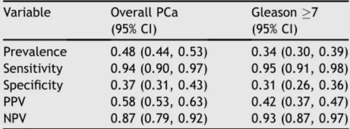
A total of 513 patients with no prior PCa diagnosis, either biopsy naı¨ve or prior negative biopsy, underwent an mpMRI and were subsequently biopsied. Of these patients, 400 (78.0%) had positive mpMRI studies. The overall preva-lence of PCa on biopsy following mpMRI was 48%, while the overall presence of Gleason score7 cancers was 34%. For overall PCa prediction of biopsy, sensitivity, specificity, positive predictive value (PPV), and negative predictive values (NPV) were 94%, 37%, 58%, and 87%, respectively. In regards to Gleason 7 cancers, sensitivity, specificity, PPV, and NPV were 95%, 31%, 42%, and 93%, respectively (Table 1).
In subgroup analyses, 282 patients had a history of negative biopsy predating mpMRI, while 231 patients had no prior biopsy history. Overall prevalence of PCa in biopsy naı¨ve patients was 55%, while Gleason score 7 cancer prevalence was 42%. In those with a history of negative
biopsy PCa prevalence was 42% for all cancers and 28% for Gleason score7 cancers. In patients with no prior biopsy history, mpMRI sensitivity, specificity, PPV and NPV were 94%, 36%, 65%, and 82%, for all cancers, and 95%, 30%, 50%, and 89% for Gleason7 lesions, respectively. In those with prior negative biopsy sensitivity, specificity, PPV and NPV were 94%, 37%, 52%, and 90% for all cancers, and 96%, 32%, 36%, and 96% for Gleason7 lesions, respectively (Table 2). 3.2. Staging
A total of 74 patients underwent RP after mpMRI. Patho-logic analyses revealed 56 patients with Gleason7 cancer, 24 with ECE, and 9 with SVI. Overall mpMRI lesion size correlated with the finding of clinically significant PCa (Gleason score 7) versus indolent disease on biopsy (pZ0.005) (Table 3). No other singular mpMRI character-istic was significant in discrimination of Gleason 7 from Gleason <7 cancer on RP specimen, although both DCE PIRADS score and overall lesion suspicion score approached statistical significance (pZ 0.054). Analysis of combined factors was limited due to low numbers. Overall sensitivity, Table 1 Predictive value of mpMRI in screening population.
Variable Overall PCa (95% CI) Gleason7 (95% CI) Prevalence 0.48 (0.44, 0.53) 0.34 (0.30, 0.39) Sensitivity 0.94 (0.90, 0.97) 0.95 (0.91, 0.98) Specificity 0.37 (0.31, 0.43) 0.31 (0.26, 0.36) PPV 0.58 (0.53, 0.63) 0.42 (0.37, 0.47) NPV 0.87 (0.79, 0.92) 0.93 (0.87, 0.97)
CI, confidence interval; mpMRI, multi-parametric magnetic resonance imaging; NPV, negative predictive value; PCa, pros-tate cancer; PPV, positive predictive value.
Table 2 Predictive value of mpMRI in biopsy naı¨ve and prior negative biopsy patients.
Variable Overall PCa (95% CI) Gleason7 (95% CI) Biopsy naı¨ve Prevalence 0.56 (0.49, 0.62) 0.42 (0.36, 0.49) Sensitivity 0.94 (0.88, 0.97) 0.95 (0.88, 0.98) Specificity 0.36 (0.27, 0.46) 0.30 (0.22, 0.38) PPV 0.65 (0.58, 0.72) 0.50 (0.42, 0.57) NPV 0.82 (0.68, 0.92) 0.89 (0.76, 0.96) Prior negative biopsy
Prevalence 0.42 (0.36, 0.48) 0.28 (0.23, 0.34) Sensitivity 0.94 (0.88, 0.98) 0.96 (0.89, 0.99) Specificity 0.37 (0.30, 0.45) 0.32 (0.26, 0.39) PPV 0.52 (0.45, 0.59) 0.36 (0.29, 0.42) NPV 0.90 (0.80, 0.96) 0.96 (0.88, 0.99)
CI, confidence interval; mpMRI, multi-parametric magnetic resonance imaging; NPV, negative predictive value; PCa, pros-tate cancer; PPV, positive predictive value.
specificity, PPV and NPV for prediction of Gleason7 can-cers was 84%, 39%, 81%, and 44%, respectively. Similarly, sensitivity, specificity, NPV and PPV were 58%, 98%, 93%, and 81%, for ECE, and 44%, 97%, 67%, and 93%, for SVI, respectively.
4. Discussion
In this study we have demonstrated the ability of MRI to predict outcomes of TRUS biopsy. Excellent negative pre-dictive capabilities have been demonstrated. Perhaps the most critical finding is 93% NPV of Gleason score7 cancers for all patients. Based on this result, an important consid-eration for the use of mpMRI in the biopsy naı¨ve and prior negative biopsy population is the value of a negative scan. Multiple studies have examined the NPV of negative mpMRI, ranging between 63% and 98%, for clinically significant disease[10,11].
Wysock et al.[12]recently reported on 75 patients with negative mpMRI prior to biopsy. In the subset of biopsy naı¨ve men, NPV was 81.3% for all cancer detection and
98.7% for Gleason sum7. Similar results were detected in the prior negative biopsy group, in which NPV was 86.2% and 100% for all cancers and Gleason sum7, respectively. Ultimately, these findings suggest negative mpMRI may negate the need for TRUS biopsy in select biopsy naı¨ve patients or those with a history of prior negative biopsy. In our subset of 282 patients with prior negative biopsies, NPV was 90% for all PCa and 96% for Gleason7 cancers. In this cohort, 24% of men had a negative MRI, indicating 24% of biopsies may have been avoided at the cost of seven missed cancer diagnoses, three of which would have been high grade (Gleason 7) lesions. Similar results were found in the biopsy naı¨ve group in which the NPVs were 82% and 89% for PCa and Gleason 7 PCa, respectively, and 19.4% of biopsy naı¨ve patients had a negative mpMRI. If biopsy was avoided in these patients, eight cancers and five Gleason
7 cancers would have been undiagnosed.
Apart from the reduction in number of biopsies per-formed and associated complications, an initial mpMRI approach may translate to cost savings. Lotan et al.[13] developed a cost comparison model comparing mpMRI versus repeat biopsy in men with prior negative biopsy. The authors found the mpMRI arm to be associated with 73 fewer biopsies per 100 men versus the TRUS arm, while diagnosing four fewer cancers. Incorporating cost of com-plications into analysis, this translated to an overall US $2700 cost savings for the mpMRI arm[13]. Use of a higher PSA threshold prior to initiation of mpMRI and subsequent fusion biopsy has been suggested to further increase the accuracy of identifying patients likely to harbor clinically significant disease[14].
Some have advocated for the use of MRI as a first line screening method. A pilot study by Nam et al. [15] demonstrated a higher odds ratio of PCa for MRI versus PSA in an unselected screening population. Additionally, a 66.7% PPV was found in those with normal PSA (<4 ng/mL) and MRI suspicion scores of 4. Similarly, in those with normal PSA and suspicion scores3, NPV was 85.7%. Find-ings in our screening cohort are similar, with few missed high grade PCa diagnoses. Although the cost of such a screening program would be astronomical, this study certainly demonstrates the predictive capability of MRI in the pre-biopsy setting. Also, despite the low likelihood, the potential for undiagnosed high grade PCa remains despite negative mpMRI in the screening population. Patients should be counseled regarding this risk if biopsy is to be avoided.
Despite these purported advantages, controversy regarding the use of mpMRI in the pre-biopsy setting con-tinues. Recent systematic review of biopsy-naı¨ve patients revealed 6%e32% false negative and 28%e79% false positive
rates of mpMRI for detection of clinically significant cancer in patients who subsequently underwent targeted biopsy [8]. These findings led the authors to not recommend the use of mpMRI in the biopsy-naı¨ve population. Additionally, benefit of mpMRI followed by targeted biopsy in the setting of previous negative systematic biopsy versus repeat sys-tematic biopsy showed statistical significance in only four included studies.
In contrast, Meng et al.[16]retrospectively found pre-biopsy mpMRI followed by targeted pre-biopsy allowed the detection of more high-grade lesions, while limiting Table 3 Prostate MRI characteristics by pathology
Glea-son score. Variable <7 7 p N(%) 18 (24.3) 56 (75.7) Number of lesions 0.192 0 8 (44.4) 11 (19.6) 1 5 (27.8) 28 (50.0) 2 5 (27.8) 12 (21.4) 3 0 (0.0) 3 (5.4) 4 0 (0.0) 2 (3.6) Primary lesion Size (mm)a 10.74.0 16.28.5 0.005 ADC (106mm2/s)a 1124.6 575.2 677.7 210.0 0.158 Zone 1.000 Central 2 (20.0) 9 (20.0) Peripheral 8 (80.0) 36 (80.0) T2 PIRADS score 0.203 3 2 (50.0) 4 (14.8) 4 2 (50.0) 15 (55.6) 5 0 (0.0) 8 (29.6)
Diffusion PIRADS score 0.598 3 0 (0.0) 1 (3.7)
4 1 (25.0) 4 (14.8) 5 3 (75.0) 22 (81.5)
Enhancement PIRADS score 0.054 1 1 (25.0) 0
2 1 (25.0) 1 3 0 (0.0) 4 4 2 (50.0) 22
Overall lesion score 0.054 3 2 (50.0) 2 (7.4)
4 2 (50.0) 16 (59.3) 5 0 (0.0) 9 (33.3)
MRI, magnetic resonance imaging; PIRADS, prostate imaginge
reporting and data system; SD, standard deviation.
detection of those Gleason6 versus that of standard sys-tematic biopsy. It was their contention that mpMRI should be considered to identify patients in whom low risk disease is likely and biopsy may not be warranted. Prospective comparison of mpMRI with guided biopsy versus TRUS biopsy in a group of 223 biopsy-naı¨ve men with elevated PSA by Pokorny and colleagues[17]found a reduction in the diag-nosis of low-risk cancer by 89.4%, and an increase in the diagnosis of intermediate and high-risk disease by 17.7%. The MRI guided biopsy pathway was associated with a reduction in the need for biopsy of 51%[17]. Similarly, in a prospective analysis, Numao et al.[18]found mpMRI valu-able in the pre-biopsy setting to reduce the number of initial prostate biopsies, particularly in the low-risk (PSA<10 ng/ mL and normal DRE) study cohort. Within this cohort, the frequency of significant cancer was 9%e13% and 43%e50%
for negative and positive mpMRI, respectively. Within the high-risk group, frequency ranged from 47% to 51% with negative mpMRI and 68%e71% with a positive study. These
findings argue for the use of risk assessment nomograms in the consideration of mpMRI prior to biopsy[18]. Mendhiratta et al.[19]found mpMRI followed by targeted biopsy had a higher rate of overall cancer (21.7%vs.18.6%) and Gleason score7 (92.3%vs.57.7%) detection in patients with one or more negative biopsies, underscoring the importance of mpMRI and potentially negating the need for additional systematic biopsy in this population.
mpMRI use in the setting of prior negative biopsy is well supported as an alternative to repeat systematic biopsy. mpMRI has been shown to diagnose and discriminate clinically significant disease in patients with negative prostate biopsies and persistently elevated PSA who sub-sequently underwent transperineal systematic biopsies, suggesting negative mpMRI may be sufficient evidence to defer additional repeat biopsy [20]. Further, a study of 117 patients, all with at least one prior negative biopsy and PSA >4.0 ng/mL, demonstrated a prostate PCa-detection rate of 41%. More significantly, the vast major-ity, 87%, was considered clinically significant by Epstein and d’Amico classification. However, of the nine patients in whom PCa was detected after negative MRI guided bi-opsy, 78% were found to have clinically significant disease, suggesting a role for continued surveillance of those with elevated PSA and negative MRI guided biopsy[21].
While the use of mpMRI in the pre-biopsy setting remains open to debate, the use of staging MRI with diagnosed PCa is better established. In this study we have demonstrated the association of tumor size on mpMRI with clinically sig-nificant disease on RP specimen. Additionally, DWI PIRADS and overall study ESUR scoring approached significance. mpMRI has been shown to improve upon risk assessment nomograms in the determination of tumor extent[3,22]. A number of studies have compared mpMRI with histopatho-logic findings after RP. De Rooij et al. [23] pooled these findings in a recent meta-analysis, demonstrating excellent specificity between 88% and 96% for detection of ECE, SVI, and overall T3 disease status. Unfortunately, it is hampered by weak sensitivity ranging between 57% and 61% for similar pathologic findings [23]. Predictive capabilities as described in this study showed similar limited sensitivity, likely owing to inability to accurately detect microscopic extraprostatic disease on mpMRI.
Functional imaging has been found to improve overall sensitivity, and the use of at least two functional modalities is recommended [9,23,24]. T2-weighted imaging, used in conjunction with functional sequences (i.e., DWI, DCE, and MR spectroscopy (MRSI)), has been shown capable of tumor detection and risk categorization [25,26]. While T2-weighted imaging versus combined T2 and DWI may be similarly capable in tumor detection, DWI may provide additional information regarding tumor aggressiveness[27]. ADC determined by DWI has been inversely correlated with biopsy Gleason score[28e30]. ADC values have been shown
able to discriminate low risk disease (Gleason <7) from intermediate-high risk cancers (Gleason7), while negative T2-weighted and DWI sequences are associated with a high rate of negative biopsy[30e33]. Interestingly, in our study
ADC values were not significantly correlated to RP specimen Gleason scores, potentially owing to low study numbers.
mpMRI may also be useful in selection of patients for active surveillance (AS) protocol. Suspicious mpMRI can demonstrate the presence of clinically significant disease amongst the AS population[27,34]. Patients with index le-sions, defined as cancer within the same sextant on biopsy in two separate surveillance biopsies, had a higher rate of biopsy reclassification. Lack of an index lesion may provide further evidence for the presence of indolent disease in the AS population, potentially negating the need for repeat biopsy [35]. Low-risk findings on ADC mapping may influ-ence the decision for repeat biopsy in the AS population.
This study has several limitations. mpMRI findings along with pathologic analyses were from a single institution’s experience and may not be replicable. Overall number of RP specimens was low, limiting statistical power, including the analysis of combined mpMRI parameters. Further research through prospective trials is necessary to deter-mine the role of mpMRI specifically within the biopsy-naı¨ve, staging population before mainstream acceptance. Addi-tional considerations such as MRI expense and patient quality of life must be included in the cost-benefit analyses prior to alteration of the current PCa-screening paradigm.
5. Conclusion
Large lesions on mpMRI were predictive of clinically signifi-cant disease at the time of RP, however variable accuracy was exhibited in the prediction of ECE, SVI, and Gleason7 disease. mpMRI may be predictive of TRUS biopsy results in a screening population, with a 93% NPV, suggesting that TRUS biopsy may be avoided in select patients with normal studies. Further large, prospective trials are needed to determine the role of mpMRI in a screening population.
Conflicts of interest
The authors declare no conflict of interest.
References
[1] American Cancer SocietyjInformation and Resources for
Can-cer: Breast, Colon, Lung, Prostate, Skin. Web. 10 May 2016. (http://www.cancer.org/cancer/prostatecancer/
[2] Delongchamps NB, Peyromaure M, Schull A, Beuvon F, Bouazza N, Flam T, et al. Prebiopsy magnetic resonance im-aging and PCa detection: comparison of random and targeted biopsies. J Urol 2013;189:493e9.
[3] Gupta RT, Faridi KF, Singh AA, Passoni NM, Garcia-Reyes K, Madden JF, et al. Comparing 3-T multiparametric MRI and the Partin tables to predict organ-confined prostate cancer after radical prostatectomy. Urol Oncol 2014;32:1292e9.
[4] Feng TS, Sharif-Afshar AR, Wu J, Li Q, Lutringer D, Saouaf R, et al. Multiparametric MRI improves accuracy of clinical no-mograms for predicting extracapsular extension of prostate cancer. Urology 2015;86:332e7.
[5] Roehl KA, Antenor JA, Catalona WJ. Serial biopsy results in prostate cancer screening study. J Urol 2002;167:2435e9.
[6] Mariotti GC, Costa DN, Pedrosa I, Falsarella PM, Martins T, Roehrborn CG, et al. Magnetic resonance/transrectal ultra-sound fusion biopsy of the prostate compared to systematic 12-core biopsy for the diagnosis and characterization of prostate cancer: multi-institutional retrospective analysis of 389 patients. Urol Oncol 2016;34:416. e9-416.e14.
[7] Haffner J, Lemaitre L, Puech P, Haber GP, Leroy X, Jones JS, et al. Role of magnetic resonance imaging before initial bi-opsy: comparison of magnetic resonance-targeted and sys-tematic biopsy for significant prostate cancer detection. BJU Int 2011;108:E171e8.
[8] Haider MA, Yao X, Loblaw A, Finelli A. Multiparametric mag-netic resonance imaging in the diagnosis of prostate cancer: a systematic review. Clin Oncol R Coll Radiol 2016;28:550e67.
[9] Barentsz JO, Richenberg J, Clements R, Choyke P, Verma S, Villeirs G, et al. ESUR prostate MR guidelines 2012. Eur Radiol 2012;22:746e57.
[10] Itatani R, Namimoto T, Atsuji S, Katahira K, Morishita S, Kitani K, et al. Negative predictive value of multiparametric MRI for prostate cancer detection: outcome of 5-year follow-up in men with negative findings on initial MRI studies. Eur J Radiol 2014;83:1740e5.
[11] Futterer JJ, Briganti A, De Visschere P, Emberton M, Giannarini G, Kirkham A, et al. Can clinically significant prostate cancer be detected with multiparametric magnetic resonance imaging? A systematic review of the literature. Eur Urol 2015;68:1045e53.
[12] Wysock JS, Mendhiratta N, Zattoni F, Meng X, Bjurlin M, Huang WC, et al. Predictive value of negative 3T multi-parametric prostate MRI on 12 core biopsy results. BJU Int 2016;118:515e20.
[13] Lotan Y, Haddad AQ, Costa DN, Pedrosa I, Rofsky NM, Roehrborn CG, et al. Decision analysis model comparing cost of multiparametric magnetic resonance imaging vs. repeat biopsy for detection of prostate cancer in men with prior negative findings on biopsy. Urol Oncol 2015;33:266. e9e16.
[14] Shakir NA, George AK, Siddiqui MM, Rothwax JT, Rais-Bahrami S, Stamatakis L, et al. Identification of threshold prostate specific antigen levels to optimize the detection of clinically significant prostate cancer by magnetic resonance imaging/ultrasound fusion guided biopsy. J Urol 2014;192: 1642e8.
[15] Nam RK, Wallis CJ, Stojcic-Bendavid J, Milot L, Sherman C, Sugar L, et al. A pilot study to evaluate the role of magnetic resonance imaging for prostate cancer screening in the gen-eral population. J Urol 2016;196:361e6.
[16] Meng X, Rosenkrantz AB, Mendhiratta N, Fenstermaker M, Huang R, Wysock JS, et al. Relationship between prebiopsy multiparametric magnetic resonance imaging (MRI), biopsy indication, and MRI-ultrasound fusion-targeted prostate bi-opsy outcomes. Eur Urol 2016;69:512e7.
[17] Pokorny MR, de Rooij M, Duncan E, Schro¨der FH, Parkinson R, Barentsz JO, et al. Prospective study of diagnostic accuracy
comparing prostate cancer detection by transrectal ultrasound-guided biopsy versus magnetic resonance (MR) imaging with subsequent MR-guided biopsy in men without previous prostate biopsies. Eur Urol 2014;66:22e9.
[18] Numao N, Yoshida S, Komai Y, Ishii C, Kagawa M, Kijima T, et al. Usefulness of pre-biopsy multiparametric magnetic resonance imaging and clinical variables to reduce initial prostate biopsy in men with suspected clinically localized prostate cancer. J Urol 2013;190:502e8.
[19] Mendhiratta N, Meng X, Rosenkrantz AB, Wysock JS, Fenstermaker M, Huang R, et al. Prebiopsy MRI and MRI-ultrasound fusion-targeted prostate biopsy in men with pre-vious negative biopsies: impact on repeat biopsy strategies. Urology 2015;86:1192e8.
[20] Abd-Alazeez M, Ahmed HU, Arya M, Charman SC, Anastasiadis E, Freeman A, et al. The accuracy of multi-parametric MRI in men with negative biopsy and elevated PSA levelecan it rule out clinically significant prostate cancer?
Urol Oncol 2014;32:45. e17e22.
[21] Hoeks CM, Schouten MG, Bomers JG, Hoogendoorn SP, Huls-bergen-van de Kaa CA, Hambrock T, et al. Three-Tesla magnetic resonance-guided prostate biopsy in men with increased prostate-specific antigen and repeated, negative, random, systematic, transrectal ultrasound biopsies: detection of clin-ically significant prostate cancers. Eur Urol 2012;62:902e9.
[22] Mullerad M, Hricak H, Kuroiwa K, Pucar D, Chen HN, Kattan MW, et al. Comparison of endorectal magnetic reso-nance imaging, guided prostate biopsy and digital rectal ex-amination in the preoperative anatomical localization of prostate cancer. J Urol 2005;174:2158e63.
[23] De Rooij M, Hamoen EH, Witjes JA, Barentsz JO, Rovers MM. Accuracy of magnetic resonance imaging for local staging of prostate cancer: a diagnostic meta-analysis. Eur Urol 2015;70: 233e45.
[24] Detection, staging, and recurrence assessment of urologic malignancy: prostate. ACR-SAR-SPR Practice parameter for the performance of magnetic resonance (MRI) of the
soft-tissue components of the pelvis. 2015.http://www.acr.org/
w/media/0249FD9C739D4AF2B3519AE5FB09E648.pdf. [25] Turkbey B, Pinto PA, Mani H, Bernardo M, Pang Y, McKinney YL,
et al. Prostate cancer: value of multiparametric MR imaging at 3 T for detectiondhistopathologic correlation. Radiology
2010;255:89e99.
[26] Tan CH, Paul Hobbs B, Wei W, Kundra V. Dynamic contrast-enhanced MRI for the detection of prostate cancer: meta-analysis. Am J Roentgenol 2015;204:W439e48.
[27] Vargas HA, Akin O, Franiel T, Mazaheri Y, Zheng J, Moskowitz C, et al. Diffusion-weighted endorectal MR imaging at 3 T for prostate cancer: tumor detection and assessment of aggressiveness. Radiology 2011;259:775e84.
[28] Haghighi M, Shah S, Taneja SS, Rosenkrantz AB. Prostate cancer: diffusion-weighted imaging versus dynamic-contrast enhanced imaging for tumor localizationea meta-analysis. J
Comput Assist Tomogr 2013;37:980e8.
[29] Shimofusa R, Fujimoto H, Akamata H, Motoori K, Yamamoto S, Ueda T, et al. Diffusion-weighted imaging of prostate cancer. J Comput Tomogr 2005;29:149e53.
[30] Faletti R, Battisti G, Discalzi A, Grognardi ML, Martinello S, Oderda M, et al. Can DW-MRI, with its ADC values, be a reli-able predictor of biopsy outcome in patients with suspected prostate cancer? Abdom Radiol 2016;41:926e33.
[31] Vargas HA, Akin O, Afaq A, Goldman D, Zheng J, Moskowitz CS, et al. Magnetic resonance imaging for predicting prostate bi-opsy findings in patients considered for active surveillance of clinically low risk prostate cancer. J Urol 2012;188:1732e8.
[32] Nowak J, Malzahn U, Baur AD, Reichelt U, Franiel T, Hamm B, et al. The value of ADC, T2 signal intensity, and a combination
of both parameters to assess Gleason score and primary Gleason grades in patients with known prostate cancer. Acta Radiol 2016;57:107e14.
[33] Li C, Chen M, Wang J, Wang X, Zhang W, Zhang C. Apparent diffusion coefficient values are superior to transrectal ultra-sound guided prostate biopsy for the assessment of prostate cancer aggressiveness. Acta Radiol 2016 Apr 6. pii: 0284185116639764. [Epub ahead of print].
[34] Schoots IG, Petrides N, Giganti F, Bokhorst LP, Rannikko A, Klotz L, et al. Magnetic resonance imaging in active surveil-lance of prostate cancer: a systematic review. Eur Urol 2015; 67:627e36.
[35] Mullins JK, Bonekamp D, Landis P, Begum H, Partin AW, Epstein JI, et al. Multiparametric magnetic resonance imaging findings in men with low-risk prostate cancer followed using active surveillance. BJU Int 2013;111:1037e45.