N o v e m b e r 2 0 1 2
SurveillaNce
hiv
This training manual was based on a manual used at the Florida Department of Health. Some sections of that document were so useful that only minor changes were made for inclusion in this manual. CSTE acknowledges the contribution of the CSTE workgroup: Marie Antoinette Bernard, Tina Brubaker, Sharon Carter, Melissa English, Douglas Frye, Kelly Gallagher, Joan Greene, Catina James, Bonnie Krampe, Benjamin Laffoon, Martin Ngokion, Luke Shouse, Stephanie Townsell, and Jeff Turner as well as CSTE National Office staff member Lauren Rosenberg. CSTE also acknowledges technical editor Karen Foster for her contributions to the project. The primary author is Michael Campsmith, CSTE consultant. This publication was supported by Cooperative Agreement Number 5U38HM000414 from CDC. Its contents are solely the responsibility of the authors and do not necessarily represent the official views of CDC.
I N T R O D U C T I O N . . . . 5
M O D U L E 1 . . . . 1 5
HIV Case Definition and the Human Immune System
M O D U L E 2 . . . . 6 5
HIV Case Report Forms
M O D U L E 3 . . . . 8 1
Managing Collection of Surveillance Data
M O D U L E 4 . . . . 1 0 1
Presenting Surveillance Data
M O D U L E 5 . . . . 1 3 3
Risk Ascertainment
M O D U L E 6 . . . . 1 5 5
eHARS
M O D U L E 7 . . . . 1 7 5
Evaluating HIV Surveillance Programs
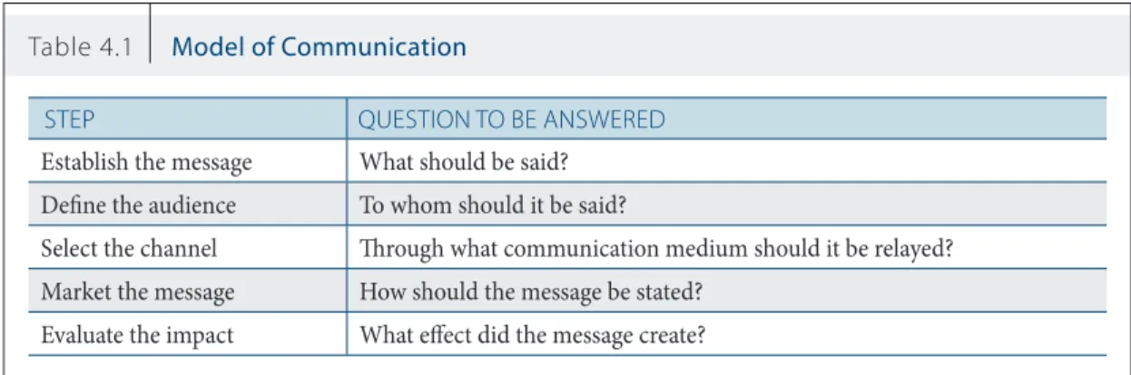
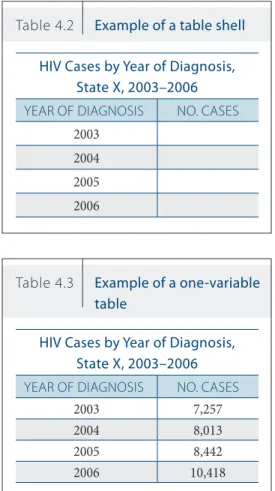
TABLE
1. Explain the spectrum of events through the course of HIV infection. 2. Describe the background and purpose of public health surveillance. 3. Outline key events in the timeline of HIV surveillance.
4. Provide information about where to find more in-depth information about topics related to HIV and HIV surveillance.
INTRODUCTION
O B J E C T I V E S :
HIV Surveillance
Introduction
The purpose of an HIV surveillance program is to promote the systematic and ongoing collection, analysis, evaluation, and dissemination of epidemiologic data of the highest possible quality. In turn, public health surveillance data are used to plan, implement, and evaluate HIV programs and interventions. Public health surveillance activities identify HIV-related conditions at various points along the spectrum of HIV disease (Table I.1)—from reporting of HIV infection in otherwise asymptomatic people to periodic clinical evaluations of immune system status (e.g., CD4 tests) to AIDS diagnosis through low CD4 value/ diagnosis of qualifying opportunistic illnesses to death certificate review for HIV-related mortality.
In the past, surveillance for HIV-related diseases focused on opportunistic infections, cancers, and conditions that are known to occur late in the course of HIV disease. When the condition that became known as AIDS was first recognized in 1981, the underlying cause of the disease was unknown, and surveillance focused on a group of specific conditions associated with severe immunosuppression (e.g.,
Pneumocystis carinii pneumonia [now known as Pneumocystis jiroveci pneumonia], Kaposi sarcoma).
In addition, persons with severe manifestations of HIV disease sought medical care and therefore came to the attention of health-care providers who could report cases to local and state health departments. Finally, because all HIV infected people eventually developed severe diseases, AIDS case finding through review of hospital records and AIDS-related deaths provided a relatively complete representation of the demographic and risk groups affected by the epidemic. National surveillance for AIDS began with identification of the initial cases in 1981. However, not until the first HIV-antibody diagnostic tests were licensed in 1985 could public health surveillance be conducted on conditions of HIV infection before a clinical diagnosis of AIDS.
To properly monitor the HIV epidemic, surveillance staff need to collect accurate information about key events from the time HIV infection is first diagnosed in a person until death. This information is collected from many sources and documents, such as health-care providers (adult and pediatric case report forms), laboratories (laboratory reports), and vital records offices (birth and death certificates). Increased use of diagnostic laboratory testing and of electronic reporting have led to an increased number of documents
Table I.1 HIV/AIDS surveillance: monitoring the spectrum of infection
HIV Exposure (children) HIV Infection AIDS-OI 1st CD4+ T-Cell <200 cells per µL 1st Viral Load Test 1st CD4+ T-Cell Count 1st Positive Confidential HIV Test Death
SENTINEL EVENTS
in the HIV surveillance system. Historically, consolidated information from all documents has been used to create a single case record for each HIV-infected person. As technology and the epidemic have evolved, the Centers for Disease Control and Prevention (CDC) has developed a document-based data management system to better track information received from HIV surveillance programs and to better monitor HIV disease progression.
Information about the spectrum of HIV disease assists CDC and state and local health departments in better understanding the direction of the epidemic, including populations most affected and in need of prevention and care services. After licensing of HIV diagnostic tests in 1985, many public health programs implemented surveillance for HIV infection (along with continuation of regular AIDS surveillance). However, several jurisdictions had concerns (e.g., confidentiality, stigma, discrimination) about surveillance for HIV infections before an AIDS diagnosis and until recently did not adopt integrated surveillance for the spectrum of HIV disease.
HIV surveillance data are used to monitor trends in the epidemic. Active case finding is conducted by state and local health departments throughout the United States, and uniform reporting methods with timely transmission of data to CDC have enabled CDC to disseminate HIV surveillance data for public health monitoring and planning purposes. Because HIV surveillance is the largest population-based system for monitoring the epidemic nationally, it has become the basis for allocation of federal, state, and local resources for prevention and patient care.
Surveillance for AIDS provides data at the late stage of HIV disease. One disadvantage of examining late-stage disease is that secondary prevention activities, such as referrals for early treatment interventions or partner notification, cannot be facilitated by health departments early in the course of infection when they are most beneficial. For this reason, as of April 2008, public health surveillance programs in all states and territories have expanded their surveillance case finding activities to also include persons in whom HIV infection is diagnosed before development of AIDS.
To ensure maximum efficiency of surveillance activities and optimum quality of resulting data, HIV surveillance program staff need to understand the concepts of public health surveillance. After that understanding, they then need to implement those concepts in a standardized manner. The goal of this training manual is to present the basic concepts of an effective HIV surveillance system in a manner that staff with various levels of experience can use in their activities. Proper application of the principles of public health surveillance will result in a successful HIV surveillance program.
By design this manual presents the basics of HIV surveillance activities. For more in-depth material, the reader is directed to the references at the end of this introduction.
In addition, this manual is designed to provide a broad overview of HIV surveillance activities and practices. Because local surveillance programs might have developed policies and procedures specific to their own programs, some of the material in this manual might not apply to individual HIV surveillance systems.
Background and Purpose
Infectious disease surveillance was initiated in the United States in the late 1800s as a method of quarantine control to prevent the spread of conditions such as cholera, smallpox, yellow and fever. In 1961, CDC assumed responsibility for the collection and publication of data on nationally notifiable diseases. However, reporting of diseases to CDC by states is voluntary; reporting is mandated (i.e., by legislation or regulation) only at the state level. Periodically, the list of nationally notifiable diseases is recommended for revision on the basis of consultation with various public health partners, including the
Council of State and Territorial Epidemiologists (CSTE).
HIV surveillance staff should contact their state department of public health or office of general council about specific laws, statutes, and regulations authorizing the collection and reporting of public health information in their jurisdiction.
H I P A A A N D P U B L I C H E A LT H S U R V E I L L A N C E
The national Health Insurance Portability and Accountability Act (HIPAA) was enacted in 1996. Title II of HIPAA, known as the Administrative Simplification provision, addresses the security and privacy of health data. The standards are intended to improve the efficiency and effectiveness of the nation’s health-care system while maintaining strict standards of data protection and confidentiality. HIPAA has a federal Privacy Regulation (45 CMR § 164.512) to allow access to health records for established public health functions—including HIV surveillance—when the public health authority is authorized by law to collect or receive such information. The Privacy Rule expressly permits disclosures without individual authorization to public health authorities authorized by law to collect or receive the information for the purpose of preventing or controlling disease, injury, or disability, including public health surveillance, investigation, and intervention.
T E C H N I C A L G U I D A N C E F O R H I V S U R V E I L L A N C E P R O G R A M S
CDC, in collaboration with CSTE and staff at the state and local levels, has prepared comprehensive material on HIV surveillance practices and recommendations. Much of the material with which new surveillance staff need to be familiar can be found in the various volumes of the Technical Guidance for HIV Surveillance Programs. The Guidance is a reference for managing state and local HIV surveillance programs in the United States. The intent of the policies and procedures is to provide the basis for maintaining a national HIV surveillance system by using a standardized framework for collecting complete, timely, and high-quality data. At the federal level, the primary functions of an HIV case surveillance system are 1) to provide accurate epidemiologic data to monitor the incidence and prevalence of HIV infection and HIV-related morbidity and mortality and 2) to use these data trends to assist in public health planning and policy.
CDC is authorized to provide federal funding to states and territories through surveillance cooperative agreements to achieve the goals of the national HIV surveillance program and to assist states in developing their own surveillance programs in accordance with state and local laws and practices. The HIV Incidence and Case Surveillance Branch (HICSB) of the Division of HIV/AIDS Prevention (DHAP),
National Center for HIV/AIDS Viral Hepatitis, STD, and TB Prevention, CDC, is responsible for national HIV surveillance. In addition to financial assistance, HICSB provides technical assistance to funded areas to ensure that HIV surveillance systems are comprehensive, timely, accurate, and up-to-date as the HIV
DHAP maintains a password-protected website called SharePoint for posting the latest versions of technical guidance volumes, case report forms, and other information for state and local HIV surveillance programs. To request a SharePoint password, or for technical questions about access to the site, send an email to the SharePoint helpdesk at [email protected]. HIV surveillance programs also can contact their CDC epidemiology consultant to discuss updates or changes to surveillance procedures, documents, and technical guidance.
C H A N G E S T O H I V A N D A I D S R E P O R T I N G
AIDS reporting began at the national level in 1981 to help monitor the scope and impact of the HIV/ AIDS epidemic. After the Food and Drug Administration approved the first HIV-antibody test in 1985, several states expanded their AIDS surveillance systems to include surveillance for HIV infection. Associated with widespread use of highly active antiretroviral therapies (HAART) that began in the mid-1990s, the progression of HIV infection to AIDS has dramatically slowed among treated persons, and an increasing number of persons with HIV infection are living longer and staying healthier. Although surveillance for late-stage HIV disease remains important because it provides information about populations for whom medical treatment is not accessed or has not succeeded, reliance on AIDS surveillance alone does not adequately describe the effects of HIV disease in the population or current trends in the epidemic. Over time, jurisdictions adopted HIV reporting in various forms (including coded identifiers and name-to-code systems), which complicated comparison of trend data. As of April 2008, all 50 states, the District of Columbia, the Commonwealth of Puerto Rico, and the U.S. Territories conduct integrated public health surveillance for HIV infection, including Stage 3 (AIDS) by using confidential name-based reporting.
An integrated HIV surveillance system, with confidential name-based reporting for the entire spectrum of HIV disease (including Stage 3 [AIDS]), allows for better collection of multiple key events during the case history of a person infected with HIV. In addition, a national, integrated HIV surveillance system is better able to monitor the evolving epidemic and to provide useful data about HIV-infected populations to enhance local, state, and federal efforts to prevent HIV transmission, improve allocation of resources for treatment services, and assist in evaluating the impact of public health interventions.
For HIV surveillance data to be comparable and valuable on a national level, all participating project areas need to collect data with a high level of accuracy and consistency. The purposes of these policies and procedures are to address the importance of maintaining a standardized framework for data collection across all surveillance project areas, to delineate the required components of an effective surveillance system, and to suggest methods and techniques designed to optimize productivity.
P R A C T I C E S A N D S T A N D A R D S
CDC and CSTE continue to recommend that all states and territories require reporting of the earliest diagnosis of HIV infection (excluding results of anonymous tests), the earliest diagnosis of HIV infection, Stage 3 (AIDS) in persons of all ages, deaths among persons with HIV infection, and all cases of perinatal HIV exposure. Recommendations represent guidance for best public health practices on the basis of scientific data. Because no single set of policies and procedures can address all of the diversity among, and local needs of, individual state and local surveillance systems, state and local programs should develop their own policies and procedures in accordance with the Technical Guidance for HIV Surveillance
Timeline of surveillance for AIDS and HIV Infection
First report published in CDC’s Morbidity and Mortality Weekly Report (MMWR) of an immunosuppressive disease condition among gay men in Los Angeles; CDC begins collecting data on cases of unexplained immunosuppression among previously healthy persons.
The term “AIDS” (acquired immune deficiency syndrome) is used for the first time; CDC develops first AIDS case definition (on the basis of presence of disease conditions associated with severe immune deficiency without other known cause).
Researchers identify HIV (human immunodeficiency virus).
The Food and Drug administration licenses the first HIV-antibody diagnostic blood test; some jurisdictions begin collecting public health surveillance data from persons with a positive HIV test result but without AIDS indicator conditions.
AIDS case definition expanded to include persons with a CD4 count <200 cells/µL or <14% of total lymphocytes (“immunological AIDS”); this expansion creates a large increase in the number of reported AIDS cases in the United States.
First large-scale release of highly active antiretroviral therapies (HAART); for many patients these drugs allow effective suppression of HIV replication and delay advancement of HIV disease.
After steep declines in the number of reported AIDS cases (1994–1997) and deaths resulting from AIDS (1995–1997), these national surveillance indicators show a general leveling off/ slow decline through 2009 (latest year of available data)
Revision incorporates HIV infection and AIDS into a single surveillance case definition. Revised case definition requires laboratory confirmation of HIV infection for inclusion as a case; HIV infection cases are classified on the basis of CD4 count, with AIDS defined as “HIV Infection, Stage 3 (AIDS).”
All 50 states, District of Columbia, and five U.S. territories conduct integrated name-based public health surveillance for HIV infection.
1982 1985 1981 1983 1993 mid 1990s 1998 1999 2008 2008
Sources of Material for Training Manual
Material in this manual was adapted primarily from existing published HIV surveillance materials. The manual structure and text were adapted from the “Florida HIV Surveillance Training Manual” developed by the Florida Department of Health.
Centers for Disease Control and Prevention and Council of State and Territorial Epidemiologists. Technical Guidance for HIV Surveillance Programs, Volume I: Policies and Procedures (http://www2a.cdc.gov/hicsb/). Centers for Disease Control and Prevention and Council of State and Territorial Epidemiologists. Technical Guidance for HIV Surveillance Programs, Volume II: Data Collection Resources and Reporting (http://www2a. cdc.gov/hicsb/).
Centers for Disease Control and Prevention and Council of State and Territorial Epidemiologists. Data Security and Confidentiality Guidelines for HIV, Viral Hepatitis, Sexually Transmitted Disease, and Tuberculosis Programs: Standards to Facilitate Sharing and Use of Surveillance Data for Public Health Action (http://www. cdc.gov/hiv/resources/guidelines/security_confidentiality_hiv.htm).
Centers for Disease Control and Prevention. Introduction to eHARS, the HIV/AIDS Reporting System. User Guide/Student Manual.v3.2.0.0, 08/2011. (Direct questions about changes to either the eHARS software or the Introduction to eHARS manual to NCHHSTP Informatics Customer Support at 1-877-659-7725 or
Centers for Disease Control and Prevention. Revised Surveillance Case Definitions for HIV Infection Among Adults, Adolescents, and Children Aged <18 Months and for HIV infection and AIDS Among Children Aged
18 Months to <13 Years—United States, 2008. MMWR 2008;57(No.RR-10)
Teutsch SM, Churchill RE, eds. Principles and Practice of Public Health Surveillance. New York, NY: Oxford University Press; 1994
Centers for Disease Control and Prevention. Principles of Epidemiology in Public Health Practice (on-line training course offered through CDC. http://www.cdc.gov/osels/scientific_edu/SS1978/).
Centers for Disease Control and Prevention. HIV publications link: www.cdc.gov/hiv/resources/index.htm Council of State and Territorial Epidemiologists, HIV-related links: www.cste.org
1. Explain the HIV case definition.
2. Explain the mechanism of action of HIV and its effect on the defenses of the human immune system.
3. Provide information about the immune system’s components and function. 4. Explain the different types of HIV tests: antibody tests, antigen tests, viral load tests. 5. Demonstrate the clinical manifestations of HIV disease.
6. Explain AIDS-defining conditions and opportunistic infections.
7. Provide sources of information about antiretroviral drugs used in managing HIV disease. Appendix A: 2008 Surveillance Case Definition for HIV Infection
Appendix B: AIDS-Defining Conditions Diagnosis Criteria
MODULE
ONE
O B J E C T I V E S :
HIV Case Definition and the
Human Immune System
HIV Case Definition
Since the illness that came to be known as AIDS was first reported in 1981, the surveillance case definitions for AIDS and HIV infection have undergone several revisions in response to diagnostic and therapeutic advances. The definitions also have been revised to improve standardization and comparability of surveillance data on all stages of disease. The surveillance case definition is intended for public health surveillance purposes only and not as a guide for clinical diagnosis or treatment. The most current surveillance case definition was implemented in 2008 (Revised Surveillance Case Definitions for HIV Infection Among Adults, Adolescents, and Children Aged <18 Months and for HIV Infection and AIDS Among Children Aged 18 Months to <13 Years —United States, 2008)1 (Appendix A at the end of this module).
The major change in the revised surveillance case definition is the requirement for laboratory-confirmed evidence of HIV infection in adults and adolescents and in children aged 18 months–12 years. The revised definition also highlights the central role of the CD4 T-lymphocyte (T-cell) counts and percentages (objective measures of immunosuppression) in staging HIV disease.
Cases of HIV infection are now classified according to CD4 count and increase in disease severity from “HIV Infection, Stage 1” to “HIV Infection, Stage 2” to “HIV Infection, Stage 3 (AIDS)”; an unknown disease stage (no CD4 information) is also included. Cases still can be classified as “HIV Infection, Stage 3 (AIDS)” on the basis of diagnosis of a recognized AIDS-defining condition regardless of the CD4 count or percentage; however, AIDS-defining conditions have been less common since the advent of highly active antiretroviral therapy (HAART) during the mid-1990s. HIV disease progression is classified from less severe to more severe; after a case is classified into a surveillance severity state, it cannot be reclassified into a less severe stage. Table 1.1 further describes the stages of HIV infection.
1 Public health case definitions are determined through consultation with CSTE. Members of CSTE propose position statements on
topics of interest; those proposals are presented to committees appropriate for the subject matter and drafted. These draft position statements are posted on the CSTE website, then presented for discussion at the CSTE Annual Conference. After appropriate input and revision, the position statements are voted on by the CSTE membership. Information about CSTE position statements is available at www.cste.org.
Table 1.1 Surveillance case definition for HIV infection among adults and adolescents (≥13 years)—United States, 2008
STAGE LABORATORY EVIDENCE* CLINICAL EVIDENCE
Stage 1 Laboratory confirmation of HIV infection and
CD4+ T-lymphocyte count ≥500 cells/µL or
CD4+ T lymphocyte percentage of ≥29%
None required (but no AIDS-defin-ing condition)
Stage 2 Laboratory confirmation of HIV infection and
CD4+ T-lymphocyte count 200–499 cells/µL or
CD4+ T lymphocyte percentage of 14%–28%
None required (but no AIDS-defin-ing condition)
Stage 3 (AIDS) Laboratory confirmation of HIV infection and
CD4+ T-lymphocyte count <200 cells/µL or
CD4+ T lymphocyte percentage of <14%†
or Documentation of an AIDS-defining condition (With laboratory confirmation of HIV infection)†
Stage unknown§ Laboratory confirmation of HIV infection and
No information about CD4+ T-lymphocyte count or percentage
and No information about pres-ence of AIDS-defining conditions
* The CD4+ T-lymphocyte percentage is the percentage of total lymphocytes. If the CD4+ T-lymphocyte count and percentage do not correspond to the same HIV infection stage, select the more severe stage.
† Documentation of an AIDS-defining condition (Module 1, page 35) supersedes a CD4+ T-lymphocyte count ≥200 cells/µL and CD4+ T-lymphocyte percentage of total lymphocytes of ≥14%. Definitive diagnostic methods for these conditions are available in Appendix C of the 1993 revised HIV classification system and the expanded AIDS case definition (CDC. 1993 Revised Classification System for HIV Infection and Expanded Surveillance Case Definition for AIDS Among Adolescents and Adults. MMWR 1992;41(No. RR-17).
§ Although cases with no information about CD4+ T-lymphocyte count or percentage or about AIDS-defining conditions can be classified as stage unknown, every effort should be made to report CD4+ T-lymphocyte count or percentage and AIDS-defining conditions at diagnosis. Additional CD4+ T-lymphocyte counts or percentages and any AIDS-defining conditions identified can be reported as recommended (Laboratory reporting of clinical test results indicative of HIV infection: new standards for a new era of surveillance and prevention [CSTE Position Statement 04-ID-07]).
Source: CDC. Revised Surveillance Case Definitions for HIV Infection Among Adults, Adolescents, and Children Aged <18 Months and for HIV Infection and AIDS Among Children Aged 18 Months to <13 Years—United States, 2008. MMWR 2008;57(No. RR-10).
2Some retroviruses can live in their hosts for years without causing any sign of illness. Retrovirus infections last for life. Outside the body, they
are inactivated when exposed to heat, alcohol, most common disinfectants, and usually by desiccation. They have high rates of mutation and tend to evolve quickly into new strains.
HIV-2 is closely related to HIV-1, the common form of HIV. Most cases of HIV-2 infection occur in West Africa, with relatively few cases reported from the United States. HIV-1 and HIV-2 both damage the immune system and make the body more vulnerable to opportunistic infection (OIs). HIV-2 infection has a longer asymptomatic period and appears to produce a less virulent course of diseas.
Figure 1.1 Parts of the human immunodeficiency virus
• Protective protein outer envelope
• Reverse transcriptase enzyme
• Two strands of RNA (ribonucleic acid)
GP120 p18 p24 lipid membrane GP41
HIV Mechanism of Action
Information in this section is intended for staff new to HIV surveillance or without clinical backgrounds.
HIV is one of a number of retroviruses.2 The genetic information of the virus is carried in two single strands of RNA (ribonucleic acid). For the retrovirus to take over a cell and produce more viruses, it must change its RNA to DNA (dioxyribonucleic acid). A unique enzyme, reverse transcriptase, allows the retrovirus to make this conversion.
HIV, comprising three main parts (Figure 1.1), is expert at evading the immune system’s defenses. It can enter the body hidden inside the cells of infected body fluids (including blood, semen, and vaginal secretions). Once inside the body, HIV binds specifically to cells bearing a particular surface marker called CD4+ (hereafter referred to as CD4). CD4 cells are part of the human immune system responsible for fighting infections; the cells are also known as T lymphocytes because they mature in the thymus gland. HIV infects and leads to the destruction of cells with CD4 surface markers (found on all CD4 T lymphocytes, some B lymphocytes and about half of all macrophages), as well as cells in the gastrointestinal and central nervous systems. CD4 lymphocytes also are called helper lymphocytes and in the laboratory are measured by a flow cytometer. The level of CD4 lymphocytes is used to divide HIV disease into various stages.
Relative concentration of HIV in infected cells with CD4 markers
The highest concentration of HIV in infected cells with CD4 markers occurs in blood and semen. An intermediate concentration of HIV occurs in vaginal secretions, breast milk, tears, and oral fluid. The lowest concentration of HIV in infected cells with CD4 markers occurs in urine, sweat, and feces.
HIV binds to the membrane of the host cell at the site of envelope glycoprotein (gp120) and cytokine receptor sites, then passes through to the glycoprotein (gp41) embedding itself into the CD4 cell membrane (Figure 1.2). The viral coat opens, and the RNA enters the cell. Using the reverse transcriptase enzyme, the RNA makes a DNA copy of itself. This “new” DNA integrates the cell’s DNA, creating a provirus. This process of viral transcription confuses the cell. It becomes no longer a pure cell but part virus, part cell— and therefore the cell acts abnormally. The HIV-infected host cell produces “new” virus particles, which travel back to the cell membrane and bud out to find other cells to infect.
Replication of the virus, which has been calculated at 10 billion copies per day, gradually exceeds the capacity of the lymphatic system to replace CD4 cells, which the virus destroys. Depletion of CD4 helper cells has the greatest impact on the immune system. A CD4 T-lymphocyte count of 800–1500 cells/μL is considered normal for a person with a healthy immune system. As CD4 counts begin to fall to <500 cells/ μL, the infected person become vulnerable to a variety of OIs.
Immunosuppression is documented when
• Absolute CD4 T-lymphocyte count is <500 cells/μL;
• Viral load testing detects HIV in cells;
• The percentage of CD4 cells among total lymphocytes is <29%; or
• The CD4:CD8 cell ratio is <1.
Many people with HIV infection also develop profound deficiencies in the complete blood count. These include red blood cell deficiencies (causing anemia), white blood cell deficiencies (causing leukopenia), platelet deficiencies (causing thrombocytopenia), and total blood component deficiencies (causing pancytopenia).
HIV-infected persons can be symptomatic from the direct effects of the virus. However, they are most susceptible to illness and death from a host of OIs and cancers that invade and weaken the body (Appendix B).
The Immune System: Basic Functions and Structure
Information in this section is intended for staff new to HIV surveillance or without clinical backgrounds.
The human immune system is a sophisticated network of organs and cells that protects us from infectious organisms, environmental toxins, and cell mutations. HIV, a retrovirus that has a special affinity for certain types of cells, directly and catastrophically affects the immune system. Once HIV enters the bloodstream, it launches an offensive against the cells responsible for activating the body’s defense mechanisms. Knowledge about the immune system has rapidly increased in recent years, partly because of intense efforts in the fields of cancer and AIDS research. Understanding the impact of HIV infection on a person requires knowledge of basic immunology.
The immune system, a complex network of cells, tissues, and organs, mobilizes the various components of the body’s defense system against attack from pathogens (disease-producing organisms). The immune system must be able to
• Distinguish “self” from “nonself” (pathogens)
• Develop a defense against pathogens
• Adapt defenses against specific invading pathogens
• “Remember” a specific response to a particular pathogen
Immune system responses fall into two basic categories: nonspecific defense mechanisms and specific defense mechanisms.
N O N S P E C I F I C D E F E N S E M E C H A N I S M S
The purpose of the nonspecific defense mechanisms—which comprise a variety of organs and cells throughout the body—is to prevent or hinder microorganisms from entering the body or to destroy microorganisms that do gain entry. The most familiar and obvious of nonspecific defense mechanisms is the external defense system. Another nonspecific defense is phagocytosis (the process by which cells take in and digest antigens [foreign proteins]).
External Nonspecific Defense System
The external defense system consists of four mechanisms:
• Skin and Eyes • Respiratory tract • Digestive tract
The skin is a first line of defense against pathogens. Although seemingly thin, intact skin is a highly effective barrier. Excretions and secretions—such as sweat—aid in repelling bacteria and fungi.
The small hairs in the lining of the nose similarly filter and prevent larger particles from entering the respiratory tract.
Mucous membranes of the respiratory and digestive tracts trap and remove many foreign particles inhaled or ingested.
The eyes are continually bathed in tears, which wash out large foreign particles and carry away the smaller particles through the tear ducts.
The digestive tract includes strongly acidic stomach fluid that can destroy most microorganisms.
The acidity of the fluids in our urinary and reproductive tracts also provides an effective barrier against some types of bacteria.
Internal Nonspecific Defense System
Phagocytosis
Phagocytes are white blood cells and scavenger cells that are the first “scouts” of the immune system to encounter, engulf, and digest antigens. Monocytes are phagocytes that circulate throughout the bloodstream. Macrophages are phagocytes in the tissues.
Monocytes and macrophages can ingest and destroy a variety of pathogens and cellular debris. A pathogen entering the body activates both. Once an infection is under control, the macrophages and monocytes carry away the particles of the destroyed invaders.
Phagocytes are involved in both the specific and nonspecific immune responses, which often work together to activate the body’s defenses. When nonspecific responses effectively eliminate the pathogen, the specific immune defenses are not activated.
Blood Components
Whole blood consists of the liquid plasma containing the formed elements. The formed elements are
• Erythrocytes (red blood cells)
• Leukocytes (white blood cells)
▶ Granular leukocytes ▷ Neutrophils ▷ Eosinophils ▷ Basophils ▶ Nongranular leukocytes ▷ Lymphocytes ◉ Macrophages ◉ T lymphocytes ◉ B lymphocytes ▷ Monocytes • Thrombocytes (platelets)
Lymphatic System
The lymphatic system is a network of vessels, lymph nodes, and organs that create, transport, and filter leukocytes. The organs of the lymphatic system are subdivided into two categories:
• Primary ▶ Thymus ▶ Bone marrow • Secondary ▶ Tonsils ▶ Lymph nodes ▶ Spleen ▶ Appendix
The primary lymphatic organs produce leukocytes, which fight disease. However, these specialized leukocytes do not begin to defend the body until they reach our secondary organs, where they first contact pathogens.
The thymus is a pyramid-shaped organ located deep beneath the breastbone. It reaches maximum size during early childhood, and then gradually shrinks as we age. The thymus produces specialized cells that play a key role in the body’s immune defenses. Bone marrow, the soft material in the hollow interior of the long bones in our arms and legs, is a crucial element of the immune defenses. The immune system begins in the bone marrow before birth, when erythrocytes and leukocytes begin early development. Approximately 99% of blood cells are erythrocytes; <1% are leukocytes.
Lymphocytes
Lymphocytes are a type of leukocyte that can move in and out of the lymphatic and blood systems. They circulate throughout blood and tissues to locate, trap, and destroy antigens. Approximately 25%–30% of total leukocytes are lymphocytes.
Some lymphocytes mature into B lymphocytes (B cells) in the bone marrow; others migrate to the thymus gland. Most T lymphocytes are produced before birth by the thymus. The remaining cells develop during the first 3–5 years of life. By age 5 years, the body’s natural immune system is developed. It peaks by our mid-20s and becomes less efficient with age. Three main types of lymphocytes are involved in the immune system’s response to antigens:
• Macrophages are produced in the bone marrow.
• T lymphocytes mature in the thymus.
• B lymphocytes mature in the bone marrow.
The secondary lymphatic organs are the information network and the transportation structure for the body’s defense forces. A network of vessels that transport lymphatic fluids from tissues connects pea-shaped lymph nodes located throughout the body. In the secondary organs, the primarily unspecialized leukocytes receive their final programming and acquire the specialized functions necessary to defend against pathogens.
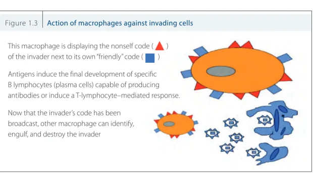
After intercepting the invader, the macrophages copy the identifying structural codes of the invading cell and display this code on the surface of their membranes. The macrophages then move about and present themselves to T lymphocytes, stimulating a specific immune response (Figure 1.3).
Figure 1.3 Action of macrophages against invading cells
This macrophage is displaying the nonself code ( ) of the invader next to its own “friendly” code ( ) Antigens induce the final development of specific B lymphocytes (plasma cells) capable of producing antibodies or induce a T-lymphocyte–mediated response. Now that the invader’s code has been
broadcast, other macrophage can identify, engulf, and destroy the invader
S P E C I F I C I M M U N E M E C H A N I S M S
The specific immune responses involve two complex processes: the cell-mediated and humoral immune responses. They are considered specific because they can identify, remember, and respond to unique patterns of antigens. Each type of lymphocyte and antibody responds only to one specific pattern.
• Cell-mediated immunity involves the interaction between antigens and specialized T lymphocytes.
• Humoral immunity involves the interaction between antigens and the antibodies produced by B lymphocytes.
Cell-Mediated Immunity
T lymphocytes circulate throughout the body. They secrete their own chemical intensification alarm (chemokines) to directly activate or inactivate other leukocytes.
T lymphocytes perform four basic functions:
• Helper T lymphocytes (T4 or CD4 cells) sound a chemical alarm that triggers T and B lymphocytes into action.
• Killer T lymphocytes (cytotoxic T lymphocytes) directly kill infected or cancerous cells by puncturing the membranes.
• Suppressor T lymphocytes (T8 or CD8 cells) turn off the immune response once the infection is brought under control.
• Inducer T lymphocytes induce the maturation of T lymphocytes and other immune system cells. In healthy persons, approximately 70% of circulating lymphocytes are T lymphocytes. T lymphocytes help provide specific immunity against fungi, viruses, parasites, and a few bacteria. T lymphocytes also play a role in destroying cancer cells and in transplant rejection.
Humoral Immunity
B lymphocytes grow rapidly and manufacture millions of antibodies when alerted by the chemical alarm of T lymphocytes. Often an antigen is carried to a lymph node, where it stimulates some of the B lymphocytes to divide and differentiate into plasma cells that produce antibodies (humoral immunity).
Only a tiny fraction of B lymphocytes will respond to any given antigen. The response time of antibodies to an antigen depends on whether they have been previously exposed to that specific antigen. On first contact with an antigen, the body may take from several hours to days before antibodies respond to the invader. Subsequent exposures usually result in a rapid and effective response because of the B lymphocytes’ ability to “remember” that specific antigen.
An antibody (immunoglobulin [Ig]) is a specialized protein capable of chemically combining with the specific antigen (i.e., to the bacteria or virus) that stimulated antibody production. A lock-and-key analogy is often used to describe the relationship: an antibody cannot enter an infected host cell, but it can mark or identify the invading cells as foreign and to guide complement components to kill the invader directly. An Ig exhibit two fundamental structural differences. Variation in the antigen-combining site, or variable
region, and outside the antigen combining site, or constant region, correlate with the different effector
functions mediated by antibodies. Through structural variation, a person’s immune system might produce >10 million antibody responses. Igs are divided into five major classes, each distinguished by certain effector functions and structural features (Kuby, J. 1997. Immunology. W.H. Freeman and Company, New York.). • IgG comprises about 70% of Ig in the tissues and is the only Ig able to cross the placenta; it is the prime
mediator of secondary immune response.
• IgA circulates in the bloodstream, killing bacteria.
• IgM is concentrated in body fluids, especially the bloodstream; it is the earliest Ig to appear after antigen challenge.
• IgE is involved in allergic reactions.
• IgD might be involved in the differentiation of B lymphocytes, but little is known about its exact function.
HIV Laboratory Testing Procedures
Three basic types of tests are currently available or under development to test for HIV:
• Antibody tests
• Antigen tests
• Viral load tests
H I V A N T I B O D Y T E S T S
An antibody test indirectly detects the virus by measuring the extent to which the body’s immune system has mobilized a response against HIV. The appearance of detectable antibodies to HIV is generally believed to occur 2 weeks to 3 months after the initial infection (and in rare cases up to 6 months after infection).
ELISA
An enzyme-linked immunosorbent assay (ELISA) or enzyme immunoassay (EIA) detects HIV antibodies, which the body starts producing 2–12 weeks after HIV exposure. These tests received FDA approval for screening the U.S. blood supply in 1985 and remain the most widely used diagnostic test for HIV antibody screening. The EIA detects specific serum antibodies (against gp120, gp41, p66, and p24 proteins) that bind to HIV antigens grown in the laboratory. Current HIV antibody tests can detect antibodies as early as 2 weeks after exposure. Current HIV antibody tests are often referred to as second generation (detecting IgG), third generation (detecting IgM and IgG), and fourth generation (detecting both HIV antibody and the p24 antigen, which comes directly from HIV). All positive HIV antibody test results should be
confirmed with a Western blot, a rapid test of a different brand than the initial test, or an HIV viral load test. Some HIV antibody tests will not detect HIV-2 and some less common strains of HIV-1. If HIV-2 infection is suspected, a test suitable to detect HIV-2 should be used for testing.
These tests can use any of three body fluids to detect antibodies to HIV:
• Blood: Drawn from a vein, blood is the most common sample used for detecting HIV antibodies. A test that returns a positive result is confirmed with a follow-up test, such as the Western blot, before the client is informed about the results (see Antibody Confirmation Tests, below).
• Oral fluid: This test uses oral fluid (not saliva) to detect HIV antibodies in cells found in the mouth along the cheeks and gums. Its reliability is similar to that of the blood test. The fluid is absorbed by a small device (about half the size of a toothbrush), which is held between the cheek and gums for a few minutes and then sent to a lab for processing. The use of oral fluid for this test does not imply transmission of HIV through oral fluids and saliva. As with all ELISAs, oral tests detect antibodies, not HIV itself. OraSure is the only oral fluid test approved by FDA in the United States. The oral test might have slightly lower sensitivity than blood-based tests (i.e., oral fluid tests may be less likely to correctly identify HIV when it is actually present; see Module 4 for definition of epidemiologic terms.)
• Urine: Some ELISAs use a urine sample to detect HIV antibodies (again, not the virus itself) in urine. Urine tests are somewhat less accurate than blood and oral fluid tests. Positive results must be confirmed with a Western blot.
Results from most ELISAs and confirmatory Western blot tests are usually available within 2–14 days. Rapid HIV Test
Using technology similar to that of an ELISA, a rapid test produces results in approximately 20 minutes. Rapid tests are available for two types of samples:
• Blood: A clinician pricks the client’s finger with a small needle and takes a few drops of blood.
• Oral fluid: The procedure is similar to the oral fluid test described above.
Rapid tests provide one of two possible outcomes: negative (meaning the test does not detect any HIV antibodies) or preliminary positive. With a preliminary positive result, the rapid HIV test shows an HIV-positive result, but as with the ELISA, that result must be confirmed with a second test, such as a Western blot or a second rapid test from a different manufacturer. If the result is preliminary positive, the provider will discuss the meaning of the result with the client, including the importance of practicing safe sex and taking other precautions until the confirmation test results are available, and will schedule a time for the client to receive confirmatory results.
Several other rapid HIV tests being used outside the United States are likely to be considered for FDA approval. Many of these tests require a single step; can be performed on whole blood, serum, plasma, oral fluid, or finger-stick blood samples; and provide results within minutes. These tests also have a high sensitivity and specificity. As these tests become available, implementation of strategies might be possible, such as one recommended by the World Health Organization, whereby specific combinations of different rapid tests might immediately confirm reactive rapid HIV test results.
HIV Antibody Confirmation Tests
The ELISA is designed to be highly sensitive, that is, to miss as few HIV infections as possible. The downside of the high sensitivity is that the ELISA can produce a small number of false-positive results, which usually are caused by antibodies to other diseases that the ELISA mistakenly recognizes as antibodies to HIV. For
this reason, positive ELISA and rapid antibody test results should be supplemented with a confirmatory test, such as a Western blot, which is less sensitive but more specific (that is, it has a lower rate of yielding a false-positive results). Sometimes a rapid antibody test is confirmed with a second rapid test from a different manufacturer.
The Western blot is the most common test used to confirm positive results from an ELISA or rapid HIV test. It generally is used only as a confirmatory test because it is difficult to perform and requires highly technical skills. Its advantage, however, is that it is less likely to give a false-positive result because it can more effectively distinguish HIV antibodies from other antibodies. However, the Western blot can yield inconclusive results in some samples.
The indirect fluorescent antibody (IFA) test also detects HIV antibodies. As with the Western blot, the IFA
is used to confirm the results of an ELISA. However, it is more expensive than a Western blot test and less likely to be conducted as a confirmatory test.
H I V A N T I G E N T E S T S
A window period exists from time of HIV exposure/infection to time an antibody test result will be positive. Testing for suspected early infections during the window period can be performed by using FDA-approved fourth-generation HIV antibody or antigen tests, which detect both HIV antibody and the p24 antigen. (Tests for HIV viral load also can detect early infection—see following section). Fourth-generation tests have the advantage of detecting early HIV infection before antibodies develop and before the antibodies indicative of chronic infection are evident.
Detecting early infection enables people to know sooner and more accurately whether they have HIV infection. Early detection also has the indirect benefit of preventing new infections because people who are aware of their HIV-positive status usually take precautions to avoid infecting their intimate partners.
M E A S U R E M E N T S O F H I V V I R A L L O A D
Beginning on the day of infection, the body produces an average of 10 billion new copies of HIV each day and about 2 billion CD4 cells to fight the virus. Tests to measure the amount of HIV in the blood are called viral load, PCR (polymerase chain reaction), or RNA tests. [In the public health community, a test to measure HIV load is called HIV NAAT (nucleic acid amplification testing). The NAAT test is normally conducted to screen for HIV in donated blood]. The viral load test usually is used by clinicians to determine whether antiretroviral medications are suppressing viral replication in HIV-infected persons taking medication. A low level indicates the disease is stable, and an increased level might mean treatment should be changed. Viral load testing also can show whether a new antiviral drug is reducing the amount of virus in the blood.
Unlike HIV antibody tests, viral load tests detect the genetic material (RNA) of the virus rather than antibodies to HIV. HIV load testing is also used to confirm infection in babies born to HIV-infected mothers because antibody testing very early in life does not accurately determine whether the infant is HIV infected.
People concerned that a recent sexual or other exposure has put them at risk for HIV infection or who have symptoms that they suspect could be caused by acute HIV infection should go to a doctor, public health clinic, or an HIV testing site to talk with a clinician. The health-care provider can determine the
risk for HIV on the basis of details of the incident. If the test is available and, in the clinician’s opinion, appropriate, a viral load test or fourth-generation HIV test can be performed to assess for very early HIV infection, in addition to an HIV antibody test. These tests to detect HIV in the window period are done on a blood sample drawn from a vein, and results can take from a few days to 2 weeks.
Some laboratories provide pooled viral load testing, a technique allowing blood that is HIV antibody negative to be tested for HIV, thus indicating early infection, without testing each individual sample. However, pooled viral load testing is not widely available.
For several reasons, viral load tests are not the standard even though they can detect HIV much earlier than antibody tests. First, in most cases, antibody tests are sufficient to detect HIV. In addition, viral load tests are expensive and so sensitive that a false-positive result is not unusual. However, the availability of the fourth-generation HIV antibody or antigen test might make simultaneous testing for very early HIV infection and chronic HIV infection much more feasible.
Technologic advances in HIV testing occur rapidly, and HIV surveillance staff are encouraged to keep abreast of advances in the field. Periodic reviews of appropriate websites, such as CDC’s HIV Testing information and the Association of Public Health Laboratories’ 2009 update on HIV testing algorithms, are recommended to learn of the latest advances in HIV testing technology.
Clinical Manifestations of HIV Disease
Information in this section is intended for staff new to HIV surveillance or without clinical backgrounds.
C L I N I C A L P R O G R E S S I O N
The CDC classification system presents a clinical view of HIV infection as a continuum of disease. After an initial acute phase of mild flu-like symptoms, most people are asymptomatic for a few months to several years. However, with the advent of intervention therapies, persons living with even advanced HIV disease can live asymptomatically for many years.
Traditional Spectrum of Clinical Progression In HIV Infection
As the immune system begins to deteriorate, the early symptoms and signs of HIV infection begin. Prolonged unexplained diarrhea, fever, or sore throat are often among the first symptoms. Persons infected with HIV may initially experience enlarged spleens, seborrheic eczema, folliculitis, and herpes zoster. The infection can progress to persistent generalized lymphadenopathy in which enlarged, firm, mostly nontender lymph nodes are palpable (>1 cm in diameter) in at least two different sites for at least 3 months without any other explanation. Persistent generalized lymphadenopathy is considered a relatively benign stage of early disease during which a person is HIV symptomatic and without any evidence of OIs or tumors. The number of CD4 cells and the ratio of CD4 to CD8 lymphocytes usually decrease.
The status of an HIV-infected person meets the HIV Infection, Stage 3 (AIDS) case definition when it includes at least one HIV-related OI with no other known underlying cause or the CD4 lymphocyte count is <200/μL or the CD4 percentage of total lymphocytes is <14%.
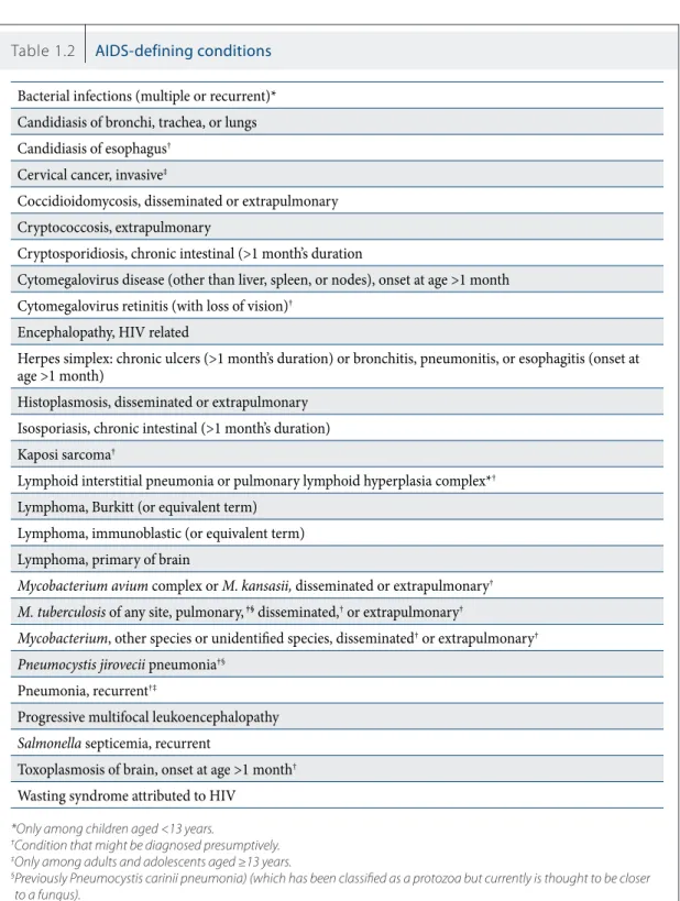
Many clinical manifestations are associated with HIV infection (Table 1.2). Most do not directly result from HIV infection itself but rather result from the consequence of the virus’ impact on the immune system. HIV affects the body in a variety of ways, primarily through OIs, cancers, and wasting syndrome.
O P P O R T U N I S T I C I N F E C T I O N S
A person with a normal immune system has a natural resistance to microorganisms. Only when the immune system is suppressed do various viruses, fungi, protozoa, and bacteria seize the opportunity to cause infection.
Four major categories of organisms cause infection.
Viruses are microorganisms that reproduce by taking over a host cell. Viruses are structurally simple. Most consist of a core RNA or DNA covered by a protein envelope. An example of a simple virus is adenovirus (which causes the common cold). Influenza is a more complex virus that frequently mutates, thereby hindering the immune system’s ability to recognize and respond to future infections. Few antiviral drugs exist. Most have side effects and are toxic. Antibiotics, which work against bacteria, are not helpful against viruses. Common AIDS-defining viral OIs include cytomegalovirus (CMV) disease (other than liver, spleen, or nodes), CMV retinitis (with loss of vision), herpes simplex (chronic ulcers >1 month’s duration), and progressive multifocal luekoencephalopathy.
Bacteria are one-celled organisms found throughout the body. Some are beneficial; others cause disease by producing poisons or toxins. Three basic forms of bacteria exist: spherical (coccus), rod-shaped (bacillus), and spiral shaped (spirillum). Common AIDS-defining bacterial OIs include
Mycobacterium avium or M. kansasii, M. tuberculosis, other Mycobacterium
species or unidentified species, and Salmonella septicemia, recurrent.
Fungi are plant-like organisms that lack chlorophyll and use organic matter as a source of food. Yeast, mold, and mildew are all capable of producing disease in humans. Fungal infections usually are spread through spores in the atmosphere or soil. Systemic fungal infections are often widely disseminated throughout the body in persons with AIDS. Candidiasis and cryptococcosis are the two fungi most frequently seen in HIV-infected persons. Other common AIDS-defining fungal OIs include coccidioidomycosis, histoplasmosis, and Pneumocystis
jiroveci pneumonia.
Protozoa belong to the animal kingdom and are microscopic one-celled parasites that depend on living creatures for food. Cryptosporidiosis and toxoplasmosis of the brain are the two most common protozoal infections associated with HIV infection. Other common AIDS-defining protozoal OIs include sosporiasis, and PCP.
Other: In addition, the following cancers and conditions are recognized as AIDS indicator diseases: invasive cervical cancer (associated with human papilloma virus), Kaposi sarcoma (associated with a variant of the herpes virus), primary lymphoma of the brain, immunoblastic lymphoma (non-Hodgkin disease), Burkitt lymphoma, HIV encephalopathy (caused by a direct effect of HIV on brain cells), and wasting syndrome attributed to HIV.
Table 1.2 AIDS-defining conditions
Bacterial infections (multiple or recurrent)* Candidiasis of bronchi, trachea, or lungs Candidiasis of esophagus†
Cervical cancer, invasive‡
Coccidioidomycosis, disseminated or extrapulmonary Cryptococcosis, extrapulmonary
Cryptosporidiosis, chronic intestinal (>1 month’s duration
Cytomegalovirus disease (other than liver, spleen, or nodes), onset at age >1 month Cytomegalovirus retinitis (with loss of vision)†
Encephalopathy, HIV related
Herpes simplex: chronic ulcers (>1 month’s duration) or bronchitis, pneumonitis, or esophagitis (onset at age >1 month)
Histoplasmosis, disseminated or extrapulmonary Isosporiasis, chronic intestinal (>1 month’s duration) Kaposi sarcoma†
Lymphoid interstitial pneumonia or pulmonary lymphoid hyperplasia complex*†
Lymphoma, Burkitt (or equivalent term) Lymphoma, immunoblastic (or equivalent term) Lymphoma, primary of brain
Mycobacterium avium complex or M. kansasii, disseminated or extrapulmonary† M. tuberculosis of any site, pulmonary, †§ disseminated,† or extrapulmonary†
Mycobacterium, other species or unidentified species, disseminated† or extrapulmonary† Pneumocystis jirovecii pneumonia†§
Pneumonia, recurrent†‡
Progressive multifocal leukoencephalopathy
Salmonella septicemia, recurrent
Toxoplasmosis of brain, onset at age >1 month†
Wasting syndrome attributed to HIV
*Only among children aged <13 years.
†Condition that might be diagnosed presumptively. ‡Only among adults and adolescents aged ≥13 years.
§ Previously Pneumocystis carinii pneumonia) (which has been classified as a protozoa but currently is thought to be closer to a fungus).
Source: CDC. Revised Surveillance Case Definitions for HIV Infection Among Adults, Adolescents, and children Aged <18 Months and for HIV Infection and AIDS Among Children Aged 18 Months to <13 Years—United States, 2008. MMWR 2008:57(No. RR-10).
HIV Disease Treatment and Drug Therapies
When AIDS and HIV were first recognized in the United States during the early 1980s, no effective medications was available to control the virus itself. Additionally, only a limited number of medications were available for treating the OIs associated with this new severe immune deficiency condition.
Antiretroviral therapy (ART) for the treatment of HIV infection has improved steadily since the advent of potent combination therapy in the mid-1990s. New drugs have been approved that offer new mechanisms of action; improvements in potency and activity, even against multidrug-resistant viruses; dosing convenience; and tolerability.
Because of the constant evolution of treatments for the prevention, prophylaxis, and treatment of HIV and associated OIs, ensuring inclusion of the latest list of available medications is beyond the scope of this manual. However, surveillance staff need to be familiar with the names of current medications used to treat HIV and related OIs because that information is vital for completing information about treatment coverage in surveillance areas.
The treatment guidelines for HIV disease and associated OIs will continue to evolve. Surveillance staff are encouraged to periodically review the literature for updates. Below are some websites with the latest information about treatments for HIV and OIs:
• National Institutes of Health
http://aidsinfo.nih.gov/guidelines/html/1/adult-and-adolescent-treatment-guidelines/0/ • Centers for Disease Control and Prevention
http://www.cdc.gov/mmwr/preview/mmwrhtml/rr5804a1.htm?s_cid=rr5804a1_e • Community Research Initiative of New England
http://www.crine.org/
This website provides a link to a full-color HIV medication chart that displays the names and pictures of current classes of antiretroviral medications.
The information in Appendix A is specific to adults and adolescents aged ≥13 years. For information about surveillance case definitions for children <13 years of age, see the 2008 Revised Surveillance Case Definition.
(The HIV surveillance case definition has been revised several times during the epidemic. State and local HIV surveillance staff are advised to monitor the CSTE position statement website for information about the most up-to-date HIV case definition.)
HIV Infection Case Definition
A reportable case of HIV infection must meet at least one of the following criteria: Laboratory Criteria
a) Positive result from an HIV antibody screening test (e.g., reactive enzyme immunoassay) confirmed by a positive result from a supplemental HIV antibody test (e.g., Western blot or indirect immunofluorescence assay).
or
b) Positive result or report of a detectable quantity (i.e., within the established limits of the laboratory test) of any of the following HIV virologic (i.e., nonantibody) tests:
• HIV nucleic acid (DNA or RNA) detection test (e.g., polymerase chain reaction [PCR])
• HIV p24 antigen test, including neutralization assay
• HIV isolation (viral culture)
OR
Other Criterion (for cases that do not meet laboratory criteria)
HIV infection diagnosed by a physician or qualified medical-care provider based on the laboratory criteria and documented in a medical record*. Oral reports of prior laboratory test results are not acceptable.
* An original or copy of the laboratory report is preferred; however, in the rare instance the laboratory report is not available, a description of the laboratory report results by a physician or qualified medical-care provider documented in the medical record is acceptable for surveillance purposes. Every effort should be made to obtain a copy of the laboratory report for documentation in the medical record.
APPENDIX A:
2008 Surveillance Case Definition for HIV Infection among
adults and adolescents
Information in this section is intended for staff new to HIV surveillance or without clinical backgrounds.
In adults and adolescents, a reportable case of AIDS (HIV infection, stage 3) must meet the following criteria:
I. A positive HIV laboratory test (or other criterion) as listed in Appendix A
AND
II. a) CD4 count <200 cells/μL or CD4 T-lymphocyte percentage of <14%
OR
b) One of the following opportunistic illnesses:
(The clinical descriptions on the following pages are adapted from material provided by the New York City Department of Health and Mental Hygiene, HIV Epidemiology and Field Services Program.)
Abbreviations frequently used in Appendix B:
• CT: computed tomography
• HAART: highly active antiretroviral therapy
• MRI: magnetic resonance imaging
• OI: opportunistic infection
• PCR: polymerase chain reaction
APPENDIX B:
Diagnostic Criteria for AIDS-Defining Conditions
Information in this section is intended for staff new to HIV surveillance or without clinical backgrounds.
Candidiasis of Bronchia, Trachea, or Lungs
DEFINITIVE
diagnosis through
Cytologic (histologic) examination of biopsy obtained through bronchoscopy, needle biopsy, open lung biopsy, or autopsy. If the histologic result is positive, it will show clusters of budding yeast or spores and hyphae (i.e., the interwoven thread-like projections that make up the mass of a fungus).
POSSIBLE SIGNS AND SYMPTOMS
Cough Fever Fatigue
Shortness of breath
CURRENT TREATMENT OR THERAPIES
Fluconazole (Diflucan)
Ketoconazole (Nizoral) for maintenance therapy Amphotericin B (Fungizone) systemic treatment
ADDITIONAL INFORMATION
These conditions can be diagnosed definitively only through cytology (histology) from biopsy as indicated above.
CONSULTATION, LABORATORY, SPECIAL REPORTS
Surgical pathology Microbiology Autopsy pathology
ICD-9-CM ICD-10-CM
Candidiasis of Esophagus
DEFINITIVE
diagnosis through
Gross inspection by endoscopy OR autopsy OR microscopy (histology or cytology) on a specimen obtained directly from the tissues affected (including scrapings from the mucosal surface) and from a culture.
OR
PRESUMPTIVE
diagnosis through
Provider diagnosed. Possible indications of the disease include 1. Recent onset of retrosternal pain on swallowing AND
2. Oral candidiasis diagnosed by the gross appearance of white patches or plaques on an erythematous base OR by the microscopic appearance of fungal mycelial filaments in an uncultured specimen scraped from the oral mucosa.
RISK FACTORS
Generally seen in patients with CD4 count <100 cells/μL
POSSIBLE SIGNS AND SYMPTOMS
Retrosternal pain or discomfort
Dysphagia and/or odynophagia, usually with absence of fever (unless co-infections are present) Oral thrush: creamy white patches on tongue, palate, or throat
CURRENT TREATMENT OR THERAPIES
Fluconazole (Diflucan) Itraconazole
Consider intravenous azoles for patients with severe manifestations of disease. Second-line therapies include voriconazole and amphotericin B (Fungizone).
ADDITIONAL INFORMATION
Definitive diagnosis can be made by gross inspection during endoscopy (esphagoscopy), autopsy, or surgery or through positive histologic report of budding yeast and/or pseudohyphae from biopsy obtained during endoscopy, autopsy, or surgery.
Presumptive diagnosis is based on the presence of oral candidiasis noted on physical examination or characteristic appearance of radiologic contrast study (barium swallow) AND the following complaints with swallowing liquids and/or solids of any temperature:
1. Pain during swallowing (bad sore throat, burning in chest or throat, chest pain (tightness or squeezing) 2. Dysphagia (awareness of difficulty in swallowing) with or without pain (food sticking in throat, food
CONSULTATION, LABORATORY, SPECIAL REPORTS
Operating room or surgery department Surgical pathology
Autopsy pathology
ICD-9-CM ICD-10-CM
112.9 Candidiasis of unspecified site B37.9 Candidiasis, unspecified site
Cervical Cancer, Invasive
DEFINITIVE
diagnosis through
Microscopy (histology or cytology)
POSSIBLE SIGNS AND SYMPTOMS
PAP test indicating the presence of class III, IV, or V cells
Presence of a friable mass or ulcer detected during physical examination
CURRENT TREATMENT OR THERAPIES
Combination therapy (can include treatments below) Radiotherapy
Chemotherapy Surgery
ADDITIONAL INFORMATION
Can be diagnosed definitively only by histologic identification of the cell type from a biopsy specimen. Invasive cervical cancer includes mico-invasive cervical cancer (stage 1A) and all more advanced stages by using the criteria of the Oncology Committee of the International Federation of Gynecologists and Obstetricians. Carcinoma in situ (stage O) is NOT included as invasive cervical cancer.
CONSULTATION, LABORATORY, SPECIAL REPORTS
Surgical pathology Autopsy pathology Radiology
ICD-9-CM ICD-10-CM
180.1 Cervical carcinoma, invasive C53 Malignant neoplasm of cervix uteri
ALSO CALLED
Coccidioidomycosis, Disseminated or Extrapulmonary
DEFINITIVE
diagnosis through
Microscopy (histology or cytology)
OR
Culture
OR
Detection of antigen in a specimen obtained directly from the tissues affected or a fluid from those tissues.
BACKGROUND AND EPIDEMIOLOGY
Coccidioides immitis lives in soil, and the vast majority of cases result from inhalation of soil or dust
containing this fungus. Many times, HIV-positive patients have positive serology without clinical disease.
RISK FACTORS
Active disease: CD4 count <250 cells/μL or diagnosis of AIDS
Disseminated disease: Elevated in pregnant women, Filipinos, blacks, and Hispanics
POSSIBLE SIGNS AND SYMPTOMS:
Fever, chills, night sweats (similar presentation to Pneumocystis jirovecii pneumonia [PCP]) Malaise
Cough Weight loss Chest pain
CURRENT TREATMENT OR THERAPIES
Fluconazole (Diflucan) Amphotericin B (Fungizone)
Ketoconazole (Nizoral) to prevent relapse
ADDITIONAL INFORMATION
Can be diagnosed through histologic identification of the fungus Coccidioides immitis from spinal fluid, biopsy specimen or blood culture OR by the presence of coccidioidomycal antigen in the tissue involved or the fluid surrounding the tissue.
CONSULTATION, LABORATORY, SPECIAL REPORTS
Microbiology Surgical pathology Clinical immunology Serology ICD-9-CM ICD-10-CM 114 Coccidioidomycosis B 38 Coccidioidomycosis
Cryptococcosis, Extrapulmonary
DEFINITIVE
diagnosis through
Microscopy (histology or cytology)
OR
Culture
OR
Detection of antigen in a specimen obtained directly from the tissues affected or a fluid from those tissues.
BACKGROUND AND EPIDEMIOLOGY
Cryptococcus is a fungus found worldwide in soil; it is a major OI in sub-Saharan Africa, Thailand, and India. In the pre-HAART era in the United States, cryptococcosis developed in 5%–8% of HIV-positive patients. Its incidence is dramatically lower in the HAART era.
POSSIBLE SIGNS AND SYMPTOMS
Fever Confusion Irritability Neck stiffness
Elevated intracranial pressure (>200 mmHg) common and might be accompanied by evidence of cerebral edema: blurred vision, diplopia (double vision), hearing loss, severe headache, confusion, and papilledema. Seizures
Loss of appetite Meningitis
Extreme bizarre behavior Depression, somnolence Nausea or vomiting
Blurred vision, photophobia (aversion to light) Uncontrolled excitement
Impaired memory or focal neurologic deficits Inappropriate speech and/or dress
CURRENT TREATMENT OR THERAPIES
Amphotericin B (Fungizone)
With or without flucytosine (5-FC), depending on response Fluconazole (Diflucan) for maintenance therapy
ADDITIONAL INFORMATION
Can be diagnosed definitively through histologic identification of Cryptococcusneoformans from spinal fluid, biopsy specimens of tissues, or blood culture OR the presence of the cryptococcal antigen in the tissue involved or the fluid surrounding the tissue.