warwick.ac.uk/lib-publications
Original citation:
Sternberg, Ulrich, Witter, Raiker, Kuprov, Ilya, Lamley, Jonathan M., Oss,
Andres, Lewandowski, Józef R. and Samoson, Ago (2018) 1H line width dependence on MAS
speed in solid state NMR – comparison of experiment and simulation. Journal of Magnetic
Resonance . doi:
10.1016/j.jmr.2018.04.003
Permanent WRAP URL:
http://wrap.warwick.ac.uk/101275
Copyright and reuse:
The Warwick Research Archive Portal (WRAP) makes this work of researchers of the
University of Warwick available open access under the following conditions.
This article is made available under the Creative Commons Attribution 4.0 International
license (CC BY 4.0) and may be reused according to the conditions of the license. For more
details see:
http://creativecommons.org/licenses/by/4.0/
A note on versions:
The version presented in WRAP is the published version, or, version of record, and may be
cited as it appears here.
1
H line width dependence on MAS speed in solid state NMR –
Comparison of experiment and simulation
Ulrich Sternberg
a,b,⇑, Raiker Witter
c,d,e, Ilya Kuprov
f, Jonathan M. Lamley
g, Andres Oss
c,e,
Józef R. Lewandowski
g, Ago Samoson
c,ea
Karlsruhe Institute of Technology (KIT), Karlsruhe, Germany
bCOSMOS GbR, Jena, Germany c
School of Information Technologies, Tallinn University of Technology, Tallinn, Estonia
d
Institute of Nanotechnology, Karlsruhe Institute of Technology (KIT), Karlsruhe, Germany
e
NMR Institute MTÜ, Tallinn, Estonia
f
School of Chemistry, University of Southampton, UK
g
Department of Chemistry, University of Warwick, Coventry, UK
a r t i c l e i n f o
Article history:
Received 30 November 2017 Revised 4 April 2018 Accepted 6 April 2018 Available online 7 April 2018
Keywords:
1H solid-state NMR
High-speed MAS MAS line widths Spinach simulation Fokker-Plank approach
a b s t r a c t
Recent developments in magic angle spinning (MAS) technology permit spinning frequencies of100 kHz. We examine the effect of such fast MAS rates upon nuclear magnetic resonance proton line widths in the multi-spin system of b-Asp-Ala crystal. We perform powder pattern simulations employing Fokker-Plank approach with periodic boundary conditions and1H-chemical shift tensors calculated using
the bond polarization theory. The theoretical predictions mirror well the experimental results. Both approaches demonstrate that homogeneous broadening has a linear-quadratic dependency on the inverse of the MAS spinning frequency and that, at the faster end of the spinning frequencies, the residual spectral line broadening becomes dominated by chemical shift distributions and susceptibility effects even for crystalline systems.
Ó2018 The Authors. Published by Elsevier Inc. This is an open access article under the CC BY license (http:// creativecommons.org/licenses/by/4.0/).
1. Introduction
The superior sensitivity of 1H detected experiments and the wealth of information on molecular structure and dynamics that they provide has always rendered observation of protons (1Hs) as
very important for the NMR spectroscopy. Without much of an exaggeration it is due to accessibility of1H NMR in liquid samples,
where anisotropic interactions such as dipolar couplings and chemical shift anisotropy (CSA) are well-averaged by overall rota-tional diffusion, solution NMR has become an extremely powerful and widely applicable approach to study molecules at atomic res-olution. In solids, however, high-resolution proton studies are hampered by the significant line-broadening effects due to the presence of strong homonuclear1H–1H dipolar couplings. Despite
this drawback, 1H NMR in the solid state has found numerous
applications, particularly in studies of small natural abundance organic molecules and their crystal polymorphs that are pharma-ceutically relevant [1] but also, increasingly, for elucidation of
structure and dynamics of biomolecules [2–7]. To combat the broadening effects of1H–1H dipolar couplings and improve
spec-tral resolution in 1H observed spectra, studies usually employ either dilution of the dense networks of protons with deuterium atoms[8–10], combined rotation and multiple pulse spectroscopy (CRAMPS) techniques to decouple the homonuclear dipolar cou-plings[11–13]or combination of these two solutions[14]. The first of these approaches is most commonly used for proteins where incorporation of2H is achieved by biosynthetic methods[15]. For
other systems where deuterated derivatives need to be synthe-sized chemically, this route is less common due to a more resource intensive nature of the approach. On the other hand, CRAMPS methods achieved a remarkable feat of efficient averaging of the
1H–1H dipolar couplings even under moderate spinning
frequen-cies [16–25]. Thanks to this, investigations on uniformly proto-nated compounds have become increasingly popular in the recent years[18,21,26–34]. However, to obtain the optimum per-formance for CRAMPS techniques effects of pulse imperfections, specifics of hardware, details of irradiation schemes, intricacies of windowed acquisition and related artifacts, etc. have to be consid-ered. Because of that homonuclear decoupling sequences typically require optimization of experimental parameters [16,35].
https://doi.org/10.1016/j.jmr.2018.04.003
1090-7807/Ó2018 The Authors. Published by Elsevier Inc.
This is an open access article under the CC BY license (http://creativecommons.org/licenses/by/4.0/).
⇑Corresponding author at: Karlsruhe Institute of Technology (KIT), Karlsruhe, Germany.
E-mail address:[email protected](U. Sternberg).
Contents lists available atScienceDirect
Journal of Magnetic Resonance
Consequently, routine implementations of CRAMPS methods require a considerable level of technical expertise from operators. In addition, because application of homonuclear decoupling scales down the chemical shifts and the theoretical scaling factors often differ from the experimental ones, proton chemical shifts need to be corrected by comparing the decoupled spectra to spectra where the dipolar coupling averaging is accomplished using only MAS
[21,29,36]. Finally, the losses of magnetization during application of CRAMPS methods mean that the full potential of1H detected
experiments is not yet fully exploited. In spite of these challenges the numerous applications enabled by CRAMPS are a testament to desirability and wide scope of applications of1H spectroscopy.
The promise to achieve similar or better resolution as the one afforded by CRAMPS methods but without the associated chal-lenges renders1H spectroscopy enabled by fast spinning highly
desirable[37]. To guide such experiments and to understand their intrinsic limitations, in this contribution we estimate the extent to which the powder lines can be narrowed by MAS.
In the absence of extensive dynamics and special cases such as e.g. in the presence of paramagnetic centers, the majority of solid state1H line widths is due to two different effects: (i) homogenous
broadening primarily due to strong dipolar couplings and to a les-ser extent1H–1H J-couplings (in the case of the dipeptide with iso-tropic values of up to 20 Hz) and (ii) inhomogenous broadening due toB0field inhomogeneities, magnetic field variation in
pow-dered samples caused by susceptibility distribution (anisotropic bulk magnetic susceptibility) due to variation of particle/crystal shapes and chemical shift dispersion caused by disorder/defects in the sample. In proteins where slow molecular motions play a much more prominent role than in small molecules, homogenous broadening from relaxation can also be a significant factor[3,4]. Inhomogeneous broadening scales linearly with magnetic field strength and is re-focused by 180°pulses[38]but stays constant when measured in ppm. Notably, this effect survives averaging by MAS (and CRAMPS as well) and will lead to residual broadening even under infinitely fast MAS. One of the goals of this investiga-tion is to provide estimainvestiga-tion for this source of broadening in a small molecule polycrystalline sample in the regime where the homogenous broadening becomes increasingly small.
Homogeneous line broadening can be separated from inhomo-geneous broadening by measuring the coherence lifetimes,T02,
dur-ing a spin echo experiment[39,40]. As discussed by Levittet al.in the context of spin pairs the MAS line width should be proportional to1/
x
MASor1/(x
MAS)2depending on the chemical shifts (CS) andthe CS tensors of the coupled nuclei[41]. Brunneret al.considered the dependence of the line width on spinning frequency in analysis that included the geometry of the network of the coupling nuclei
[42]. Since the Hamiltonian of a dipolar network of like spins such as protons does not, in general, commute with itself at different times, the analytical treatment of the homogeneous broadening effect is mostly limited to simple manageable cases. Here we were able to perform rigorous simulations of homogenous proton broad-ening on a multiple spin system consisting of a much more realistic number of spins compared to studies found in the literature. Our simulations are based on the Fokker-Plank equations applied to a crystal lattice with periodic boundary conditions and calculated
1H chemical shift anisotropy[43]. A direct comparison with
exper-imental results allows the extraction of simple rules for the line narrowing effect due to fast MAS. Our simulations avoid most lim-itations that hampered numerical simulations in the past [44]. These investigations aid in guiding the relatively straightforward and time-efficient ‘‘fast MAS only” method that is appropriate for the characterization of small organic molecules at natural abun-dance (i.e. without any isotopic enrichment) in the solid state.
2. Experimental
NMR experiments were performed on a 850 MHz Bruker Avance III spectrometer using a triple resonance 0.81 mm probe (Fig. 1) developed in the Samoson laboratory. The rotor size was carefully optimized for the sensitivity. In the final design sample with volume of700–800 nL could spin at100 kHz. Powdered natural abundance b-L-Asp-L-Ala dipeptide was purchased from Bachem and packed, without further re-crystallization, into a 0.81 mm rotor. The adjustment of the magic angle was carried out at 80 kHz spinning speed via line width observation and min-imization for 1H spectra. 1D 1H spectra of the dipeptide were
obtained over a range of spinning frequencies (
x
r/2p
) between15 and 100 kHz (±50 Hz). A spin-echo experiment was employed to improve the spectral baseline, with a total echo length (2
s
) of 24 rotor periods[32]. Spin-echo experiments were also performed at different spinning frequencies in the range between 15 and 100 kHz to measure, for each proton in the dipeptide, the transverse dephasing time in the absence of inhomogeneous broadening (T20) [40]. These experiments were repeated to account for theeffects of sample temperature on the line widths. A Bruker BCU-X cooling unit was used with the target temperature set to80
°C and the input nitrogen gas pressure set to 0.2 bar for relevant spinning frequencies (leading to different internal sample temper-atures at different spinning frequencies). While the exact sample temperature was not known, application of cooling compensated for the frictional sample temperature rise and no difference in line-width was observed consistent with a lack of extensive molecular motions in the molecule. In the subsequent experiments on pro-teins with internal chemical shift reference and water chemical shift as the indicator of sample temperature, we learned that in a 0.81 mm probe the frictional heating varies between 5 and 10°C at 90 kHz and reaches over 20°C at 100 kHz (resulting in the overall sample temperature of45–50°C if no cooling is applied). Spectral line widths were obtained by fitting the 1H spectra
with ACD/NMR Processor. Each line width presented corresponds to the mean value of five independent fits (for which the peak
height, width, position and Lorentzian/Gaussian fraction were opti-mized) of the same spectrum, with varying starting fitting condi-tions. The standard deviations of the resulting width calculations were interpreted as the experimental errors. It should be noted that at lower spinning frequencies, these uncertainties were mostly larger due to the unresolved nature of the spectra, which meant that the same overall spectrum could be recreated from a range of close to equivalent solutions. T0
2 values were estimated
by fitting in Origin 8.5[45]the signal intensity decay curves from spin echo experiments. For each curve, the points were obtained by taking the intensities of the relevant deconvoluted resonances of the spectra. The spin-echo line widths - homogeneous line-widths - were calculated as1/(
p
T20), with errors propagated by fitvariations from relatedT02calculations.
3. Simulations
3.1. Structure modeling and NMR calculations
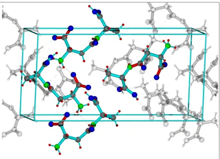
The crystal structure of a b-L-Asp-L-Ala dipeptide [46] was obtained from the Cambridge Crystallographic Data Centre [47]
(CSD refcode FUMTAI). The original CIF-file (space group P212121) was transformed into a COSMOS[48] coordinate coo-file. The symmetry operations of the space group P212121 were executed on the non equivalent sites using COSMOS to generate all atoms within the unit cell. The cell contained 4 zwitterions with 4 equivalent sets of 12 protons (seeFig. 1 in Supplementary Mate-rials) giving finally a spin system with 48 protons. By applying periodic boundary conditions on this spin matrix (33348 = 1296 spins) we obtained one of the most realistic model of1H network used for simulations to date (also see state of the art by Hodgkinsonet al.[49,50]). For protons of the CH3and NH3groups
which experimentally provide only single resonances, we distin-guished all 8 different signals by the simulations (seeTable 1 in Supplementary Materials). All dipolar couplings and chemical shift tensors were considered.1H shielding tensors have to be taken into
account because their orientation dependences in respect to dipo-lar interactions are not negligible (e.g. level crossing during MAS)
[51]. All nuclear shielding tensors were calculated using the bond polarization theory[52–54](BPT). Due to the zwitterionic charac-ter of the crystal molecules within the unit cell, wide-ranging polarization effects had to be considered by taking periodic bound-ary conditions into account. By these means atomic charges and shielding tensors were calculated and included in the spin matrix (seeSupplementary Materials for details).
BPT calculations are well suited for shielding tensor predictions (seeSupplementary Materials Figs. 2a and 2b: R > 0.992 and error RMDS < 1.6 ppm). However, the accuracy of the isotropic values was further improved by shifting the tensor traces to the mean experimental values. The proton sites of the unit cell including the CS tensors were saved to a COSMOS coordinate file as an input for the simulations with Spinach.
3.2. Spectra simulations
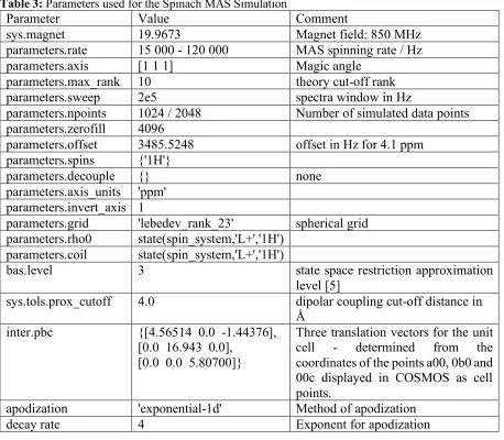
The MAS powder pattern simulations were performed using the Spinach[55]simulation package written as a set of MATLAB[56]
functions. The routines for the Fokker-Plank calculations[57] of the Spinach library were executed from a program controlling the input of the COSMOS coordinate files, data handling, powder averaging which also allowed to set the parameters of the NMR experiment. The simulation routine was complemented by a script for exporting graphics and spectra. A special routine was included that allowed to sample separate FIDs of all 48 proton sites to
ana-lyze allT0
2decay times. The unbiased 48 complex FIDs were stored
in a file for further analysis.
Spinach simulations were carried out as batch processes on a high-performance computing cluster of Tallinn University of Tech-nology. The powder averaging was simultaneously performed with parallelized algorithms on 12 processors. The simulations were performed using ‘‘spinach_1.4.2313” requiring at least MATLAB version R2013b (64-bit). Related Spinach version facilitates the usage of crystallographic periodic boundary conditions. The atoms of a unit cell must be provided as an input and then the periodic continuation is generated automatically. A single simulation with such unit cell consumed between 6 (15 kHz MAS) and 10 days (120 kHz MAS) of total computating time.
Spinach read directly Cartesian coordinates and calculated all dipolar interactions of the spin system. The CS tensors were imported from COSMOS coordinate file. Periodic boundary condi-tions were applied to the unit cell with a dimension of a = 4.845 Å which has been only slightly larger than the used dipolar cut off radius of 4 Å. Since proton interactions between connected unit cells were considered, realistic NMR signals can be expected.
Under experimental conditions (300 K) the NH3 and CH3
groups undergo rotational jumps or diffusion which partially aver-ages out their dipolar interactions on the NMR time scale. As an approximation for the influence of this effect, we set all dipolar coupling values of the protons within these groups to zero.
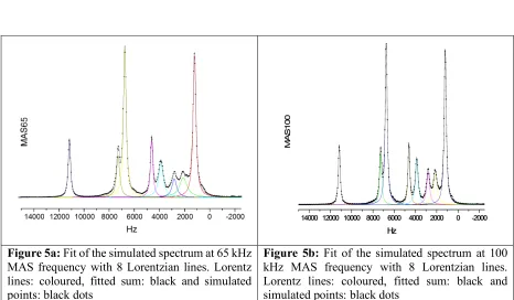
Spectra were obtained by fast Fourier transformation of the original simulated FIDs. FIDs were preprocessed using zero filling and gentle exponential apodization. The simulated FIDs did not decay to zero (seeFig. 3 is Supporting Materials). We increased the exponent for the apodization until the Fourier transform arti-facts from the step contribution just started to disappear. Despite the fact that the apodization added 82 Hz to the line widths the simulated spectra display sharper lines than the experimental ones (seeFig. 2) because the applied line broadening is still smaller than inhomogeneous line width in the sample used in the experiments (seeTable 2). Resulting spectra could be analyzed by fitting reso-nances to Lorentzian line-shapes (seeFigs. 5a and 5b in Supple-mentary Materials) but spectra cannot be expected to provide the quantitative identity of the influence of MAS on the residual line widths which are dominated at high speed by susceptibility and disorder effects. Only the homogeneous part of the line widths can be compared and interpreted. This part is related toT0
2decay
time measured during spin echo experiments. We decided to determine T02 directly from simulated FIDs. In a
model-free-approach of the form of decay only absolute values of the 48 com-plex FIDs were used. TheT0
2timings were obtained by searching for
the time interval where the intensity dropped to1/eof the values with respect to the zero-time-point. This procedure can be per-formed for all kinds of decaying functions disregarding the special functional forms. From all 48 individual positions, similar protons were averaged to obtain the 8 detectable meanT0
2values (for the
complete setup of the simulations seeSupplementary Materials).
4. Results and discussion
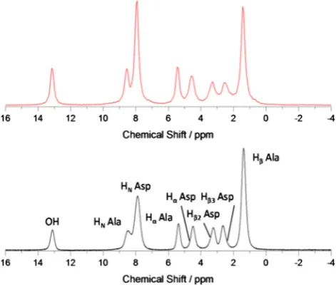
To observe the effect of increasing MAS on averaging of proton line widths, we measured 1D1H spectra on the dipeptideb
-Asp-Ala at spinning frequencies from 15 to 100 kHz at 8501H Larmor
frequency, without homonuclear decoupling but with13C
decou-pling (Figs. 2aand2b).
is increased; with the two Asp CH2 proton resonances becoming
resolved at around 65 kHz (seeSupplementary Materials Fig. 6) where corresponding resonances have line widths of 418 ± 5 and 351 ± 1 Hz (0.49 ppm and 0.41 ppm, Hb2and Hb3Asp respectively).
The distance between the two protons of CH2groups give rise to a
dipolar coupling of 64.3 kHz (seeTable 1) and obviously we have to spin at least with a similar minimum frequency to separate the lines of the sites. At 850 MHz1H Larmor frequency and 100 kHz
spinning the line width of the CH2 protons becomes 0.34 ppm
(Hb2: 292 ± 1 Hz) and 0.32 ppm (Hb3: 274 ± 2 Hz, seeTable 2).
These are comparable to the 0.36 ppm and 0.34 ppm improved line widths that had been achieved using eDUMBO-PLUS-1 homonuclear decoupling scheme at a similar field strength (800 MHz)[18]. Thus line narrowing at 100 kHz spinning is comparable to the state of the art high power homonuclear decoupling at lower
spinning frequencies. One of the obvious benefits of the approach based on fast magic spinning is its simplicity. At a given tempera-ture, the extent of obtained line-narrowing only depends on the precision of the magic angle setting and quality of shimming, both of which would be required for any other type of experiment setup, e.g. CRAMPS. Another major advantage is that intensities of chem-ical shift resonances which are reflecting certain molecular envi-ronments are directly proportional to a number of related proton spins:1H concentrations can be directly extracted from such
spec-tra without any special manipulation or rescaling.
The contributions to the proton line widths can be grouped into two categories: homogeneous broadening and inhomogeneous boradening. Homogenous broadening is dominated by the incom-plete averaging of homonuclear dipolar couplings, which can be reduced by MAS, with contribution from transverse relaxation. Inhomogeneous broadening due to field alternations and disorder within the sample cannot be eliminated by spinning (or CRAMPS) without removing chemical shift information. As such it is useful to separate the two broadening components and compare experi-ments with realistic calculations. In order to remove the inhomo-geneous part, we measured the transverse dephasing time of protons during the spin-echo experiments, T0
2[40], as a function
of spinning frequency, in the 30–100 kHz range. For 100 kHz spin-ning the values are compiled inTable 1(andFig. 7 of Supplemen-tary Materials) which shows the MAS frequency dependence of the total and spin-echo line widths (equal to1/(
p
T20)) of the protons inb-Asp-Ala. It has been found in numerous other studies that line widths decrease with increasing
x
r since the dipolar couplingsare averaged out more effectively. The offset between the two sets of data for each proton (total broadening and homogeneous broad-ening) represent the inhomogeneous contribution of the line width, which is not refocused in aT0
2measurement. Although the
inhomogeneous contribution is approximately constant with vary-ing
x
r,[38]the offset is site-specific. The amount of suchinhomo-geneous broadening in this particular polycrystalline b-Asp-Ala sample is typically > 125 Hz (> 0.15 ppm). The absolute spin-echo line widths should, however, be considered with some degree of caution, as systematic errors can arise in cases where a single exponentials fit the T0
2 data rather poorly (we recognized such
deviations in our simulation results)[38].
The line width dependency on
x
rhas been investigatedprevi-ously. For instance, Zorin et al. [44] used a semi-analytical approach of dipolar interacting spin systems to show a 1/
x
rdepen-dency of line width. This analysis disregards the influences of chemical shift tensors. Brunneret al.[42]assumed that the1H CS
tensors should be axially symmetric (skew +1 or1) which doesn’t reflect the results of our COSMOS BPT-calculations (see evaluation results inTable 2 of Supplementary Materials). Since our simula-tions of spectra included realistic1H CS tensor values for of each
site their relative influence can be understood in detail. First of all, it is clear that the influence of rotational resonances[41]even at lowest spinning frequency of 15 kHz can be excluded: such con-ditions may occur if the differences of isotropic shifts of two sites are equal to a multiple (n) of the rotational frequency (D
x
ison
x
r). In our experiments, the largest possible CS difference of11.7 ppm yieldsD
x
iso/2p
of 10.5 kHz.When discussing the
x
rdependency of the line width threecases have to be distinguished: (i) case of negligible small CS differ-ence (D
x
iso0), (ii) case where the chemical shift difference is onthe same order of magnitude as the dipolar coupling (D
x
isox
dip)and (iii) case where the chemical shift difference exceeds the dipo-lar coupling (D
x
iso»x
dip). In the limit of case (iii) the flip-flop term [image:5.595.51.286.69.258.2]of the interaction Hamiltonian can be neglected [41] and the Hamiltonian has then a similar form as a hetero-nuclear spin inter-action. Such an operator is self-commuting and behaves like an
Fig. 2a.Simulated (top panel, red) and experimental (bottom panel, black) 1D1
H NMR spectra ofb-Asp-Ala at 15 kHz MAS frequency. (For interpretation of the references to color in this figure legend, the reader is referred to the web version of this article.)
Fig. 2b.Simulated (top panel, red) and experimental (bottom panel, black) 1D1
[image:5.595.50.286.323.523.2]inhomogeneous contribution under MAS, i.e. produces sharp split-ting in the spectrum but cannot be re-focused with a 180° spin-echo. In our simulations, we frequently encountered the interme-diate scenario (ii) whereD
x
isoandx
dipare in the same order of magnitude. In this case, we should observe a decrease of the line width with the inverse of the MAS rate squared[41]:1/(x
r)2. Sincethe Hamiltonian for this case does not commute with itself at dif-ferent times, the interaction is called homogeneous and is not re-focused in spin-echo experiments. If the coupling nuclei have neg-ligible CS differences (i) we have the case of so-called ‘‘n0 rota-tional resonances” [41]. Since in our simulations also the CS anisotropy is present this effect does occur if the principal axes of the CS tensors of the coupling nuclei do not coincide. This effect
has essentially the same behavior as a case (ii) and leads to a homogeneous broadening of the MAS line with a narrowing related to1/(
x
r)2. These cases that are discussed above are strictly validonly for isolated spin pairs but the theory [41] provides some guidelines to tackle the case of an interacting multi-spin system.
For the simulated spectra, we needed to introduce apodization which introduced artificial broadening mostly reflecting the inho-mogeneous contributions but not the influence of MAS. Therefore, only the homogeneous part of the line widths is considered under thorough theoretical investigations involving magnetization decay timeT0
2in spin echo experiments. A direct determination ofT02from
the calculated FID was obtained avoiding any data processing. The collected theoretical data ofT0
[image:6.595.34.553.95.190.2]2 as a function of MAS frequency
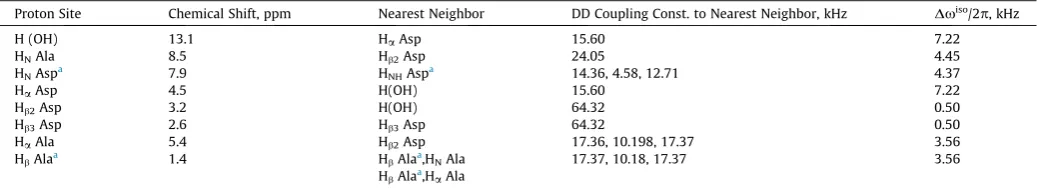
Table 1 Observed1
H chemical shift differences and calculated dipolar couplings to the nearest neighbor proton sites. The shift difference in frequency units is displayed in the last column. The1
H dipolar couplings within the CH3and NH3groups are not displayed.
Proton Site Chemical Shift, ppm Nearest Neighbor DD Coupling Const. to Nearest Neighbor, kHz Dxiso
/2p, kHz
H (OH) 13.1 HaAsp 15.60 7.22
HNAla 8.5 Hb2Asp 24.05 4.45
HNAspa 7.9 HNHAspa 14.36, 4.58, 12.71 4.37
HaAsp 4.5 H(OH) 15.60 7.22
Hb2Asp 3.2 H(OH) 64.32 0.50
Hb3Asp 2.6 Hb3Asp 64.32 0.50
HaAla 5.4 Hb2Asp 17.36, 10.198, 17.37 3.56
HbAlaa 1.4 HbAlaa,HNAla 17.37, 10.18, 17.37 3.56
HbAlaa,HaAla
a
[image:6.595.34.551.259.329.2]The chemical shifts are mean values of 3 protons of a CH3or NH3group resp.
Table 2
Total, homogeneous and inhomogeneous limit1H line widths ofb-Asp-Ala, measured at 100 kHz MAS spinning and 850 MHz1H Larmor frequency. Inhomogeneous limit values
indicate the minimum line width that may be measured on this sample at an infinite spinning frequency which reflects the contributions from inhomogeneous broadening,1H–1H
J-couplings (here up to 20 Hz) and relaxation.
Peak H(OH) HNAla HNAsp HaAla HaAsp Hb2Asp Hb3Asp HbAla
Line width/Hz (ppm) 229 ± 1 339 ± 5 325 ± 2 211 ± 0.5 295 ± 1 292 ± 0.5 274 ± 2 269 ± 0.5
(0.27) (0.40) (0.38) (0.25) (0.30) (0.34) (0.32) (0.32)
Spin-echo line width/Hz (ppm) 78 ± 9 146 ± 38 174 ± 15 71 ± 10 138 ± 25 290 ± 130 240 ± 120 136 ± 6
(0.09) (0.17) (0.20) (0.08) (0.16) (0.34) (0.28) (0.16)
Inhomogenous limit line width/Hz (ppm) 147 ± 23 170 ± 10 164 ± 16 135 ± 5 84 ± 8 142 ± 29 163 ± 12 166 ± 12
(0.17) (0.20) (0.19) (0.16) (0.10) (0.17) (0.19) (0.20)
Fig. 3.SimulatedT0
2times at spinning frequencies from 30 to 100 kHz. The lines are obtained by fitting the data to a linear-quadratic dependence. Values ofaandb
[image:6.595.111.477.511.730.2]m
r=x
r/2p
,can be seen inFig. 3. Strikingly, all simulatedT02-curvesfollow a linear-quadratic model of the following form:
T02¼a
m
MASþbm
2MASIt is not possible to derive the linear-quadratic trend ofT02on the
MAS frequency directly from the line widths dependence on1/
x
rand1/(
x
r)2but we represent two limiting cases -T02proportionalto
m
MAS if the 1/(x
r)2 term is vanishing and T02 proportional to(
m
MAS)2if the1/x
rterm is zero. The linear-quadratic dependenceofT0
2on the MAS frequency should, therefore, be regarded as the
first two-term of a power series ofT0
2with respect to
m
MAS. Thetwo limiting cases can be readily identified in our simulations
(seeFig. 3andTable 3):T0
2of HaAla has a nearly linear dependence
on
m
MASand for Hb2and Hb3of Asp the linear term is approximatelyzero and only the quadratic term remains. For HaAla echo decay times are dominated by a1/
x
rdependence, which is reflected bythe parametersaandb(seeTable 3): HaAsp has a much smaller quadratic term compared to the other proton sites. Hb2Asp and
Hb3Asp have the lowest linear contribution and follow dependency
closer to1/(
x
r)2in accordance with the findings of Levittet al.[41].The obvious differences between the proton sites display some reg-ular patterns when looking at the nearest neighbors (seeTable 1). The CH2 protons have the largest dipolar interaction (64.32 kHz)
and the smallest CS difference (0.5 kHz) such that they have the smallest linear terma. The proton of the OH-site has a small quad-ratic termband a large linear termacorresponding to CS differ-ence to Ha Asp with 7.22 kHz and a dipolar coupling of only 15.6 kHz. But these rules are to be taken with care since they involve only isolated spin pairs. Until now there is no theory for the parametersaandbof the linear-quadratic dependency ofT0
2
on
m
MASbut some trends can be observed howaandbdepend onthe NMR parametersD
x
isandx
dip. For NMR experimentalists, itis encouraging that proton lines with a small difference in CS but a large dipolar interaction are narrowed in a quadratic manner with the MAS frequency.
Having considered extended Spinach simulations the question arises whether calculated trends are also consistent with experi-ments.Fig. 4shows plots correlating the theoretical and
experi-Table 3
Best fit parameters for the fit of theT0
2times to a linear-quadratic model of the form:
T02¼amMASþbm2MAS(mMASin kHz, seeFig. 3).
Proton resonance Parameter a105
Parameter b107
HbAla (CH3) 4.158 3.151
H(OH) 5.848 1.519
HNAsp (NH3) 2.997 3.279
HNAla 0.814 4.988
HaAsp 1.395 1.052
Hb2Asp (0.0)-0.147a 1.710
Hb3Asp (0.0)-0.633a 2.298
HaAla 5.243 0.126
[image:7.595.41.294.237.324.2]a Within the error limits these values should be zero.
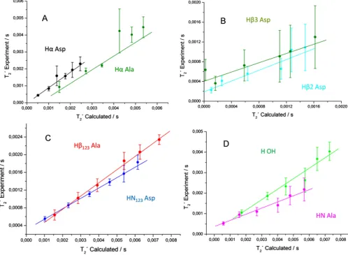
Fig. 4.Correlation plots for the simulated and experimental1HT0
2times inb-L-Asp-L-Ala. The color code is the same as inFig. 3. The lines represent linear fits of the
[image:7.595.52.551.358.723.2]mentalT0
2times, which exhibit linear correlation within the
exper-imental error bounds. Ideally, the slope of the linear correlations should be close to 1.0 and the lines crossing with the ordinate at zero point. However, such behavior is only met for Ha Ala and HaAsp (panel A inFig. 3 and Table 5 in Supplementary Materials). All other proton sites theoretically provide a slope smaller than 1.0 which indicates a lower simulated homogeneous line width than observed in the experiment. The main reason for that might be the neglecting of homonuclear scalar J couplings (up to 20 Hz) and disregarding of relaxation due to molecular motion. Rotation or fast jumps of the CH3and NH3groups not only influence
corre-sponding protons of the groups but also scale down the dipolar interactions to protons in the neighborhood. The dipolar interac-tions within those groups were set to zero but this treatment might need an improvement in future simulations. In particular, for the NH3group, the considered approximation yields a slope
of only 0.24. It is known that NH3protons are forming hydrogen
bonds to carbonyl oxygen atoms (see Fig. 1 in Supplementary Materials) that will slow down the motion of this group, with the result of a larger line width than in the case of CH3protons which
are much less affected by hydrogen bonds.
We could use the determined trends of1H line widths as a
func-tion of the spinning frequency to evaluate benefits of developing even faster spinning probes than currently available. The broadest
1H resonances in the considered here dipeptide and probably also
in most other organic compounds originate from the CH2protons
with a large dipolar interaction of more than 60 kHz. Based on the determined in our work trends to reduce homogeneous line widths by a factor of 2 one has to raise the spinning by a factor ofp2. Thus even for CH2protons inb-L-Asp-L-Ala an increase of
the spinning rate from 100 to 141 kHz would result in a homoge-nous line broadening of 0.2 ppm (170 Hz) which becomes on the order of inhomogeneous line broadening of powdered polycrys-talline samples. Considering the minimum inhomogeneous contri-butions reported in the literature for single crystals of glycine, the total line width can be around 120 Hz[58]for CH2, i.e. a further
increase in MAS spinning speed up to 200 kHz for strongly coupled protons and beyond for other 1H sites might still considerably
improve the resolution.
5. Conclusions
The Spinach Fokker-Plank approach using PBC with COSMOS/ BPT chemical shift tensor values and crystal coordinates of b-L-Asp-L-Ala dipeptide has been successfully applied for MAS spin-echo simulations from 15 to 100 kHz spinning frequencies. Subtle details of spectra were faithfully reproduced by simulation, validating our computational approach to a complicated many-spin and time-dependent problem. The 1H homogeneous line
broadening had been extracted and provided as simplest descrip-tion a linear-quadratic dependency ofT0
2on
m
MAS, i.e. the two termsrepresent the limiting cases with vanishing1/
x
r-term or vanishing1/(
x
r)2-term. At spinning frequencies on the order of 100 kHz,inhomogeneous broadening starts becoming a dominant or at least non-negligible factor even in crystalline systems but the coherence lifetimes are increasing dramatically, in fact, faster than previously thought. The quadratic improvement of1H coherence lifetimes as a
function of spinning frequency will be particularly beneficial for applications involving scalar couplings for polarization transfer, which have a theoretical 100% efficiency. The simple linear-quadratic dependence ofT0
2on the MAS frequency allows an easy
estimate of the1H line widths at higher spinning speed from
pre-liminary experiments.
We show that at 100 kHz MAS frequencies and 850 MHz1H Lar-mor frequency, high-quality proton spectra can be obtained on
powdered samples with1H line widths in the same order as those
achievable with state of the art homonuclear decoupling schemes under optimal conditions. This work and other impressive exam-ples reported recently in the literature suggest that the approach relying on1H detected spectroscopy at100 kHz spinning should
be widely applicable both to small as well as large molecules espe-cially for material, medical and biological applications.
Acknowledgments
We are grateful to Prof. Steven Brown for providing theb -Asp-Ala sample. JML acknowledges support from the EPSRC. The UK 850 MHz solid-state NMR Facility used in this research was funded by EPSRC and BBSRC, as well as the University of Warwick, includ-ing part fundinclud-ing through Birminclud-ingham Science City Advanced Mate-rials Projects 1 and 2 supported by Advantage West Midlands (AWM) and the European Regional Development Fund (ERDF). JRL acknowledges funding from Royal Society Grant RG130022, EPSRC Grant EP/L025906/1, ERC Starting Grant 639907, BBSRC Grant BB/L022761/1 and Gates Foundation OPP1160394. RW is gratefully acknowledging Tallinn University of Technology and Karlsruhe Institute of Technology. Funding from ESF and ETAG for project PUT126 as well as PUT1534 is acknowledged.
Appendix A. Supplementary material
Supplementary data associated with this article can be found, in the online version, at https://doi.org/10.1016/j.jmr.2018.04.003. Raw 1H spectra are available on Mendeley Data at https://doi.
org/10.17632/rrc9pzc4wk.1.
References
[1]S.P. Brown, Applications of high-resolution H-1 solid-state NMR, Solid State Nucl. Mag. 41 (2012) 1–27.
[2]L.B. Andreas, K. Jaudzems, J. Stanek, D. Lalli, A. Bertarello, T. Le Marchand, D.C. D. Paepe, S. Kotelovica, I. Akopjana, B. Knott, S. Wegner, F. Engelke, A. Lesage, L. Emsley, K. Tars, T. Herrmann, G. Pintacuda, Structure of fully protonated proteins by proton-detected magic-angle spinning NMR, P. Natl. Acad. Sci. USA 113 (2016) 9187–9192.
[3]J.M. Lamley, C. Oster, R.A. Stevens, J.R. Lewandowski, Intermolecular interactions and protein dynamics by solid-state NMR spectroscopy, Angew. Chem. Int. Ed. 54 (2015) 15374–15378.
[4]J.M. Lamley, D. Iuga, C. Oster, H.J. Sass, M. Rogowski, A. Oss, J. Past, A. Reinhold, S. Grzesiek, A. Samoson, J.R. Lewandowski, Solid-state NMR of a protein in a precipitated complex with a full-length antibody, J. Am. Chem. Soc. 136 (2014) 16800–16806.
[5]E. Barbet-Massin, C.T. Huang, V. Daebel, S.T.D. Hsu, B. Reif, Site-specific solid-state NMR studies of ‘‘Trigger Factor” in complex with the large ribosomal subunit 50S, Angew. Chem. Int. Ed. 54 (2015) 4367–4369.
[6]J.P. Demers, V. Chevelkov, A. Lange, Progress in correlation spectroscopy at ultra-fast magic-angle spinning: basic building blocks and complex experiments for the study of protein structure and dynamics, Solid State Nucl. Mag. 40 (2011) 101–113.
[7]C. Oster, S. Kosol, C. Hartlmuller, J.M. Lamley, D. Iuga, A. Oss, M.L. Org, K. Vanatalu, A. Samoson, T. Madl, J.R. Lewandowski, Characterization of protein-protein interfaces in large complexes by solid-state NMR solvent paramagnetic relaxation enhancements, J. Am. Chem. Soc. 139 (2017) 12165–12174. [8]A.E. Mcdermott, F.J. Creuzet, A.C. Kolbert, R.G. Griffin, High-resolution
magic-angle-spinning nmr-spectra of protons in deuterated solids, J. Magn. Reson. 98 (1992) 408–413.
[9]L. Zheng, K.W. Fishbein, R.G. Griffin, J. Herzfeld, 2-Dimensional solid-state H-1-Nmr and proton-exchange, J. Am. Chem. Soc. 115 (1993) 6254–6261. [10]B. Reif, C.P. Jaroniec, C.M. Rienstra, M. Hohwy, R.G. Griffin, 1H–1H MAS
correlation spectroscopy and distance measurements in a deuterated peptide, J. Magn. Reson. 151 (2001) 320–327.
[11]B.C. Gerstein, R.G. Pembleton, R.C. Wilson, L.M. Ryan, High-resolution Nmr in randomly oriented solids with homonuclear dipolar broadening - combined multiple pulse nmr and magic angle spinning, J. Chem. Phys. 66 (1977) 361– 362.
[12]L.M. Ryan, R.E. Taylor, A.J. Paff, B.C. Gerstein, Experimental-study of resolution of proton chemical-shifts in solids - combined multiple pulse Nmr and magic-angle spinning, J. Chem. Phys. 72 (1980) 508–515.
[14]J.R. Lewandowski, J.N. Dumez, U. Akbey, S. Lange, L. Emsley, H. Oschkinat, Enhanced resolution and coherence lifetimes in the solid-state NMR spectroscopy of perdeuterated proteins under ultrafast magic-angle spinning, J. Phys. Chem. Lett. 2 (2011) 2205–2211.
[15]M. Hologne, V. Chevelkov, B. Reif, Deuterated peptides and proteins in MAS solid-state NMR, Prog. Nucl. Mag. Res. Sp. 48 (2006) 211–232.
[16]M. Mehring, J.S. Waugh, Magic-angle Nmr experiments in solids, Phys. Rev. B 5 (1972) 3459–4000.
[17]M.H. Levitt, A.C. Kolbert, A. Bielecki, D.J. Ruben, High-resolution 1H NMR in solids with frequency-switched multiple-pulse sequences, Solid State Nucl. Magn. Reson. 2 (1993) 151–163.
[18]E. Vinogradov, P.K. Madhu, S. Vega, High-resolution proton solid-state NMR spectroscopy by phase-modulated Lee-Goldburg experiment, Chem. Phys. Lett. 314 (1999) 443–450.
[19]E. Vinogradov, P.K. Madhu, S. Vega, Phase modulated Lee-Goldburg magic angle spinning proton nuclear magnetic resonance experiments in the solid state: a bimodal Floquet theoretical treatment, J. Chem. Phys. 115 (2001) 8983–9000.
[20]D. Sakellariou, A. Lesage, P. Hodgkinson, L. Emsley, Homonuclear dipolar decoupling in solid-state NMR using continuous phase modulation, Chem. Phys. Lett. 319 (2000) 253–260.
[21]A. Lesage, D. Sakellariou, S. Hediger, B. Elena, P. Charmont, S. Steuernagel, L. Emsley, Experimental aspects of proton NMR spectroscopy in solids using phase-modulated homonuclear dipolar decoupling, J. Magn. Reson. 163 (2003) 105–113.
[22]J.P. Amoureux, B. Hu, J. Trebosc, Enhanced resolution in proton solid-state NMR with very-fast MAS experiments, J. Magn. Reson. 193 (2008) 305–307. [23]J.P. Amoureux, B.W. Hu, J. Trebosc, Q. Wang, O. Lafon, F. Deng, Homonuclear
dipolar decoupling schemes for fast MAS, Solid State Nucl. Mag. 35 (2009) 19– 24.
[24]M.E. Halse, L. Emsley, Improved phase-modulated homonuclear dipolar decoupling for solid-state NMR spectroscopy from symmetry considerations, J. Phys. Chem. A 117 (2013) 5280–5290.
[25]E. Salager, J.N. Dumez, R.S. Stein, S. Steuernagel, A. Lesage, B. Elena-Herrmann, L. Emsley, Homonuclear dipolar decoupling with very large scaling factors for high-resolution ultrafast magic angle spinning H-1 solid-state NMR spectroscopy, Chem. Phys. Lett. 498 (2010) 214–220.
[26]A. Lesage, L. Duma, D. Sakellariou, L. Emsley, Improved resolution in proton NMR spectroscopy of powdered solids, J. Am. Chem. Soc. 123 (2001) 5747– 5752.
[27]S.P. Brown, A. Lesage, B. Elena, L. Emsley, Probing proton-proton proximities in the solid state: high-resolution two-dimensional 1H–1H double-quantum CRAMPS NMR spectroscopy, J. Am. Chem. Soc. 126 (2004) 13230–13231. [28]M. Leskes, P.K. Madhu, S. Vega, Supercycled homonuclear dipolar decoupling
in solid-state NMR: toward cleaner 1H spectrum and higher spinning rates, J. Chem. Phys. 128 (2008) 052309.
[29]M. Leskes, S. Steuernagel, D. Schneider, P.K. Madhu, S. Vega, Homonuclear dipolar decoupling at magic-angle spinning frequencies up to 65 kHz in solid-state nuclear magnetic resonance, Chem. Phys. Lett. 466 (2008) 95–99.
[30]S. Paul, R.S. Thakur, P.K. Madhu, H-1 homonuclear dipolar decoupling at high magic-angle spinning frequencies with rotor-synchronised symmetry, Chem. Phys. Lett. 456 (2008) 253–256.
[31]E. Salager, R.S. Stein, S. Steuernagel, A. Lesage, B. Elena, L. Emsley, Enhanced sensitivity in high-resolution H-1 solid-state NMR spectroscopy with DUMBO dipolar decoupling under ultra-fast MAS, Chem. Phys. Lett. 469 (2009) 336– 341.
[32]S. Paul, R.S. Thakur, M. Goswami, A.C. Sauerwein, S. Mamone, M. Concistre, H. Forster, M.H. Levitt, P.K. Madhu, Supercycled homonuclear dipolar decoupling sequences in solid-state NMR, J. Magn. Reson. 197 (2009) 14–19.
[33]Z.H. Gan, P.K. Madhu, J.P. Amoureux, J. Trebosc, O. Lafon, A tunable homonuclear dipolar decoupling scheme for high-resolution proton NMR of
solids from slow to fast magic-angle spinning, Chem. Phys. Lett. 503 (2011) 167–170.
[34]M.E. Halse, L. Emsley, A common theory for phase-modulated homonuclear decoupling in solid-state NMR, PCCP 14 (2012) 9121–9130.
[35]B. Elena, G. de Paepe, L. Emsley, Direct spectral optimisation of proton-proton homonuclear dipolar decoupling in solid-state NMR, Chem. Phys. Lett. 398 (2004) 532–538.
[36]E. Vinogradov, P.K. Madhu, S. Vega, Proton spectroscopy in solid state nuclear magnetic resonance with windowed phase modulated Lee-Goldburg decoupling sequences, Chem. Phys. Lett. 354 (2002) 193–202.
[37]Y. Nishiyama, Fast magic-angle sample spinning solid-state NMR at 60–100 kHz for natural abundance samples, Solid State Nucl. Mag. 78 (2016) 24–36. [38]A. Samoson, T. Tuherm, Z. Gan, High-field high-speed MAS resolution
enhancement in 1H NMR spectroscopy of solids, Solid State Nucl. Magn. Reson. 20 (2001) 130–136.
[39]E.L. Hahn, Spin Echoes, Phys. Rev. 80 (1950) 580–594.
[40] G. De Paepe, N. Giraud, A. Lesage, P. Hodgkinson, A. Bockmann, L. Emsley, Transverse dephasing optimized solid-state NMR spectroscopy, J. Am. Chem. Soc. 125 (2003) 13938–13939.
[41]M.H. Levitt, D.P. Raleigh, F. Creuzet, R.G. Griffin, Theory and simulations of homonuclear spin pair systems in rotating solids, J. Chem. Phys. 92 (1990) 6347–6364.
[42]E. Brunner, D. Freude, B.C. Gerstein, H. Pfeifer, Residual linewidths of Nmr-spectra of spin-1/2 systems under magic-angle spinning, J. Magn. Reson. 90 (1990) 90–99.
[43]L.J. Edwards, D.V. Savostyanov, A.A. Nevzorov, M. Concistre, G. Pileio, I. Kuprov, Grid-free powder averages: on the applications of the Fokker-Planck equation to solid state NMR, J. Magn. Reson. 235 (2013) 121–129.
[44]V.E. Zorin, S.P. Brown, P. Hodgkinson, Origins of linewidth in H-1 magic-angle spinning NMR, J. Chem. Phys. 125 (2006).
[45] Origin, in, OriginLab, Northampton, MA.
[46]C.H. Gorbitz, Crystal and molecular-structures of the isomeric dipeptides alpha-L-aspartyl-L-alanine and beta-L-aspartyl-L-alanine, Acta Chem. Scand. B 41 (1987) 679–685.
[47] C.R. Groom, I.J. Bruno, M.P. Lightfoot, S.C. Ward, The Cambridge Structural Database.
[48] U. Sternberg, F.-T. Koch, P. Losso, COSMOS Pro, COSMOS-Software, Jena, Germany.
[49]P. Hodgkinson, D. Sakellariou, L. Emsley, Simulation of extended periodic systems of nuclear spins, Chem. Phys. Lett. 326 (2000) 515–522.
[50] P. Hodgkinson, L. Emsley, Numerical simulation of solid-state NMR experiments, Prog. Nucl. Mag. Res. Sp. 36 (2000) 201–239.
[51]A. Lesage, M. Bardet, L. Emsley, Through-bond carbon-carbon connectivities in disordered solids by NMR, J. Am. Chem. Soc. 121 (1999) 10987–10993. [52]I. Jakovkin, M. Klipfel, C. Muhle-Goll, A.S. Ulrich, B. Luy, U. Sternberg, Rapid
calculation of protein chemical shifts using bond polarization theory and its application to protein structure refinement, PCCP 14 (2012) 12263–12276. [53]R. Witter, L. Seyfart, G. Greiner, S. Reissmann, J. Weston, E. Anders, U.
Sternberg, Structure determination of a pseudotripeptide zinc complex with the COSMOS-NMR force field and DFT methods, J. Biomol. Nmr 24 (2002) 277– 289.
[54]U. Sternberg, Theory of the influence of the 2nd coordination sphere on the chemical-shift, Mol. Phys. 63 (1988) 249–267.
[55]H.J. Hogben, M. Krzystyniak, G.T.P. Charnock, P.J. Hore, I. Kuprov, Spinach - A software library for simulation of spin dynamics in large spin systems, J. Magn. Reson. 208 (2011) 179–194.
[56] MATLAB, The MathWorks, Inc., Natick, Massachusetts, United States. [57]I. Kuprov, Fokker-Planck formalism in magnetic resonance simulations, J.
Magn. Reson. 270 (2016) 124–135.
Supplementary Materials
1
H Line Width Dependence on MAS Speed in Solid State NMR - Comparison of
Experiment and Simulation
Ulrich Sternberg, Raiker Witter,
Ilya Kuprov
, Jonathan Lamley, Andres Oss, Józef R. Lewandowski
and Ago Samoson
1. Structure Modeling
Figure 1:
Structure of ß-L-Aspartic acid-L-Ala dimer. The four molecules of the unit cell are
displayed in color and the other molecules are displayed in transparent grey. The 48
1H sites are shown
in red. The hydrogen bridges of the charged NH
3groups are displayed as dotted lines
2.
1H Shielding Tensor Calculations using the Bond Polarization Theory (BPT) [1, 2]
Table 1:
Parameter for BPT
1H shielding tensor calculations*
Bond
Type
Shielding of Isolated Bond in ppm
Contribution of Polarization in
ppm/Hartree
σ
xxσ
yyσ
zzA
xxA
yyA
zzH-C(sp3)
27.5576
24.9134
30.7793
56.9518
-64.264
12.5664
[image:10.595.74.526.225.551.2]Figure 2a:
Correlation of
1H
α
nuclear shielding
tensor components calculated on the MP2-TZVPP
level with BPT values. Correlation coefficient
R=0.995 and RMS deviation of 1.4 ppm [3].
Figure 2b:
Correlation of
1H
N

![Table 2: Principal components of the 1H chemical shift (CS) tensors of ß-L-Aspartic acid-L-Alanine dimer [4] calculated using the BPT](https://thumb-us.123doks.com/thumbv2/123dok_us/9432085.449179/11.595.77.487.490.686/table-principal-components-chemical-tensors-aspartic-alanine-calculated.webp)