CRYPTIC CYSTIC PRESENTATION: A CASE REPORT
Dr. Asha M. L.1, Dr. Laboni Ghorai2*, Dr. Basetty Neelakantam Rajarathnam3, Dr. Mahesh Kumar H. M.4, Dr. Lekshmy J.5 and Dr. Reenu Jose6
1
MDS, Professor and Head of the Department; Department of Oral Medicine & Radiology, Dr. Syamala Reddy Dental College, Hospital & Research Centre, Bangalore.
2
PG Student, Department of Oral Medicine & Radiology, Dr. Syamala Reddy Dental College, Hospital & Research Centre, #111/1, SGR College Main Road, Munnekolala, Marathahalli
(Post), Bangalore- 560037. 3
MDS, Senior Lecturer; Department of Oral Medicine & Radiology, Dr. Syamala Reddy Dental College, Hospital & Research Centre, Bangalore.
4
MDS, Senior Lecturer; Department of Oral Medicine & Radiology, Dr. Syamala Reddy Dental College, Hospital & Research Centre, Bangalore.
5
MDS, Senior Lecturer; Department of Oral Medicine & Radiology, Dr. Syamala Reddy Dental College, Hospital & Research Centre, Bangalore.
6
PG Student, Department of Oral Medicine and Radiology, Dr. Syamala Reddy Dental College, Hospital and Research Centre, Bangalore.
ABSTRACT
Dentigerous cyst is the second most common odontogenic cyst, caused
by alteration of reduced enamel epithelium. It shows a male
predominance and is reported predominantly associated with unerupted
third molars. The most common clinical feature is painless swelling.
Radiographic examination reveals a unilocular radiolucent lesion in
association with the crown of an unerupted tooth. Treatment of
dentigerous cyst often includes enucleation of the cyst. Presentation of
dentigerous cyst with pain only is unusual as in our case. Presented
here is a case report of 31-years-old male patient who complained of
pain in the left lower back tooth region. A detailed clinical,
radiographic and histopathological analysis aided in the diagnosis of
infected dentigerous cyst.
KEYWORDS: Dentigerous cyst, odontogenic cyst, Enucleation.
INTRODUCTION
Dentigerous cyst can be defined as an odontogenic cyst that surrounds the crown of an
impacted tooth; caused by fluid accumulation between
Volume 4, Issue 11, 1549-1556. Case Report ISSN 2277– 7105
Article Received on 09 Sep 2015,
Revised on 29 Sep 2015, Accepted on 19 Oct 2015
*Correspondence for
Author
Dr. Laboni Ghorai
(MDS), Department of
Oral Medicine &
Radiology, Dr. Syamala
Reddy Dental College,
Hospital & Research
Centre, #111/1, SGR
College Main Road,
Munnekolala,
Marathahalli (Post),
the reduced enamel epithelium and the enamel surface, resulting in a cyst in which crown is
located within the lumen.[1] Dentigerous cyst is the most common developmental cyst.[2] The
exact cystogenesis of this cyst remains indefinite, but the most probable aetiology is its
developmental origin from a tooth follicle.[3] Dentigerous cysts are almost always associated
with the crowns of unerupted teeth and are seen attached to the cemento-enamel junction.[4]
Less frequently, they can be found in relation with supernumerary teeth,[5] odontomas[6] or
unerupted deciduous teeth.[7] Dentigerous cyst is usually painless and delayed eruption of
tooth may be first or only clinical sign. The most common clinical feature is painless
swelling. Here, we have reported a case of dentigerous cyst where pain was the chief
complaint. Careful clinical, radiological and histological findings led to the diagnosis of an
occult dentigerous cyst.
CASE REPORT
A 31-years-old male patient reported to the Department of Oral Medicine and Radiology, Dr.
Syamala Reddy Dental College, Hospital and Research Centre, Bangalore with the chief
complaint of pain in the left lower back teeth region since four days. Pain was sudden in
onset, sharp, severe in intensity, continuous in nature with postural variation. Pain aggravated
on mastication and relieved on taking medication. The patient also gave the history of
associated swelling three days back, which subsided on taking medication. The past dental,
medical and surgical history was not contributory.
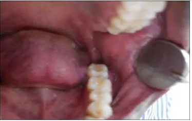
On extra-oral examination, nothing significant was found. Intra-oral examination revealed
partially erupted tooth with inflamed, soft, oedematous pericoronal flap in relation to 38 and
there was discharge of pus in relation to the pericoronal flap. The regional lymph node was
tender and palpable. With the above findings, it was provisionally diagnosed as acute
pericoronal abscess.
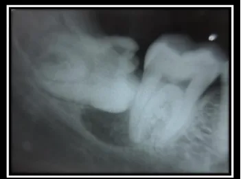
Routine radiographic examinations were carried out. Intra-Oral Periapical Radiograph in
relation to 38 revealed a well-defined radiolucency along the mesial aspect of the coronal
portion extending approximately 2mm below the cemento-enamel junction up to the mesial
cusp tip in relation to horizontally impacted 38. The radiolucency was singular, unilocular
with roughly oval shape and a corticated boundary. The internal structure was totally
radiolucent. Narrowing of inferior dental canal was noted below the radiolucency. However,
confirmed no cortical expansion in relation to 38 region and orthopantomograph was advised
to rule out multiple similar lesions of the jaws.
Extraction of the impacted tooth followed by the surgical enucleation of the cyst, was done
under antibiotic coverage and the cystic lining was sent for histopathological evaluation.
Histology revealed an odontogenic cystic lining which confirmed the diagnosis of a
dentigerous cyst.
[image:3.595.156.440.224.348.2]Figure 1 &2: Extra-Oral photograph of the patient.
Figure 3: Intra-Oral Photograph showing partially erupted 38 with inflamed pericoronal flap.
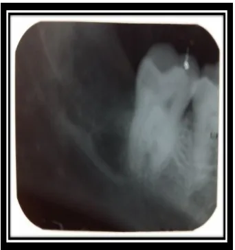
[image:3.595.222.374.390.526.2] [image:3.595.210.387.583.713.2]Figure 5: Mandibular Lateral Occlusal Radiograph showing no cortical expansion.
Figure 6: OPG showing the impacted third molar with the cyst.
Figure 7: Photomicrograph showing the cystic lesion lined by non-keratinised squamous epithelium.
[image:4.595.204.392.436.556.2] [image:4.595.205.392.614.732.2]
Figure 9: 1 month post-operative Intra-Oral Periapical Radiograph.
DISCUSSION
Dentigerous cyst is the most common pathologic pericoronal radiolucency in the jaws.[8]
They are mostly discovered by routine radiographic examinations or by enlargement of the
affected region of the jaw.
The pathogenesis of dentigerous cyst is still controversial. Three feasible mechanisms have
been proposed for histogenesis of the cyst by Benn and Altini. They proposed that
developmental dentigerous cyst might form a dental follicle and might become secondarily
inflamed, the source of inflammation being a non-vital tooth. The second mechanism they
proposed was formation of a radicular cyst at an apex of a non-vital deciduous tooth followed
by eruption of its permanent successor into the radicular cyst resulting in a dentigerous cyst
of extrafollicular origin. They also suggested that follicle of permanent successor may get
secondarily infected from either periapical inflammation of a non-vital predecessor or other
source leading to a dentigerous cyst formation.[3]
Dentigerous cyst accounts for 20% of all the jaw cysts. 10% of impacted teeth form
dentigerous cyst and they occur in 8.1% of impacted third molar tooth.[9] They are reported to
be significantly more common in males than in females.[10] Shear (1985) reported that
dentigerous cyst is more common in whites than black people in Africa and Cabini and
colleagues found a male to female ratio of 2:1.[8] The lesion occurs most often in second and
third decades of life. Regarding the site, the teeth most frequently affected are mandibular
third molars, maxillary canine, mandibular premolar and maxillary third molar in that
This case shows a solitary cyst which is a usual finding. Bilateral and multiple cysts are
usually found in association with a number of syndromes such as Maroteaux-Lamy syndrome
and Cleidocranial dysplasia.
The cysts vary greatly in size, from less than 2cm to massive expansion of the jaws. Cysts
which extend from one angle of the jaw to the other, there may be one tooth in the cavity or
there may be two or more, usually greatly displaced from their normal position and often
lying on the floor of the cavity, in the site (as in our case) or even the roof of the cyst. It is
common for the tooth to be horizontal,[11] as in our case.
Sapphire (2009) reported that dentigerous cyst with large extension may have the potential
for expansion of the cortical bone, which can lead to paraesthesia of the inferior alveolar
nerve, when present in the mandibular region.[8] Costal et al. stated that development of
dentigerous cyst has serious complications such as tooth impaction, ectopic eruption, facial
asymmetry, displacement and root resorption of teeth[12] and even facial oedema due to
cortical expansion by large cyst.[13]
Clinically, dentigerous cyst occurs most often as painless alveolar swelling.[3] Sometimes, the
cyst is associated with pain.[14] Pain usually indicates presence of infection. It rarely expands
rapidly to produce pain by pressure on sensory nerve. Our case also presented with pain due
to infection.
Radiologically, dentigerous cyst classically consists of a well corticated pericoronal
radiolucency, which exceeds 3 mm when measured from the edge of the crown to the
periphery of the lesion on panoramic radiograph and 2.5 mm on periapical radiograph. The
cystic cavity often originates from the cervical region of the tooth.[8] There are three
radiologic variants of Dentigerous cyst: Central, Lateral and Circumferential. The dentigerous
cysts that are eccentric and develop from the lateral aspect of the follicle, occupy an area
beside crown instead of above the crown,[15] as in our case.
Histologically, dentigerous cyst consists of a fibrous wall that may contain young fibroblasts
widely separated by stroma and ground substance rich in mucopolysaccharides. The cyst
lining is non-keratinized stratified squamous epithelium.[16,17] The histology of our case
Treatment option of dentigerous cyst includes enucleation via Caldwell-Luc procedure under
local or general anaesthesia which has been the standard treatment, along with extraction of
the associated tooth. In large cysts, an initial marsupialization to diminish the size of the
osseous defect, followed by enucleation and tooth extraction, has been advocated.
Marsupialization might also offer an opportunity for the permanent tooth to erupt in the oral
cavity. Endoscopic approach for management of dentigerous cyst of the maxilla is also
described in the literature.[18] Our case was treated by enucleation of the cyst with removal of
the impacted tooth under antibiotic coverage, as it was hopelessly malposed and had no
chance to erupt into the oral cavity.
CONCLUSION
Dentigerous cysts are benign odontogenic cysts associated with the crowns of unerupted
permanent teeth and have more affinity for mandibular third molar region. Despite their
benign nature, it must be kept in mind that some untreated dentigerous cyst may grow large
and may have the potential to develop into an odontogenic tumour like ameloblastoma or
become malignant as oral squamous cell carcinoma and mucoepidermoid carcinoma.[19] Thus,
early recognition of the entity and removal of the occult cyst is important to reduce
morbidity.
REFERRENCES
1. Shafer WG, Hine MK, Levy BM. A Textbook of Oral Pathology, 7th ed.; Elsevier: India,
2012; 259-263.
2. Yazdani J, Kahnamouii SS. Developmental Odontogenic cysts of Jaws: A clinical study
of 245 cases. J Dent Res Dent Clin Dent Prospects, 2009; 3(2): 64-66.
3. Benn A, Altini M. Dentigerous cysts of inflammatory origin: A clinicopathologic study.
Oral Surg Oral Med Oral Pathol Oral Radiol Endod, 1996; 81(2): 203-9.
4. Saluja JS et al. Multiple dentigerous cysts in a non-syndromic minor patient: Report of an
unusual case. Natl J Maxillofac Surg, 2010; 1(2): 168-172.
5. Jiang Q et al. Dentigerous cysts associated with impacted supernumerary teeth in the
anterior maxilla. Exp Ther Med, 2011; 2(5): 805-9.
6. Sales MA, Cavaleani MG. Complex odontoma associated with dentigerous cyst in
maxillary sinus: case report and computed tomography features. Dentomaxillofac Radiol,
7. Kusukawa J et al. Dentigerous cyst associated with a deciduous tooth: A Case Report.
Oral Surg Oral Med Oral Pathol, 1992; 73(4): 415-8.
8. Langlais RP, Langland OE, Nortje CJ. Diagnostic Imaging of the Jaws, 1st ed.; William &
Wilkins: USA, 1995; 286-91.
9. Wood NK, Goaz PW. Differential Diagnosis of Oral & Maxillofacial Lesions, 5th ed.;
Elsevier: India, 2013; 283-5.
10.Maximiw WG, Wood RE. Carcinoma arising in a dentigerous cyst: A case report and
review of literature. J Oral Maxillofac. Surg, 1991; 49: 639-43.
11.Worth HM. Principles and Practice of Oral Radiologic Interpretation, 1st ed.; Year book
medical publishers Inc: Chicago, 1969; 466-72.
12.Costa DD et al. Cisto Dentigero associado a canino incluso em maxila. ClipeOdonto,
2011; 3(1): 32-6.
13.Vaz LGM, Radrigues M, Ferreira OJ. Cisto Dentigero: Caracteristicas clinicas,
radiograficase criterios para o plano de tratamento. Porto Alegre, 2010; 58(1): 127-30.
14.Main DMG. Epithelial Jaw Cyst: 10 years of WHO classification. J Oral Pathol, 1985;
14: 1-7.
15.White SC, Pharoah MS. Oral Radiology Principles and Interpretation, 6th ed.; Elsevier:
India, 2009; 338-43.
16.Boyczuk MP, Berger JR. Identifying a dentigerous cyst. J Am Dent Assoc, 1995; 126:
643-4.
17.Holmlund A et al. Ameloblastoma originating from odontogenic cyst. J Oral Pathol Med,
1991; 20(7): 318-21.
18.Kasat VO et al. Dentigerous cyst associated with an ectopic third molar in the maxillary
sinus: A case report and review of literature. Contemp Clin. Dent, 2012; 3: 373-6.
19.Slootweg PJ. Carcinoma arising from reduced enamel epithelium. J Oral Pathol, 1987;