b
-
D-Gulose
Tomohiko Ishii,a* Shunsuke Ohga,aKazuhiro Fukada,b Kenji Morimotoband Genta Sakanec
a
Department of Advanced Materials Science, Faculty of Engineering, Kagawa University, 2217-20 Hayashi-cho, Takamatsu, Kagawa 761-0396, Japan, bDepartment of Applied Biological Science, Faculty of Agriculture, Kagawa University, 2393 Ikenobe, Kagawa 761-0795, Japan, andcDepartment of Chemistry, Faculty of Science, Okayama University of Science, 1-1 Ridaicho, Kita-ku, Okayama 700-0005, Japan
Correspondence e-mail: [email protected]
Received 23 March 2014; accepted 10 April 2014
Key indicators: single-crystal X-ray study;T= 294 K; mean(C–C) = 0.004 A˚; Rfactor = 0.035;wRfactor = 0.073; data-to-parameter ratio = 11.7.
The title compound, C6H12O6, a C-3 position epimer of d
-galactose, crystallized from an aqueous solution, was confirmed as-d-pyranose with a4C1(C1) conformation. In the crystal, O—H O hydrogen bonds between the hydroxy groups at the C-1 and C-6 positions connect molecules into a tape structure with anR3
3
(11) ring motif running along the
a-axis direction. The tapes are connected by further O—H O hydrogen bonds, forming a three-dimensional network.
Related literature
For related structures. see: Fukada et al. (2010). For the chemical synthesis of the title compound, see: Morimotoet al.
(2013). For hydrogen-bonding networks, see: Jeffrey & Saenger (1994); Jeffrey & Mitra (1983).
Experimental
Crystal data
C6H12O6 Mr= 180.16
Orthorhombic,P212121 a= 7.0800 (3) A˚
b= 9.8644 (3) A˚ c= 10.6156 (4) A˚ V= 741.39 (4) A˚3 Z= 4
CuKradiation
= 1.28 mm1 T= 294 K
0.100.100.10 mm
Data collection
Rigaku R-AXIS RAPID II diffractometer
Absorption correction: multi-scan (ABSCOR; Higashi, 1995) Tmin= 0.645,Tmax= 0.879
7803 measured reflections 1358 independent reflections 1199 reflections withF2> 2(F2) Rint= 0.070
Refinement
R[F2> 2(F2)] = 0.035 wR(F2) = 0.073 S= 1.05 1358 reflections
116 parameters
H-atom parameters constrained
max= 0.14 e A˚
3
min=0.14 e A˚
3
Table 1
Hydrogen-bond geometry (A˚ ,).
D—H A D—H H A D A D—H A
O1—H1A O6i
0.82 1.93 2.736 (3) 168
O2—H2A O3ii 0.82 2.12 2.785 (3) 139
O3—H3A O4iii
0.82 1.91 2.722 (3) 173
O4—H4A O6iv
0.82 2.10 2.915 (3) 173
O6—H6A O1v
0.82 1.99 2.805 (3) 177
Symmetry codes: (i)xþ1;y;z; (ii)xþ1 2;yþ
3
2;zþ2; (iii)xþ1;yþ 1 2;zþ
3 2; (iv)
xþ1
2;yþ1;z 1 2; (v)x
1 2;yþ
1 2;zþ2.
Data collection:RAPID-AUTO (Rigaku, 2009); cell refinement:
RAPID-AUTO; data reduction:RAPID-AUTO; program(s) used to solve structure:SIR2008inIl Milione(Burlaet al., 2007); program(s) used to refine structure:SHELXL2013(Sheldrick, 2008); molecular graphics:CrystalStructure(Rigaku, 2010); software used to prepare material for publication:CrystalStructure.
Supporting information for this paper is available from the IUCr electronic archives (Reference: IS5352).
References
Burla, M. C., Caliandro, R., Camalli, M., Carrozzini, B., Cascarano, G. L., De Caro, L., Giacovazzo, C., Polidori, G., Siliqi, D. & Spagna, R. (2007).J. Appl. Cryst.40, 609–613.
Fukada, K., Ishii, T., Tanaka, K., Yamaji, M., Yamaoka, Y., Kobashi, K. & Izumori, K. (2010).Bull. Chem. Soc. Jpn,83, 1193–1197.
Higashi, T. (1995).ABSCOR. Rigaku Corporation, Tokyo, Japan. Jeffrey, G. A. & Mitra, J. (1983).Acta Cryst.B39, 469–480.
Jeffrey, G. A. & Saenger, W. (1994). Hydrogen Bonding in Biological Structures, Study Edition. New York: Springer-Verlag.
Morimoto, K., Shimonishi, T., Miyake, S., Takata, G. & Izumori, K. (2013). Biosci. Biotechnol. Biochem.77, 253–258.
Rigaku (2009).RAPID-AUTO. Rigaku Corporation, Tokyo, Japan. Rigaku (2010).CrystalStructure. Rigaku Corporation, Tokyo, Japan. Sheldrick, G. M. (2008).Acta Cryst.A64, 112–122.
Acta Crystallographica Section E Structure Reports Online
supporting information
Acta Cryst. (2014). E70, o569 [doi:10.1107/S1600536814008046]
β
-
D-Gulose
Tomohiko Ishii, Shunsuke Ohga, Kazuhiro Fukada, Kenji Morimoto and Genta Sakane
S1. Comment
The crystal system (orthorhombic), space group (P212121), and number of molecules in the unit cell (Z = 4) of the title
compound are the same as for the typical hexose (C6H12O6) monosaccharides (Fukada et al., 2010). There is a difference
in the hydrogen bonding patterns, having a circular chain network returning to the same molecule, and the intermolecular
interactions between two adjacent β-D-gulose molecules in the crystal.
In an equatorial OH group at C-2 position, the hydrogen bond can be confirmed as a donor, which connects to the OH
group at C-3 position of the neighboring molecule. However, for the axial OH groups at C-3 and C-4 positions, each has
hydrogen bonds both as a donor and an acceptor to the OH groups at either the C-2 and C-4, or the C-3 and C-6
positions, respectively. In the OH group at the C-6 position, there is an intermolecular hydrogen bond between the OH
group at C-4 position of the neighboring molecule, and there are two additional hydrogen bonds with the OH groups at
different C-1 positions in these two different D-gulose molecules. There is an infinite hydrogen bonding chain along to
the a-axis (···O1—H1A···O6—H6A···O1—H1A···), which is connecting to a finite chain (O2—H2A···O3—H3A···O4—
H4A···O6—H6A). Therefore, the hydrogen bonding network can be categorized as Jeffrey's class (iv) (Jeffrey & Saenger,
1994; Jeffrey & Mitra, 1983). There is a step for returning to the same gulose molecule in an infinite chain (···gulose O1
—H1A···O6—H6A···O1—H1A···gulose O6—H6A···). Such a significant circular hydrogen bonding ring should be
treated differently from the typical infinite chain.
S2. Experimental
D-Gulose was prepared from disaccharide lactitol by a combination of microbial and chemical reactions. 3-Ketolactitol,
oxidized from lactitol by Agrobacterium tumefaciens, was reduced by chemical hydrogenation. The resulting product, D
-gulosyl-(β-1, 4)-D-sorbitol containing D-gulose, was hydrolyzed by acid hydrolysis, and its subsequent hydrolysates were
separated by chromatography. Lastly, a crude crystal from the concentrated D-gulose syrup was recovered by ethanol
precipitation, and then its aqueous solution was recrystallized, resulting in pure D-gulose. The D-gulose was concentrated
to a brix value in a range of approximately 85–90%. Ethanol (twice the volume of the resulting syrup) was added and the
resulting solution was mixed vigorously. The resulting crystals were dissolved in ultrapure water and then concentrated
and crystallized at room temperature. The specific optical rotation of D-gulose was analyzed using a polarimeter (JASCO
P-1030 Tokyo). An optical rotation was also performed, providing [α]20D = -24.10 (authentic sample = -24.74). The 13
C-NMR spectra of the isolated D-gulose was measured at 600 MHz in D2O using an ALPHA 600 system (Jeol Datum,
Tokyo). All spectra were collected at 30 °C using trimethylsilyl propanoic acid as internal reference. All of the chemical
shifts [δ = 94.6 (C1), 74.5 (C5), 71.9 (C3), 70.2 (C4), 69.8 (C2), 61.7 (C6)] corresponded well with an authentic D-gulose
sample. These results indicate that the isolated material was D-gulose and that the current study was successful in
preparing D-gulose. The gulose is a specialized member of the rare sugar family, therefore, the details regarding the
S3. Refinement
H atoms bounded to methine-type C (H1B, H2B, H3B, H4B, H5A) were positioned geometrically and refined using a
riding model with C—H = 0.98 Å and Uiso(H) = 1.2Ueq(C). H atoms bounded to methylene-type C (H6B, H6C) were
positioned geometrically and refined using a riding model with C—H = 0.97 Å and Uiso(H) = 1.2Ueq(C). H atoms
bounded to O (H1A, H2A, H3A, H4A, H6A) were positioned geometrically and refined using a riding model with O—H
[image:3.610.125.492.163.425.2]= 0.82 Å and Uiso(H) = 1.2Ueq(O), allowing for free rotation of the OH groups.
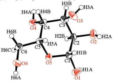
Figure 1
ORTEP view of the title compound with the atom-labeling scheme. The thermal ellipsoids of all non-hydrogen atoms are
drawn at the 50% probability level. H atoms are shown as small spheres of arbitrary radius.
Figure 2
Part of the crystal structure of the title compound with hydrogen-bonding network represented as light blue dashed lines,
[image:3.610.128.482.475.651.2]β-D-Gulose
Crystal data
C6H12O6
Mr = 180.16
Orthorhombic, P212121
Hall symbol: P 2ac 2ab
a = 7.0800 (3) Å
b = 9.8644 (3) Å
c = 10.6156 (4) Å
V = 741.39 (4) Å3
Z = 4
F(000) = 384.00
Dx = 1.614 Mg m−3
Cu Kα radiation, λ = 1.54187 Å Cell parameters from 7124 reflections
θ = 4.2–68.2°
µ = 1.28 mm−1
T = 294 K Block, colorless 0.10 × 0.10 × 0.10 mm
Data collection
Rigaku R-AXIS RAPID II diffractometer
Detector resolution: 10.000 pixels mm-1
ω scans
Absorption correction: multi-scan (ABSCOR; Higashi, 1995)
Tmin = 0.645, Tmax = 0.879
7803 measured reflections
1358 independent reflections 1199 reflections with F2 > 2σ(F2)
Rint = 0.070
θmax = 68.2°
h = −8→8
k = −11→11
l = −12→12
Refinement
Refinement on F2
R[F2 > 2σ(F2)] = 0.035
wR(F2) = 0.073
S = 1.05 1358 reflections 116 parameters 0 restraints
Primary atom site location: structure-invariant direct methods
Secondary atom site location: difference Fourier map
Hydrogen site location: inferred from neighbouring sites
H-atom parameters constrained
w = 1/[σ2(F
o2) + (0.0261P)2]
where P = (Fo2 + 2Fc2)/3
(Δ/σ)max < 0.001
Δρmax = 0.14 e Å−3
Δρmin = −0.14 e Å−3
Extinction correction: SHELXL2013 (Sheldrick, 2008)
Extinction coefficient: 0.0063 (12)
Special details
Refinement. Refinement was performed using all reflections. The weighted R-factor (wR) and goodness of fit (S) are based on F2. R-factor (gt) are based on F. The threshold expression of F2 > 2.0 σ(F2) is used only for calculating R-factor
(gt).
Fractional atomic coordinates and isotropic or equivalent isotropic displacement parameters (Å2)
x y z Uiso*/Ueq
O1 0.6475 (3) 0.4070 (3) 1.02633 (16) 0.0367 (6)
O2 0.7927 (3) 0.6435 (3) 0.89378 (15) 0.0385 (6)
O3 0.4219 (3) 0.7683 (2) 0.87238 (18) 0.0369 (6)
O4 0.3674 (3) 0.49846 (18) 0.64349 (14) 0.0295 (5)
O5 0.3828 (2) 0.41908 (19) 0.90855 (15) 0.0259 (5)
O6 −0.0053 (3) 0.35032 (19) 0.92624 (16) 0.0304 (5)
C1 0.5316 (4) 0.4977 (3) 0.9615 (2) 0.0270 (7)
C2 0.6282 (4) 0.5724 (3) 0.8553 (2) 0.0257 (6)
C4 0.3165 (4) 0.5860 (3) 0.7452 (2) 0.0255 (7)
C5 0.2385 (4) 0.5021 (3) 0.8521 (2) 0.0234 (6)
C6 0.0815 (4) 0.4071 (3) 0.8155 (3) 0.0277 (7)
H1A 0.7468 0.3971 0.9876 0.0441*
H1B 0.4786 0.5635 1.0210 0.0324*
H2A 0.7726 0.6811 0.9614 0.0462*
H2B 0.6682 0.5042 0.7937 0.0308*
H3A 0.4827 0.8379 0.8608 0.0443*
H3B 0.5483 0.7077 0.7161 0.0319*
H4A 0.3971 0.5441 0.5821 0.0354*
H4B 0.2191 0.6495 0.7164 0.0306*
H5A 0.1902 0.5641 0.9167 0.0281*
H6A 0.0367 0.2740 0.9386 0.0364*
H6B −0.0125 0.4559 0.7669 0.0333*
H6C 0.1315 0.3348 0.7634 0.0333*
Atomic displacement parameters (Å2)
U11 U22 U33 U12 U13 U23
O1 0.0251 (10) 0.0477 (13) 0.0374 (11) 0.0010 (11) 0.0001 (9) 0.0157 (11) O2 0.0320 (11) 0.0493 (15) 0.0341 (11) −0.0151 (10) −0.0014 (9) −0.0041 (11) O3 0.0461 (13) 0.0227 (12) 0.0420 (11) −0.0050 (10) 0.0113 (10) −0.0076 (10) O4 0.0416 (12) 0.0273 (12) 0.0196 (9) −0.0012 (10) 0.0020 (9) 0.0013 (8) O5 0.0241 (9) 0.0231 (10) 0.0305 (10) −0.0011 (9) −0.0031 (8) 0.0033 (9) O6 0.0276 (10) 0.0251 (11) 0.0384 (10) −0.0001 (9) 0.0042 (9) 0.0057 (9) C1 0.0263 (14) 0.0289 (17) 0.0258 (13) 0.0003 (13) −0.0030 (13) 0.0012 (12) C2 0.0230 (13) 0.0283 (16) 0.0257 (13) −0.0054 (13) −0.0010 (12) −0.0013 (13) C3 0.0302 (15) 0.0242 (17) 0.0253 (13) −0.0006 (13) 0.0060 (13) −0.0002 (12) C4 0.0284 (14) 0.0251 (15) 0.0231 (13) 0.0058 (14) −0.0004 (12) 0.0016 (13) C5 0.0225 (13) 0.0247 (15) 0.0231 (12) 0.0041 (11) 0.0019 (12) 0.0011 (12) C6 0.0265 (14) 0.0320 (17) 0.0248 (13) 0.0019 (14) 0.0006 (11) 0.0001 (13)
Geometric parameters (Å, º)
O1—C1 1.396 (4) O1—H1A 0.820
O2—C2 1.420 (3) O2—H2A 0.820
O3—C3 1.424 (4) O3—H3A 0.820
O4—C4 1.429 (3) O4—H4A 0.820
O5—C1 1.424 (3) O6—H6A 0.820
O5—C5 1.440 (3) C1—H1B 0.980
O6—C6 1.439 (3) C2—H2B 0.980
C1—C2 1.510 (4) C3—H3B 0.980
C2—C3 1.527 (4) C4—H4B 0.980
C3—C4 1.513 (4) C5—H5A 0.980
C4—C5 1.510 (4) C6—H6B 0.970
C5—C6 1.505 (4) C6—H6C 0.970
O4···H5A 3.2256 H1A···O6ii 1.9284
O1···H6Ai 1.9853 H2A···O3ix 2.1169
O2···H6Bii 2.6723 H3A···O4iii 1.9072
O2···H6Ciii 2.5757 H3B···O5iii 2.5179
O3···H2Aiv 2.1169 H4A···O6vi 2.0992
O4···H3Av 1.9072 H5A···O4viii 2.5185
O4···H5Avi 2.5185 H6A···O1x 1.9853
O5···H3Bv 2.5179 H6B···O2vii 2.6723
O6···H1Avii 1.9284 H6C···O2v 2.5757
C1—O5—C5 112.3 (2) C6—O6—H6A 109.480
O1—C1—O5 106.3 (2) O1—C1—H1B 109.359
O1—C1—C2 114.5 (2) O5—C1—H1B 109.362
O5—C1—C2 107.82 (18) C2—C1—H1B 109.363
O2—C2—C1 113.41 (19) O2—C2—H2B 107.098
O2—C2—C3 111.9 (3) C1—C2—H2B 107.103
C1—C2—C3 109.9 (2) C3—C2—H2B 107.097
O3—C3—C2 110.75 (19) O3—C3—H3B 109.292
O3—C3—C4 107.5 (2) C2—C3—H3B 109.290
C2—C3—C4 110.7 (3) C4—C3—H3B 109.286
O4—C4—C3 110.1 (2) O4—C4—H4B 109.124
O4—C4—C5 109.2 (3) C3—C4—H4B 109.127
C3—C4—C5 110.15 (19) C5—C4—H4B 109.123
O5—C5—C4 111.4 (2) O5—C5—H5A 108.140
O5—C5—C6 106.1 (3) C4—C5—H5A 108.132
C4—C5—C6 114.7 (2) C6—C5—H5A 108.139
O6—C6—C5 110.29 (19) O6—C6—H6B 109.601
C1—O1—H1A 109.471 O6—C6—H6C 109.595
C2—O2—H2A 109.468 C5—C6—H6B 109.603
C3—O3—H3A 109.466 C5—C6—H6C 109.595
C4—O4—H4A 109.475 H6B—C6—H6C 108.127
C1—O5—C5—C4 61.5 (3) C1—C2—C3—C4 −55.0 (3)
C1—O5—C5—C6 −173.06 (15) O3—C3—C4—O4 168.99 (18)
C5—O5—C1—O1 172.54 (16) O3—C3—C4—C5 −70.5 (3)
C5—O5—C1—C2 −64.2 (3) C2—C3—C4—O4 −69.9 (3)
O1—C1—C2—O2 −55.5 (3) C2—C3—C4—C5 50.5 (3)
O1—C1—C2—C3 178.41 (18) O4—C4—C5—O5 68.1 (3)
O5—C1—C2—O2 −173.59 (19) O4—C4—C5—C6 −52.5 (3)
O5—C1—C2—C3 60.4 (3) C3—C4—C5—O5 −53.0 (3)
O2—C2—C3—O3 −62.8 (3) C3—C4—C5—C6 −173.52 (19)
O2—C2—C3—C4 178.08 (16) O5—C5—C6—O6 66.8 (3)
C1—C2—C3—O3 64.1 (3) C4—C5—C6—O6 −169.7 (2)
Symmetry codes: (i) x+1/2, −y+1/2, −z+2; (ii) x+1, y, z; (iii) −x+1, y+1/2, −z+3/2; (iv) x−1/2, −y+3/2, −z+2; (v) −x+1, y−1/2, −z+3/2; (vi) −x+1/2, −y+1,
Hydrogen-bond geometry (Å, º)
D—H···A D—H H···A D···A D—H···A
O1—H1A···O6ii 0.82 1.93 2.736 (3) 168
O2—H2A···O3ix 0.82 2.12 2.785 (3) 139
O3—H3A···O4iii 0.82 1.91 2.722 (3) 173
O4—H4A···O6vi 0.82 2.10 2.915 (3) 173
O6—H6A···O1x 0.82 1.99 2.805 (3) 177