Original Paper Veterinarni Medicina, 54, 2009 (7): 324–332
Supported by the Ministry of Education, Youth, and Sports (Grant No. MSM 6215712403).
Effect of a long-term peroral supplementation
with sodium selenite and selenium lactate-protein
complex on selenium status in goats and their kids
L. Misurova, L. Pavlata, A. Pechova, R. Dvorak
Faculty of Veterinary Medicine, University of Veterinary and Pharmaceutical Sciences, Brno, Czech Republic
AbstrAct: The aim of this study was to evaluate the effect of a long-term peroral selenium supplementation in the form of sodium selenite and selenium lactate-protein complex by comparing selenium concentrations and glutathione peroxidase activity in blood of goats and their kids as well as comparing selenium concentrations in goat colostrums. For the study, a total of 27 clinically healthy pregnant white shorthair goats were used. They were divided to three groups, i.e., the control group (C) without any selenium supplementation, sodium selenite group (E1) and selenium lactate-protein complex group (E2). For four months, experimental goats received 0.43 mg of selenium per animal per day in diet; goats from the control group were given 0.15 mg of selenium per animal per day. At the beginning of the experiment, goats of all groups showed an average selenium concentration of 96 μg/l in whole blood. On the parturition day, samples of first colostrum from goats and heparinized blood from goats and kids were taken. In the control group (C), average blood selenium concentrations of 111.4 ± 33.5 μg/l were observed on the parturition day. In both experimental groups, selenium concentrations were significantly higher (P < 0.05). Average selenium concentration in the sodium selenite group (E1) was 177.2 ± 34.8 μg/l and in the group supplemented with selenium lactate-protein complex (E2) 159.0 ± 28.5 μg/l. Average glutathione peroxidase (GSH-Px) activity in blood of control goats (C) was 581.9 ± 99.2 μkat/l, in group E1 1 154.6 ± 156.2 μkat/l and in group E2 1 011.6 ± 153.6 μkat/l. GSH-Px activity in experimental groups was significantly higher (P < 0.05) as compared with the control group. Average selenium concentrations in colostrum was in the control group 40.1 ± 12.8 μg/l, in E1 99.0 ± 29.9 μg/l and in group E2 79.0 ± 17.7 μg/l. Colostral selenium concentrations in experimental groups were significantly higher (P < 0.05) as compared with the control group. No significant difference in the monitored parameters was found between experi-mental groups. In kids of control mothers (kC), average selenium concentrations in blood on the parturition day were 62.4 ± 22.9 μg/l; kids of mothers supplemented with sodium selenite (kE1) showed average selenium levels of 100.0 ± 31.2 μg/l, and the average selenium concentration in kids of mothers receiving lactate-protein complex was 83.4 ± 20.1 μg/l (kE2). Average GSH-Px activity in control kids (kC) was 402.1 ± 153.9 μkat/l. Kids from kE1 showed average activity of GSH-Px 806.1 ± 254.9 μkat/l and kids from group kE2 529.9 ± 119.8 μkat/l. Statistically significant difference (P < 0.05) was found only between kC and kE1 which showed significantly higher selenium concentration and GSH-Px activity. The results of this study confirm that both forms of selenium administered in experimental groups (i.e., sodium selenite and selenium lactate-protein complex) had similar biological effect in goats. However, results obtained in kids indicate a better effect of supplementation with sodium selenite.
Keywords: mother-kid relationship; trace element; glutathione peroxidase; colostrum; organic selenium; inorganic selenium
Selenium (Se) saturation in newborn kids fed on mother’s milk depends on Se levels in the mother’s organism. Although Se passes both placental and mammary barriers, placental transfer is more
per-oxidase (GSH-Px) activity in whole blood of their mothers.
In areas with low Se content in soil, to which belongs the Czech Republic, it is important to monitor Se levels in animals and, if necessary, consider Se supplementation (Pavlata et al., 2000, 2002; Ludvikova et al., 2005; Podhorsky et al., 2007). Selenium is supplemented either in in-organic forms such as sodium selenite, sodium selenate, barium selenate, or, more recently, in organic forms such as selenomethionine and se-lenium yeast (which contain Se bound primarily as selenomethionine), selenium chelates (where selenium is part of a chelate complex with pro-teins or amino acids), Se-methylselenocysteine contained in Se-enriched plants such as onion, garlic, broccoli, cabbage or radish (Wrobel et al., 2004; Pedrero et al., 2006; Kapolna and Fodor, 2006; Yamanoshita et al., 2007). In human nutri-tion, also other foodstuffs are increasingly used for their anticarcinogenic and antioxidative
ef-fects. They include Se-enriched green tea (Yu et al., 2007; Xu et al., 2007; Li et al., 2008), Se-en-riched eggs and meat (Pan et al., 2007; Fisinin et al., 2008, 2009), Se-enriched mushrooms (Zhao et al., 2008) or Se-enriched Lactobacillus (Calomme et al., 1995; Xia et al., 2007).
As far as the impact of various Se forms in rumi-nants is concerned, the majority of studies focus on cattle or sheep. Studies on Se in goats are quite rare. Various Se forms in goat’s milk were studied by Aspila (1991) and Pechova et al. (2008). Studies on health problems caused by selenium deficiency or a combined selenium and vitamin E deficiency in goats (Tontis, 1984; Bickhardt et al., 1999; Culjak et al., 1999; Ramirez-Bribiesca et al., 2001) resulted in efforts to understand the effect of Se supplementa-tion on increased Se levels in blood and its impact on animal health (Hayashida et al., 2003; Ramirez-Bribiesca et al., 2005; Sanchez et al., 2007).
In ruminants, Se is supplemented either parenter-ally or perorparenter-ally in feed, mainly in the form of licks,
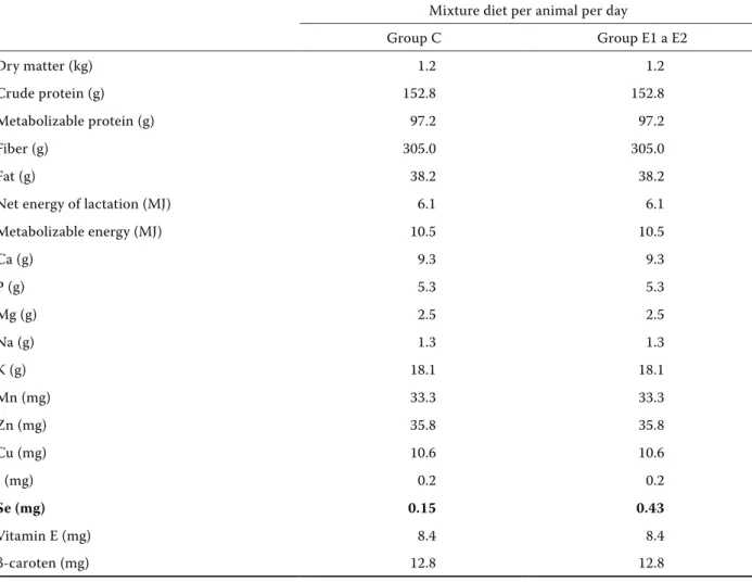
Table 1. Nutrient content in diet of individual groups of goats with expected intake of 1.2 kg meadow hay and 350 g pelleted grain mixture diet
Mixture diet per animal per day
Group C Group E1 a E2
Dry matter (kg) 1.2 1.2
Crude protein (g) 152.8 152.8
Metabolizable protein (g) 97.2 97.2
Fiber (g) 305.0 305.0
Fat (g) 38.2 38.2
Net energy of lactation (MJ) 6.1 6.1
Metabolizable energy (MJ) 10.5 10.5
Ca (g) 9.3 9.3
P (g) 5.3 5.3
Mg (g) 2.5 2.5
Na (g) 1.3 1.3
K (g) 18.1 18.1
Mn (mg) 33.3 33.3
Zn (mg) 35.8 35.8
Cu (mg) 10.6 10.6
I (mg) 0.2 0.2
se (mg) 0.15 0.43
Vitamin E (mg) 8.4 8.4
Original Paper Veterinarni Medicina, 54, 2009 (7): 324–332
intraruminal boluses (Wichtel et al., 1994; Grunder and Auer, 1995; Krys et al., 2009), or in drinking water (Lokajova et al., 2004). Currently, the bio-logical availability of individual Se forms is being investigated. This study will focus on a comparison between sodium selenite and the rarely adminis-tered selenium lactate-protein complex.
MAtEriAl And MEthods
A total of 27 clinically health pregnant white shorthair goats were used for the study. The goats were kept on the premises of the Ruminant Clinic, Faculty of Veterinary Medicine, University of Veterinary and Pharmaceutical Sciences Brno. They were divided into three groups (nine animals in each group). The first group was a control (C) without any selenium supplementation. Animals of the second group (E1) were given sodium selenite. In the third group (E2), goats received selenium lactate-protein complex (Selene Chelate Agrobac, Czech Republic). This form of selenium complex was produced by cultivation of Lactobacillus acido-philus on a substrate containing sodium selenite.
The experiment was started four months before expected parturitions. Goats of all groups showed average Se levels in whole blood of 96 μg/l. They were fed twice a day with granules (350 g per ani-mal) and meadow hay. Granules intended for ex-perimental goats were enriched with 0.9 Se per 1 kg of feeding mixture (Table 1). Goats received drinking water and salt lick was ad libitum. On the parturition day, heparinized blood was obtained by puncturing the vena jugularis of mothers and their kids in order to determine selenium concentration and glutathione peroxidase activity. Blood samples
of kids were taken immediately after parturition before they sucked the first colostrum. In twins and triplets, mixed samples from all kids were used for analyses. First colostrum (150 ml) was taken by milking mothers; 10 ml of this volume was used to determine selenium concentrations. Blood and colostrum samples were stored frozen at -20°C until use.
For selenium concentration tests, all samples were mineralized in a closed system by microwave diges-tion equipment in the presence of HNO3 and H2O2 (Milestone Ethos TC by Milestone, Italy). The min-eralized sample was prepared for the determina-tion of selenium by evaporadetermina-tion and dissoludetermina-tion in water to which 20% HCl was added. These samples were then tested for selenium concentrations by the AAS hydride technique using the AAS Solaar M6 (Unicam, Great Britain) (Pechova et al., 2005).
GSH-Px activity was established by the Paglia and Valentine method (1967) using the Ransel Set by Randox (Great Britain) and the Cobas Mira au-tomatic analyzer (Roche, Switzerland).
Basic statistical parameters of results (means, standard deviations) in individual groups and a comparison between the results of groups (using the two-tailed Student t-test after F-test for equal-ity of variations) were computed using Microsoft Excel XP.
rEsults
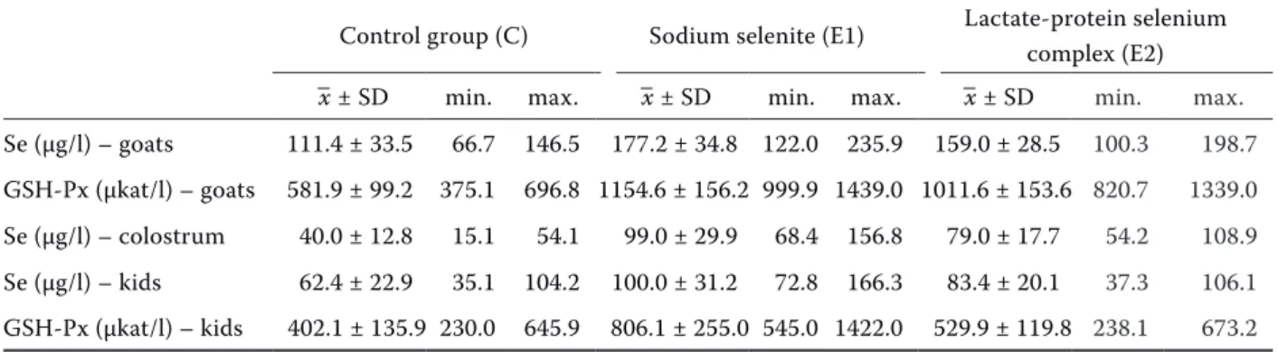
Means, standard deviations and minimum and maximum of identified parameters are presented in Table 2 and Figures 1 to 5 where the statisti-cal significance of differences between individual groups is also shown.
Table 2. Basic statistical characteristics (mean ± standard deviation, minimum, and maximum) of selenium (Se) concentration and glutathione peroxidase activity (GSH-Px) in whole blood of goats and kids and selenium con-centration in colostrum of goats in individual groups on the parturition day
Control group (C) Sodium selenite (E1) Lactate-protein selenium complex (E2)
–x ± SD min. max. –x ± SD min. max. –x ± SD min. max.
Se (μg/l) – goats 111.4 ± 33.5 66.7 146.5 177.2 ± 34.8 122.0 235.9 159.0 ± 28.5 100.3 198.7
GSH-Px (μkat/l) – goats 581.9 ± 99.2 375.1 696.8 1154.6 ± 156.2 999.9 1439.0 1011.6 ± 153.6 820.7 1339.0
Se (μg/l) – colostrum 40.0 ± 12.8 15.1 54.1 99.0 ± 29.9 68.4 156.8 79.0 ± 17.7 54.2 108.9
Se (μg/l) – kids 62.4 ± 22.9 35.1 104.2 100.0 ± 31.2 72.8 166.3 83.4 ± 20.1 37.3 106.1
Selenium levels and glutathione peroxidase ac-tivity in mother’s blood was in both experimental groups (E1 and E2) statistically significantly (P < 0.05) higher than in the control group (C) (Figure 1 and 2). However, no statistically significant dif-ference was found between the sodium selenite group (E1) and the group supplemented with se-lenium lactate-protein complex (E2), though the values tended to increase in the sodium selenite group. The same results were found in colostrum (Figure 3): both experimental groups showed sig-nificantly higher selenium levels as compared to the control group (P < 0.05). Again, no statistically sig-nificant difference was found between the sodium selenite group (E1) and the group supplemented with selenium lactate-protein complex (E2), though
* * 0 20 40 60 80 100 120 140 160 180 200
Figure 1. Selenium concentration (μg/l) in whole blood of goats from individual groups on the parturition day, and statistical significance of differences between the groups – *E1, E2 > C (P < 0.05)
* * 0 200 400 600 800 1000 1200 1400
Figure 2. Glutathione peroxidase activity (μkat/l) in who-le blood of goats from individual groups on the partu-rition day, and statistical significance of differences between the groups – *E1, E2 > C (P < 0.05)
* 0 20 40 60 80 100 120 140 160 controlȱgroupȱ(C) sodiumȱseleniteȱ(E1) seleniumȱlactateȬprotein complexȱ(E2) * 0 20 40 60 80 100 120 140
160 controlȱgroupȱ(C)
sodiumȱseleniteȱ(E1)
seleniumȱlactateȬprotein complexȱ(E2)
* * 0 20 40 60 80 100 120 Seȱ(ΐg/l)ȱinȱwholeȱbloodȱofȱgoats * * 0 20 40 60 80 100 120 140 160 180 200 controlȱgroupȱ(C) sodiumȱseleniteȱ(E1) seleniumȱlactateȬprotein complexȱ(E2)
Figure 3. Selenium concentration (μg/l) in colostrum of goats from individual groups on the parturition day, and statistical significance of differences between the groups – *E1, E2 > C (P < 0.05)
* 0 20 40 60 80 100 120 * 0 100 200 300 400 500 600 700 800 900
Seȱ(ΐg/l)ȱinȱwholeȱbloodȱofȱkids
* 0 20 40 60 80 100 120
controlȱgroupȱ(kC) sodiumȱseleniteȱ(kE1) seleniumȱlactateȬprotein complexȱ(kE2)
Seȱ(ΐg/l)ȱinȱwholeȱbloodȱofȱkids * 0 20 40 60 80 100 120 controlȱgroupȱ(kC) sodiumȱseleniteȱ(kE1) seleniumȱlactateȬprotein complexȱ(kE2)
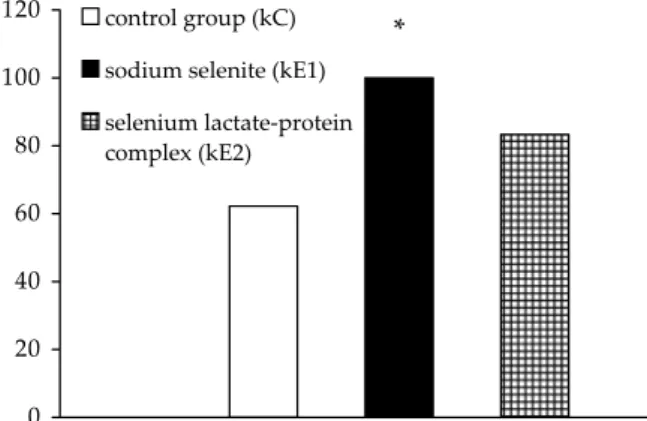
Figure 5. Glutathione peroxidase activity (μkat/l) in who-le blood of kids from individual groups on the parturition day, and statistical significance of differences between the groups – * kE1 > kC (P < 0.05), kE1 > kE2 (P < 0.05)
Original Paper Veterinarni Medicina, 54, 2009 (7): 324–332
the values tended to rise in the sodium selenite group.
In kids from the control group (kC), average se-lenium levels of 62.4 ± 22.9 μg/l were observed. Kids of sodium selenite mothers (kE1) had aver-age selenium values of 100.0 ± 31.2 μg/l, and kids of lactate-protein mothers (kE2) had average sele-nium concentrations of 83.4 ± 20.1 μg/l (Figure 4). Compared to the control group, statistically sig-nificantly higher (P < 0.05) selenium levels were found only in kids of mothers that received sodium selenite.
The following average activity of glutathione per-oxidase was identified in individual groups: 402.1 ± 153.9 μkat/l in the control group (kC), 806.1 ± 254.9 μkat/l in the sodium selenite group (kE1) and 529.9 ± 119.8 μkat/l in the lactate-protein group (kE2) (Figure 5). Compared to the control group, statistically significantly higher (P < 0.05) GSH-Px activity was observed only in kids supplemented with sodium selenite (kE1). Furthermore, a statisti-cally significant difference in GSH-Px activity was found also between kE1 and kE2 groups. Therefore, the average glutathione peroxidase activity in the blood of kids of mothers supplemented with sodi-um selenite was higher than in those supplemented with the selenium lactate-protein complex.
discussion
The results of this study indicate that both se-lenium forms enhanced sese-lenium levels in the blood of experimental goats as compared with the control group and that no significant differences were found between experimental groups. Similar results were reported by Travnicek et al. (2007) who compared the effect of supplementation with sodium selenite vs. selenium bound in the biomass of the alga Chlorella on selenium levels in the blood of ewes and their lambs. In their study, selenium levels in experimental groups were significantly higher than in the control group, but no signifi-cant difference between experimental groups was found in blood selenium levels. Baumgartner et al. (1998) reported similar results in dairy cows that received selenium in the form of selenomethionine and sodium selenite. In their study, no difference in blood selenium levels between experimental groups was observed, but statistically significantly higher selenium levels were found in the milk of cows that received selenium bound in selenomethionine. In
the present study, no differences in colostral se-lenium levels were found between experimental groups. However, experimental groups showed statistically significantly higher colostral selenium levels as compared with the control group.
Selenium levels observed in newborn kids dur-ing this study indicate that supplementation with sodium selenite is more effective as compared with supplementation with a lactate-protein complex. Blood selenium levels and GSH-Px activity were statistically significantly higher in kids of moth-ers that received sodium selenite. Similar results that suggested a better effect of selenite on GSH-Px activity as compared with selenium yeast were re-ported by Knowles et al. (1999) who performed ex-periments on dairy cows. They found that GSH-Px activity was lower by 15% in the group supplement-ed with Se-enrichsupplement-ed yeast as comparsupplement-ed with the selenite group. In contrast, milk selenium levels were twice to three times as high as in dairy cows that received Se-enriched yeast.
However, the results of the present study differ from certain other studies that compared inorganic selenium forms and organically bound selenium, mainly in the form of Se-enriched yeast. Guyot et al. (2007) compared Se-enriched yeast and sodium selenite administered perorally to pregnant Belgian Blue cows two months before and after parturition. Plasmatic levels of selenium in dams and newborn calves as well as selenium levels in colostrum and milk were higher in the group supplemented with Se-enriched yeast. Pehrson et al. (1999) made similar findings in Hereford cows. Ortman and Pehrson (1999) compared the effect of selenate, selenite and selenium yeast in dairy cows. Blood and milk selenium levels significantly increased in dairy cows supplemented with Se-enriched yeast. No difference was found between selenate and se-lenite groups. Although GSH-Px activity signifi-cantly increased in all groups as compared with the control group, no statistically significant difference in GSH-Px activity was found among experimental groups. Nicholson et al. (1991) reported similar data in calves that were supplemented with sodium selenite and Se-enriched yeast for four months – or-ganic selenium statistically significantly increased selenium levels and GSH-Px activity in the blood of animals receiving yeast.
un-ambiguously confirm a better biological effect of one of these forms are still missing. There is com-paratively little data in the literature on the sele-nium lactate-protein complex used in the present study. Pechova et al. (2008) monitored selenium concentration in milk after peroral supplemen-tation of goats with Se-enriched yeast, selenium lactate-protein complex and selenium protein-ate. They observed significantly higher selenium levels in milk of goats supplemented with Se-en-riched yeast as compared with the control group, lactate-protein complex and selenium proteinate group. Pavlata et al. (2007) compared the effect of 5-month supplementation of goats with selenite and lactate-protein complex on Se levels and GSH-Px activity in blood. They observed a significant increase in selenium concentration and GSH-Px activity in the blood of all experimental goats as compared with the control group. However, they found no significant differences in Se levels and GSH-Px activity between the selenite group and lactate-protein group. Nevertheless, they noted a significant growth in the blood GSH-Px activity in the selenite group during the first month of the ex-periment, whereas GSH-Px activity in the lactate-protein group increased as late as three months after the beginning of supplementation.
Based on data supplied by the manufacturer and information obtained from various studies, the lac-tate-protein complex used in the present study was expected to contain organically bound selenium, as this form of selenium was produced by cultivation of Lactobacillus acidophilus on substrate contain-ing sodium selenite. However, it is not completely clear how much and what form of organically bound selenium is contained in this preparation because no precise specification of the lactate-protein com-plex is available.
The assumption that the lactate-protein com-plex contains organically bound selenium is sup-ported by, e.g., the study of Calomme et al. (1995) who found that various species of lactobacilli can concentrate selenium intracellularly in the form of selenocysteine in biomass. This could constitute a potential source of organically bound selenium. They did not confirm that the microorganisms incorporate Se into their proteins in the form of selenomethionine. Alzate et al. (2007) detected se-lenocysteine and Se-methylsese-lenocysteine in their speciation studies of Se-enriched yogurt after en-zymatic hydrolysis, the concentration of which in-creased with the amount of supplemented selenite.
When they used selenate, biotransformation was very poor. Selenomethionine was found at the same level in both unenriched and enriched yogurt, indi-cating that Lactobacillus does not metabolize Se4 to
selenomethionine. Zhang et al. (2009) reported that Bifidobacterium animalis 01 could absorb 16.7– 39.6% of inorganic selenium in the medium and trans-form most of it into organic selenium. Most of organic selenium (57–63%) was found in the pro-tein fraction, 9.6–18.7% in the polysaccharide frac-tion, 0.3–0.8% in the nucleic fraction and 21–31% in other components. Based on chemical analysis, they determined selenomethionine as the main se-lenium component of the total protein. Alzate et al. (2008) compared selenium forms that are pro-duced from lactic fermentation with two different types of microorganism, bacteria (Lactobacillus) and yeast (Saccharomyces). While Lactobacillus is responsible for yogurt fermentation, synergy of bacteria and the yeast Saccharomyces causes ke-fir fermentation. Lactic fermentation for yogurt elicited an incremental rise in selenocysteine and Se-methylselenocysteine, while fermentation to produce kefir also increased the selenomethionine concentration. Besides these studies that confirm the transformation of inorganic selenium to organi-cally bound selenium, there are other studies that prove that, e.g., Lactobacillus bulgaricus is able to reduce selenite to insoluble elemental selenium, electro-dense Se0 granules, thereby depositing in
both the cytoplasm and the extracellular space (Xia et al., 2007). Alzate et al. (2008) and Zhang et al. (2009) reported as well that at higher selenite con-centrations, Se4 microorganisms are reduced to Se0
as a result of detoxication mechanisms. Considering the abovementioned facts, it should be stated that further research into the lactate-protein complex must be better specified in terms of representation of various selenium forms that are the result of the culturing of Lactobacillus acidophilus in the pres-ence of selenite. If it turns out that the greater part of selenium is transformed to elementary selenium Se0, then using this preparation would practically
Original Paper Veterinarni Medicina, 54, 2009 (7): 324–332
selenium levels and GSH-Px activity in the blood of these three animal species. Grace et al. (1995) compared selenium oxide and sodium selenate and came to the conclusion that both these forms in-crease blood Se level in dairy cows similarly.
conclusion
Both selenium forms, i.e., sodium selenite and sele-nium lactate-protein complex, significantly increased Se levels in the whole blood and colostrum of adult goats as compared with the control group. In new-born kids, however, a significant increase in blood Se levels and GSH-Px activity was observed only in the kids of mothers supplemented with sodium selenite. This indicates that the biological efficiency of sodium selenite in goats in terms of selenium status of their newborn kids seems to be higher as compared with the selenium lactate-protein complex.
Acknowledgements
Many thanks to, among others, the laboratory staff (Katerina Hrdlickova, Stanislava Hruba, Ivana Pohlova, Alena Kaluzova, Nasta Vankova, Lubomira Zedkova) and technicians (Matej Sidlo, Zdenka Kopeckova, Milan Stanek) of the Ruminant Clinic, Faculty of Veterinary Medicine Brno.
rEfErEncEs
Alzate A., Canas B., Perez-Munguia S., Hernandez-Men-doza H., Perez-Conde C., Gutierrez A.M., Camara C. (2007): Evaluation of the inorganic selenium biotrans-formation in selenium-enriched yogurt by HPLC-ICP-MS. Journal of Agricultural and Food Chemistry, 55, 9776–9783.
Alzate A., Fernandez-Fernandez A., Perez-Conde M.C, Gutierrez A.M., Camara C. (2008): Comparison of biotransformation of inorganic selenium by Lactoba-cillus and Saccharomyces in lactic fermentation proc-ess of yogurt and kefir. Journal of Agricultural and Food Chemistry, 56, 8728–8736.
Aspila P. (1991): Metabolism of selenite, selenomethio-nine and feed-incorporated selenium in lactating goats and dairy-cows. Journal of Agricultural Science in Finland, 63, 9–73.
Baumgartner W., Matzka B., Scherr E., Tataruch F. (1998): Comparison of anorganic and organically
bound selenium-supplementation in dairy cows. Prak-tische Tierarzt, 79, 76–77.
Bickhardt K., Ganter M., Sallmann P., Fuhrmann H. (1999): Investigation on manifestation of vitamin E and selen deficiency in sheep and goat. Deutsche Tier-arztliche Wochenschrift, 106, 242–247.
Calomme M.R, Vandenbranden K., Berghe D.A.V. (1995): Selenium and Lactobacillus species. Journal of Applied Bacteriology, 79, 331–340.
Culjak K., Sabocanec R., Karacic V., Vranesic D., Pre-varek B., Rinck J., Rinck R., Rinck M. (1999): Con-genital myopathy and cardiomyopathy in goat kids. Magyar Allatorvosok Lapja, 121, 85–87.
Enjalbert F., Lebreton P., Salat O., Meschy F., Schelcher F. (1999): Effect of pre- or postpartum selenium sup-plementation on selenium status in beef cows and their calves. Journal of Animal Science, 77, 223–229. Fisinin V.I., Papazyan T.T., Surai P.F. (2008): Producing
specialist poultry products to meet human nutrition requirements: Selenium enriched eggs. Worlds Poul-try Science Journal, 64, 85–97.
Fisinin V.I., Papazyan T.T., Surai P.E. (2009): Producing selenium-enriched eggs and meat to improve the se-lenium status of the general population. Critical Re-views in Biotechnology, 29, 18–28.
Grace N.D., Venning M., Mills A.R., Death A.F. (1995): The efficacy of selenium dioxide as a selenium sup-plement for dairy-cattle. New Zealand Veterinary Jour-nal, 43, 77–78.
Grunder H.D., Auer S. (1995): Selenium status of cattle in Hessian herds and possible preventive measures. Tierarztliche Umschau, 50, 250–255.
Guyot H., Spring P., Andrieu S., Rollin F. (2007): Com-parative responses to sodium selenite and organic selenium supplements in Belgian Blue cows and calves. Livestock Science, 11, 259–263.
Hayashida M., Orden E.A., Cruz E.M., Cruz L.C., Fuji-hara T. (2003): Effect of intraruminal soluble glass bolus on blood selenium and plasma mineral level of grazing does under backyard conditions in selected area in Nueva Ecija, Philippines. Asian-Australasian Journal of Animal Sciencies, 16, 189–197.
Kapolna E., Fodor P. (2006): Speciation analysis of sele-nium enriched green onions (Alium fistulosum) by HPLC-ICP-MS. Microchemical Journal, 84, 56–62. Knowles S.O., Grace N.D., Wurms K., Lee J. (1999): Sig-nificance of a mount and form of dietary selenium on blood, milk and casein selenium concetrations in graz-ing cows. Journal of Dairy Science, 82, 429–437. Krys S., Lokajova E., Podhorsky A., Pavlata L. (2009):
Li F., Wang F.F., Yu F., Fang Y., Xin Z.H., Yang F.M., Xu J., Zhao L.Y., Hu Q.H. (2008): In vitro antioxidant and anticancer activities of ethanolic extract of selenium-enriched green tea. Food Chemistry, 111, 165–170. Lokajova E., Pavlata L., Podhorsky A. (2004): Dealing
with selenium deficit in Limousine breed by oral ap-plication of selenium to pregnant cows. In: The effect of herd health of cattle, sheep and goat on production efficiency. The 5th Middle-European Buiatrics
Con-gress, Hajduszoboszlo, Hungary, 384–386.
Ludvikova E., Pavlata L., Vyskocil M., Jahn P. (2005): Selenium status of horses in Czech Republic. Acta Veterinaria Brno, 74, 369–375.
Misurova L., Pavlata L., Pechova A., Dvorak R. (2009): Selenium metabolism in goats – maternal transfer of selenium to newborn kids. Veterinarni Medicina, 54, 125–130.
Nicholson J.W.G., McQeen R.E., Bush R.S. (1991): Re-sponse of growing cattle to supplementation with or-ganically bound or inorganic sources of selenium or yeast cultures. Canadian Journal of Animal Science, 71, 803–811.
Ortman K., Pehrson B. (1999): Effect selenate as a feed supplement to dairy cows in comparison to selenite and selenium yeast. Journal of Animal Science, 77, 3365–3370.
Paglia D.E., Valentine W.N. (1967): Studies on quantita-tive and qualitaquantita-tive characterization of erytrocyte glutathione peroxidase. Journal of Laboratory and Clinical Medicine, 70, 158–169.
Pan C.L., Huang K.H., Zhao Y.X., Qin S.Y., Chen F., Hu Q.H. (2007): Effect of selenium source and level in hen´s diet on tissue selenium deposition and egg se-lenium concentrations. Journal of Agricultural and Food Chemistry, 55, 1027–1032.
Pavlata L., Pechova A., Illek J. (2000): Direct and indirect assessment of selenium status in cattle – a comparison. Acta Veterinaria Brno, 69, 281–287.
Pavlata L., Illek J., Pechova A., Matejicek M. (2002): Se-lenium status of cattle in the Czech Republic. Acta Veterinaria Brno, 71, 3–8.
Pavlata L., Prasek J., Podhorsky A., Pechova A., Haloun T. (2003): Selenium metabolism in cattle: maternal transfer of selenium to newborn calves at different selenium concentrations in dams. Acta Veterinaria Brno, 72, 639–646.
Pavlata L., Misurova L., Pechova A., Dvorak R. (2007): Comparison of the effect of sodium selenate and se-lenium lactate supplementation in goats. Revista Ro-mana de Medicina Veterinara, 17 Supl. Ia, 38–39. Pechova A., Pavlata L., Illek J. (2005): Blood and tissue
selenium determination by hydride generation atomic
absorption spectrophotometry. Acta Veterinaria Brno, 74, 483–490.
Pechova A., Misurova L., Pavlata L., Dvorak R. (2008): Monitoring of changes in selenium concentration in goat milk during short-term supplementation of var-ious forms of selenium. Biological Trace Element Re-search, 121, 180–191.
Pedrero Z., Madrid Y., Camara C. (2006): Selenium spe-cies bioaccessibility in enriched radish (Raphanus sativus): A potential dietary source of selenium. Jour-nal of Agricultural and Food Chemistry, 54, 2412– 2417.
Pehrson B., Ortman K., Madjid N., Trafikowska U. (1999): The influence of dietary selenium as selenium yeast or sodium selenite on the concentration of selenium in milk of suckler cows and on selenium status of their calves. Journal of Animal Science, 77, 3371–3376. Podhorsky A., Pechova A., Dvorak R., Pavlata L. (2007):
Metabolic disorders in dairy calves in postpartum pe-riod. Acta Veterinaria Brno, 76, S45–S53.
Podoll K.L. Bernard J.B., Ullrey D.E., Debar S.R., Ku P.K., Magee W.T. (1992): Dietary selenate versus selenite for cattle, sheep and horses. Journal of Animal Science, 70, 1965–1970.
Ramirez-Bribiesca J.E., Tortora J.L., Hernandez L.M., Huerta M. (2001): Main causes of mortalities in dairy goat kids from Mexican plateau. Small Ruminant Re-search, 41, 77–80.
Ramirez-Bribiesca J.E., Tortora J.L., Huerta M,. Hernan-dez L.M., Lopez R., Cosby M.M. (2005): Effect of sele-nium-vitamin E injection in selenium-deficient dairy goats and kids on Mexican plateau. Arquivo Brasileiro De Medicina Veterinaria E Zootecnia, 57, 77–84. Sanchez J., Montes P., Jimenez A., Andres S. (2007):
Pre-vention of clinical mastitis with barium selenate in dairy goats from a selenium – deficient area. Journal of Dairy Science, 90, 2350–2354.
Tontis A. (1984): Occurrence of Nutritional Myodystro-phy (NMD) in goat kids in Switzerland. Schweizer Archiv fur Tierheilkunde, 126, 41–46.
Travnicek J., Pisek L., Herzig I., Doucha J., Kvicala J., Kroupova V., Rodinova H. (2007): Selenium content in the blood serum and urine of ewes receiving sele-nium-enriched unicellular alga Chlorella. Veterinarni medicina, 52, 42–48.
Wichtel J.J., Craigie A.L., VarelaAlvarez H., Williamson N.B. (1994): The effect of intraruminal selenium pel-lets on growth-rate, lactation and reproductive effi-ciency in dairy cattle. New Zealand Veterinary Journal, 42, 205–210.
Original Paper Veterinarni Medicina, 54, 2009 (7): 324–332
(2004): HPLC-ICP-MS speciation of selenium in en-riched onion leaves a potential dietary source of Se-methylselenocysteine. Food Chemistry, 86, 617–623. Xia S.K., Chen L., Liang J.Q. (2007): Enriched selenium
and its effect on growth and biochemical composition in Lactobacillus bulgaricus. Journal of Agricultural and Food Chemistry, 55, 2413–2417.
Xu J., Yang F.M., An X.X., Hu Q.H. (2007): Anticarcino-genic activity of selenium-enriched green tea extracts
in vivo. Journal of Agricultural and Food Chemistry, 55, 5349–5353.
Yamanoshita O., Ichihara S., Hama H., Ichihara G., Chiba M., Kamijima M., Takeda I., Nakajima T. (2007): Che-mopreventive effect of selenium-enriched Japanese radish sprout against breast cancer induced by 7,12-dimethylbenz[a]antracene in rats. Tohoku Journal of Experimental Medicine, 212, 191–198.
Corresponding Author:
Doc. MVDr. Leos Pavlata, Ph.D., Dipl. ECBHM, University of Veterinary and Pharmaceutical Sciences, Faculty of Veterinary Medicine, Ruminant Clinic, Palackeho 1–3, 612 42 Brno, Czech Republic
Tel. +420 541 562 407, +420 602 501 562, Fax +420 541 562 407, E-mail:[email protected]
Yu F., Sheng J.C., Xu J., An X.X., Hu Q.H. (2007): Anti-oxidant activities of crude tea polyphenols, polysac-charides and proteins of selenium-enriched tea and regular green tea. European Food Research and Tech-nology, 225, 843–848.
Zhang B.W., Zhou K., Zhang J.L., Chen Q., Liu G.R., Shang N., Quin W., Li P.L., Lin F.X. (2009): Accumula-tion and species distribuAccumula-tion of selenium in Se-en-riched bacterial cells of Bifidobacterium animalis 01. Food Chemistry, 115, 727–734.
Zhao L., Zhao G.H., Du M., Zhao Z.D., Xiao L.X., Hu X.S. (2008): Effect of selenium on increasing free rad-ical scavenging activities of polysaccharide extracts from a Se-enriched mushroom species of genus Ga-noderma. European Food Research and Technology, 226, 499–505.