ORIGINAL ARTICLE
Iran J Allergy Asthma Immunol February 2020; 19(1):1-8. Doi:10.18502/ijaai.v19i1.2409
Analysis of Methylation and Expression Profile of
Foxp3
Gene
in Patients with Behçet’s Syndrome
Jafar Farhadi1,2, Mohammad Nouri3, Alireza Khabbazi2, Nasser Samadi4, Zohreh Babaloo5, Mahdi Azad6, Somayeh Abolhasani7, Shahriar Alipour7, Golamreza Jadideslam1,2, Sam Seydi Shirvani1,2, and Ebrahim Sakhinia8
1
Molecular Medicine, Faculty of Advanced Medical Sciences, Tabriz University of Medical Sciences, Tabriz, Iran
2
Connective Tissue Diseases Research Center, Tabriz University of Medical Sciences, Tabriz, Iran
3
Department of Biochemistry, Faculty of Medicine, Tabriz University of Medical Sciences, Tabriz, Iran
4
Cancer Biochemistry, Cancer Biotechnology, Faculty of Medicine, Tabriz University of Medical Sciences, Tabriz, Iran
5
Department of Immunology, Faculty of Medicine, Tabriz University of Medical Sciences, Tabriz, Iran
6
Department of Medical Laboratory Sciences, Faculty of Allied Medicine, Qazvin University of Medical Sciences, Qazvin, Iran
7
Department of Biochemistry, Faculty of Medicine, Urmia University of Medical Sciences, Urmia, Iran
8
Department of Genetic, Faculty of Medicine, Tabriz University of Medical Sciences, Tabriz, Iran
Received: 26 December 2018; Received in revised form: 12 February 2019; Accepted: 24 February 2019
ABSTRACT
Forkhead box P3 (Foxp3) gene is an important means in the Treg cells function, in both maintenances of immune tolerance and regulation of response. Epigenetic modifications of the foxp3 gene at its regulatory regions control the chromatin accessibility for the transcription factors and other transcriptional regulators in order to control Foxp3 expression. In addition, the methylation status of CpG islands within the Foxp3 promoter and regulatory elements regulate the expression of Foxp3. This study was performed to assess the role of the foxp3 gene in patients with Behçet’s syndrome (BS).
Venous blood samples were collected from all participants and peripheral blood mononuclear cells (PBMC) were extracted through Ficoll-Hypaque method. Genomic DNA was randomly sheared by sonication and immunoprecipitated with a monoclonal antibody. The status methylation of the foxp3 gene was estimated in 108 blood samples of active BS patients and healthy individuals (controls); using methylation DNA immunoprecipitation (MeDIP) technique. Expression analysis was carried out; using Real-time PCR.
The expression of foxp3 gene in the patients' group (mean±SD: 1.79±1.12) was significantly lower than the healthy group (mean±SD: 2.73±1.33) (p<001). Also, the methylation levels of Foxp3 promoter showed that its level in patients (mean±SD: 2.3±1.16) was higher than the healthy group (mean±SD: 1.85±0.59). However, this increase was not statistically significant (p>0.05). Also, these results indicated that increasing the amount of methylation of the foxp3 gene by reducing its expression leads to an increase and intensifying of the disease.
The decrease in Foxp3 expression is possibly associated with hypermethylation of the gene, and it can be considered as a risk factor for BS. Future studies may be needed to identify the capability of specific DNA methylation alterations in this syndrome.
Keywords: Behçet’s syndrome; DNA methylation; Foxp3; DIP
Corresponding Author: Ebrahim Sakhinia, PhD:
Department of Genetics, Faculty of Medicine, Tabriz University of
INTRODUCTION
Behçet’s syndrome (BS) is defined by frequent aphthous stomatitis, uveitis, genital ulcers, and skin lesions. Since vascular appearances are prevalent in the syndrome, BS is categorized as vasculitis.1 In addition, other characteristics of the syndrome include the arterial, cardiac, central nervous system, gastrointestinal, pulmonary, articular, and genitourinary manifestations.2 BS is named after the Turkish physician, Hulusi Behcet, in 1937. The prevalence of the syndrome varies in males and females from region to region.3 BS is more prevalent along the ancient ‘Silk Road’ from Japan to the Mediterranean area.1 Although the pathogenesis of BS is still unknown, immune factors and environmental features like viral and bacterial agents may trigger the development and recurrence of the disease in a genetically susceptible host.3,4 HLA-B51 is, to a large extent, related to the disease among different ethnicities.1 Natural CD4+CD25+ FOXP3+ regulatory (or suppressor) Treg cells have been the focus of numerous studies concerning suppressor CD4+ T cells. Such cells are implicated in immune response regulation, allograft rejection, and sensitivity. In addition, they are vital in preventing effector functions through infections and cancers. The deletion or functional deficiency of Treg cells in mice models resulted in the development of some autoimmune diseases.5 A pivotal development in the understanding of Treg cell biology resulted in the identification of the X chromosome-encoded members of the forkhead/winged-helix family of transcription regulators Foxp3 (also known as scurfin or forkhead box P3).6 It demonstrated that a loss of function mutation in the foxp3 gene is affected by fatal lymphoproliferative immune-mediated disease IPEX (immune dysregulation, polyendocrinopathy, enteropathy, X-linked) syndrome in humans, and the scurfy phenotype in mice.7-10 Foxp3 appears to be involved in the development, maintenance, and suppressor function of Treg cells.6,11 Furthermore, the reduced expression of Foxp3 in Treg cells contributes to the acquisition of effector T cell properties like the generation of non-Treg cell-specific cytokines.12,13 The foxp3 gene is an important agent in T-regs cells function, in both maintenances of immune tolerance and regulation of response. Hence, this transcription factor plays a crucial role in the generation of Treg phenotype. The human Foxp3 is located at the small arm of the X-chromosome (Xp11.23) and is 1296 bp in size, consisting of 11 different exons.9 In humans, 1 to
3% of total T cells are CD4+CD25+ FOXP3+ T-regs.14 Foxp3 is also associated with proteins that epigenetically modulate the transcriptional activity of target gene loci via changing DNA methylation, transcription factors, and histone post-translational modifications.15 The differentiation of T helper cells is controlled by epigenetic regulation through CpG methylation at specific sites in T cells.14 Epigenetic modifications of the foxp3 gene at its regulatory regions regulate the chromatin accessibility for the transcription factors and other transcriptional regulators in order to control Foxp3 expression and Treg cell stability.16 In addition, the methylation status of CpG islands within the Foxp3 promoter and regulatory elements regulate the expression of Foxp3 in Treg cells.14,16 From 10 to 45% of the CpG sites in the Foxp3 proximal promoter (-250 to +1) are methylated in naive CD4-CD25- T cells.14
The aim of this study was to evaluate the methylation of foxp3 gene and the role of this gene in patients with BS.
MATERIALS AND METHODS
The present study was approved by the ethical committee of the Tabriz University of Medical Sciences, Iran. This study was approved by the medical ethics committee of the university under No. TBZMED.REC.1394.310. A total of 47 patients with BS (18 females, 29 males) along with 61 healthy normal volunteers (25 females, 36 males) comprised the participants of the study.
Venous blood samples (5 mL) were collected from all participants; using anti-coagulant tubes. Peripheral blood mononuclear cells (PBMC) were extracted through a Ficoll-Hypaque density gradient centrifugation (Sigma). We measured BS activity by the Iranian Bechet’s disease Dynamic Activity Measure (IBDDAM) and Total Inflammatory Activity Index (TIAI).17,18
RNA Extraction, RT PCR and Real-time PCR
RNA extraction was conducted; using TRIZOL reagent (Invitrogen). Complementary DNA (cDNA) synthesis was performed; using random hexamer primers and RNase H-reverse transcriptase (Thermo Fisher Scientific, USA). The reaction mechanism included 0.3 µL of cDNA template, 7 µL of SYBR Green mix (amplicon, USA), and 6 µL of distilled H2O, 0.15 µL (4 pM) of each forward
(GGGATTTGGGAAGGTGCAGA) primer set. The Real-time PCR detection system (MIC, BioMolecular Systems, AUSTRALIA) was used for the amplification of cDNA through the working cycling platform. To do so, denaturation was conducted at 94°C for 3 min followed by 40 cycles of denaturation for 10 seconds at 94°C. The annealing stage was performed for 35 seconds at 54°C. Subsequently, the extension stage was carried out for 20 seconds at 72°C followed by the final extension at 72°C for 10 min. Dissociation curve plots were used to measure the pureness of PCR products. In addition, amplification plots were used to assign values to the cycle threshold (Ct); using SLAN software. To calculate the differences in gene expression in each sample, 2-∆∆Ct method was used.
DNA Extraction, Shearing, and MeDIP-QPCR
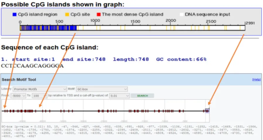
The RGDE (Rapid Genomic DNA Extraction) protocol informed the DNA extraction process. To ensure the quality and concentration of nucleic acid, nanodrop (Thermo Fisher) was employed. Genomic DNA was randomly sheared by sonication and immunoprecipitated with a monoclonal antibody to anticipate the foxp3 gene Promoter CpG islands, Methyl Primer Express (Applied Biosystems, CA, USA) was employed. Two pairs of primers were designed; using Primer Quest Tool and methMarker (PREMIER Biosoft, CA, USA) (Figure 1). Sonicate 3-4 pulses of 10-15 seconds each, followed by 30-3-40 seconds rest on ice between each pulse. The conditions of DNA shearing optimized based on the sonication instrument. After sonication, parts of 300 to 1000 bp of DNA was observed in the gel electrophoresis.
The EpiQuik™ MeDIP Ultra Kit (Epigentek, Farmingdale, NY) was used to conduct Methylated DNA immunoprecipitation (MeDIP). MeDIP reactions were set up in a 0.5 ml vial by adding 84 µL of MeDIP buffer, 10 µL of sample DNA (ng/µL), 5 µL of BS (Blocker Solution), and 1 µL of 5-mC Ab. Then, the content of the vial was incubated at room temperature for 60 min on a rolling shaker for 1 hour. The same steps were followed for negative controls; using 1 µL of non-immune IgG. The content was then transferred
from the vial to the strip-wells and incubated for 60 minutes at room temperature. Next, the immunoprecipitated methylated DNA in each well was washed three times; using 200 µL of WB (Wash Buffer) and released by adding 40 µL of the DRB-PK solution after incubation at 60°C for 20 min. Then, the DNA solution was transferred from the well to a 0.2 ml strip PCR tube. The PCR tubes containing DNA solution were incubated at 95°C for 5 min in a thermal cycler. The eluted DNA was stored at –20°C for later use in the real-time PCR.
To perform real-time PCR reactions, 1 µL of eluted DNA, 1 µl from each one forward and reverse primer (0.5 µM), 10 µl of the master mix, and 7 µL of DNA/RNA-free water were used. The real-time PCR detection system19 was used for the amplification and working cycling platform as follows: activation was conducted at 95°C for 2 min followed by denaturation at 95°C for 5 min. Subsequently, 40 cycles of denaturation were carried out for 10 seconds at 95°C; followed by annealing for 10 seconds at 55°C and extension for 10 seconds at 72°C. The final extension was performed at 72°C for 1 min.
For the negative control, DNA/RNA-free water was used instead of the DNA template. Fold enrichment characterized the amount of recovered DNA and was calculated according to the real-time PCR Ct value. The specificity of the 5-mC antibody and low background for non-immune IgG was calculated; using the following formula:
FE % =2 (IgG CT- Sample CT)x 100
Statistical Analysis
To analyze the data, SPSS 20 was used. The association between foxp3 gene methylation and risk of BS was examined by estimating the odds ratio (OR) and 95% confidence interval (95%CI); using binary logistic regression analysis. The Mann–Whitney test was used to compare the expression data from BS samples and health control. The level of statistical significance was set at p=0.05.
Table 1. Comparison of relative gene expression for the foxp3 gene in patients with BS and normal controls
Sample No Mean±SD p-value*
BS Patients 47 1.79±1.12 <0.001
Controls 61 2.73±1.33
Figure 1. Foxp3promoter methylation. CpG site and island around the TSS (transcription start site) were predicted by EPD (Eukaryotic Promoter Database) and CpG finder software.
RESULTS
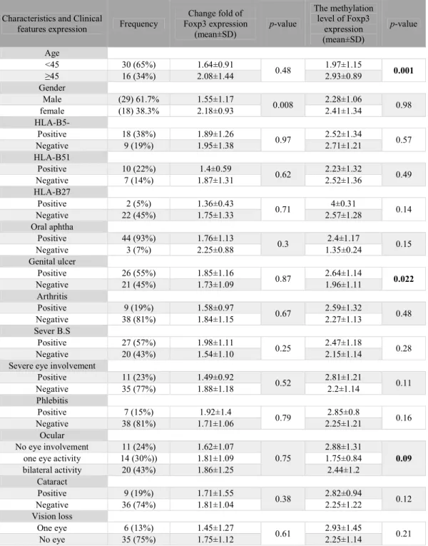
The demographic, pathological, genetical, and medical features of the contributors are obtainable in Table 3. The gender composition is as follows: 29 men and 18 women with a mean±SD age of 38.02 ± 10.25 years. The control group included 36 males and 25 females with a mean±SD age of 37.4±8.5 years. No significant differences were found between the control group and the patients in terms of sex and age.
Differences in Gene Expression and Methylation Levels
As the results show, the expression of Foxp3 was significantly different between control and BS groups (p<0.05) but the amount of methylation does not show any significant difference (p>0.05). The expression of foxp3 gene in the patient group (n=47, mean±SD:
1.79±1.12) was significantly lower than the healthy group (n=61, mean±SD: 2.73±1.33, p<001) (Table 1). Results showed that Foxp3 hypermethylation is related to a rise in the BS risk compared to the normal control, however; its methylation level was not significant despite the increase in the patient group (p=0.132) (Table 2).
Furthermore, we analyzed the relationship between methylation level and clinical features in the patient group. As the results showed, the methylation levels were significantly different in the age and genital ulcer subgroups. So that the methylation level of Foxp3 was increased in genital ulcer subgroups (p-value=0.004). Also, the change fold of Foxp3 expression was significantly different in the gender subgroups (p -value=0.008). So that expression levels of Foxp3 was increased in females rather than males.
Table 2. Fold change Foxp3 methylation in patients with BS and normal controls.
Sample No Mean±SD p-value*
BS Patients 47 2.3±1.16 <0.05
Controls 61 1.85±0.59
*Mann-Whitney test
Table 3. Demographic and medical features of patients with Behçet’s Syndrome (BS)
Characteristics and Clinical
features expression Frequency
Change fold of Foxp3 expression
(mean±SD)
p-value
The methylation level of Foxp3
expression (mean±SD)
p-value
Age
<45 30 (65%) 1.64±0.91
0.48 1.97±1.15 0.001
≥45 16 (34%) 2.08±1.44 2.93±0.89
Gender
Male (29) 61.7% 1.55±1.17
0.008 2.28±1.06 0.98
female (18) 38.3% 2.18±0.93 2.41±1.34
HLA-B5-
Positive 18 (38%) 1.89±1.26
0.97 2.52±1.34 0.57
Negative 9 (19%) 1.95±1.38 2.71±1.21
HLA-B51
Positive 10 (22%) 1.4±0.59
0.62 2.23±1.32 0.49
Negative 7 (14%) 1.87±1.31 2.52±1.36
HLA-B27
Positive 2 (5%) 1.36±0.43
0.71 4±0.31 0.14
Negative 22 (45%) 1.75±1.33 2.57±1.28
Oral aphtha
Positive 44 (93%) 1.76±1.13
0.3 2.4±1.17 0.15
Negative 3 (7%) 2.25±0.88 1.35±0.24
Genital ulcer
Positive 26 (55%) 1.85±1.16
0.87 2.64±1.14 0.022
Negative 21 (45%) 1.73±1.09 1.96±1.11
Arthritis
Positive 9 (19%) 1.58±0.97
0.67 2.59±1.32 0.48
Negative 38 (81%) 1.84±1.15 2.27±1.13
Sever B.S
Positive 27 (57%) 1.98±1.11
0.25 2.47±1.18 0.28
Negative 20 (43%) 1.54±1.10 2.15±1.14
Severe eye involvement
Positive 11 (23%) 1.49±0.92
0.52 2.81±1.21 0.11
Negative 35 (77%) 1.88±1.18 2.2±1.14
Phlebitis
Positive 7 (15%) 1.92±1.4
0.79 2.85±0.8 0.16
Negative 38 (81%) 1.71±1.06 2.25±1.21
Ocular
No eye involvement 11 (24%) 1.62±1.07
0.75
2.88±1.31
0.09
one eye activity 14 (30%)) 1.81±1.09 1.75±0.84
bilateral activity 20 (43%) 1.86±1.25 2.44±1.2
Cataract
Positive 9 (19%) 1.71±1.55
0.38 2.82±0.94 0.12
Negative 36 (74%) 1.81±1.04 2.25±1.22
Vision loss
One eye 6 (13%) 1.45±1.27
0.61 2.93±1.45 0.21
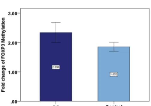
Figure 3. Methylation levels of Foxp3 (forkhead box P3). Regarding the average changes in the methylation of the foxp3
gene in BS and the control groups, the amount of it is comparable to that of the BS (Behçet’s Syndrome) group in the patient group, which shows that Foxp3 methylation was increased among the patients but this increase was not significant (p=0.132).
DISCUSSION
According to the results of the current investigation, the expression of the foxp3 gene in the patients' group was significantly lower than the healthy group; however, DNA methylation patterns in the promoter regions did not show any significant difference. Our study results showed that the methylation pattern of the foxp3 gene is related to a rise in the BS risk. Also, the results showed a higher expression of the foxp3 gene in individuals with genital ulcer subgroups as well as in females rather than males.
It has been reported that Foxp3+CD4+ Tregs are a very heterogeneous population in mice20 and humans21 and different subsets of Tregs possess different levels of CpG DNA methylation at the Foxp3 locus.21 The role of Foxp3 in immunoregulation has been extensively investigated. In both autoimmune diseases and cancer, Foxp3 may play a role in immunopathology due to potent suppressive T-cell activation and effector function.22 An experimental study by Polansky et al in the murine model showed that DNA methylation in the T-regulatory cell-specific demethylated region not only regulates foxp3 gene transcription but also is critically involved in maintaining stable Foxp3 expression.23
Lal et al show that the expression of Foxp3 in Treg is regulated by DNA methylation. They showed that increased methylation of CpG nucleotides at the Foxp3 locus was linked with less Foxp3 expression, decreased Treg stability, and reduced suppressive Treg function.24 Kim et al pointed out that DNA methylation was probably a vital control mechanism in expressing Foxp3.25 Foxp3 expression exhibits a reverse correlation with the methylation status of the intronic CpG island.25 In addition, they demonstrated that DNA methylation can influence long-term changes in Foxp3 expression during Treg cell development.25 Janson et al
demonstrated that total demethylation of the conserved Foxp3 promoter region contributes to a stable long-term Foxp3 expression and a committed Treg phenotype in humans.26
Analyzing the demethylation of the Foxp3 locus exhibits quantitative defects in regulatory T cells in the IPEX-like syndrome.27 In a recent study, analyzing MeDIP-Seq identified differentially methylated regions (DMRs); addressing almost the whole genome with sufficient depth and high resolution. Results showed a considerably higher number of hypermethylated DMRs than that of hypomethylated DMRs in psoriatic skin samples.28
It has been shown that the expression of Foxp3 is associated with activated, rather than regulatory T cells in humans.29 Valencia et al showed that Foxp3 mRNA expression was reduced in CD4CD25hi Tregs of patients with active RA (29). Long et al demonstrated that preserving Foxp3 expression in CD4+CD25+T-cells is reduced in type 1 diabetic patients.30 Preserving Foxp3 expression is impaired in Systemic lupus erythematosus (SLE).31 Previous studies suggested that several factors (i.e. genetic, epigenetic, and environmental) could be involved in BS progress.
Regarding the low expression of Foxp3 in patients with BS, we could not find a significant association between methylation patterns and the expression level of the foxp3 gene. It means that lower expression of Foxp3 in BS is possibly associated with hypermethylation of Foxp3 DNA. Future studies may be needed to identify the capability of specific DNA methylation alterations in this syndrome. It seems that recognizing other genetic and epigenetic modifications and affecting factors in the incidence of this syndrome could be a contributing asset in early detection and treatment of BS.
analyzed the methylation of Foxp3 DNA. Other epigenetic mechanisms may have been involved in the lower expression of the Foxp3 in the BS. So, we performed the foxp3 gene DNA methylation study in BS. In addition, we examined the relationship between the level of methylation of this gene and its expression in these patients and in healthy matched controls.
We have shown that DNA methylation could play an important role in the regulation of Foxp3. Also, the current study showed that gene silencing and a decrease in Foxp3 expression are possibly associated with hypermethylation of the gene and it can be considered as a risk factor for BS. These findings increase the likelihood of the foxp3 gene to be considered as a potential therapeutic approach for BS. To the best of our knowledge, this study is the first investigation in addressing this area of science.
ACKNOWLEDGEMENTS
The authors wish to thank Tabriz University of Medical Sciences and Connective Tissue Research Center for financially supporting this study. In addition, the authors would like to thank the patients and healthy subjects who willingly participated in the study.
REFERENCES
1. Hirohata S, Kikuchi H. Behçet's disease. Arthritis Res
Ther 2003; 5(3):139.
2. Saadoun D, Wechsler B. Behçet's disease. Orphanet J Rare Dis 2012; 7(1):20.
3. Demirelli S, Degirmenci H, Inci S, Arisoy A. Cardiac manifestations in Behcet's disease. Intractable Rare Dis Res 2015; 4(2):70-5.
4. Wang C, Ye Z, Kijlstra A, Zhou Y, Yang P. Decreased expression of the aryl hydrocarbon receptor in ocular Behcet’s disease. Mediators Inflamm 2014; 2014:195994. 5. Lourenço EV, La Cava A. Natural regulatory T cells in
autoimmunity. Autoimmunity 2011; 44(1):33-42. 6. Hori S, Nomura T, Sakaguchi S. Control of regulatory T
cell development by the transcription factor Foxp3. Science 2003; 299(5609):1057-61.
7. Bennett CL, Christie J, Ramsdell F, Brunkow ME, Ferguson PJ, Whitesell L, et al. The immune dysregulation, polyendocrinopathy, enteropathy, X-linked syndrome (IPEX) is caused by mutations of FOXP3. Nat Genet 2001; 27(1):20-1.
8. Wildin RS, Ramsdell F, Peake J, Faravelli F, Casanova J-L, Buist N, et al. X-linked neonatal diabetes mellitus, enteropathy and endocrinopathy syndrome is the human equivalent of mouse scurfy. Nat Genet 2001; 27(1):18.
9. Oda JMM, Hirata BKB, Guembarovski RL, Watanabe MAE. Genetic polymorphism in FOXP3 gene: imbalance in regulatory T-cell role and development of human diseases. J Genet 2013; 92(1):163-71.
10. Morikawa H, Ohkura N, Vandenbon A, Itoh M, Nagao-Sato S, Kawaji H, et al. Differential roles of epigenetic changes and Foxp3 expression in regulatory T cell-specific transcriptional regulation. Proc Natl Acad Sci U S A 2014; 111(14):5289-94.
11. Gavin M, Rudensky A. Control of immune homeostasis by naturally arising regulatory CD4+ T cells. Curr Opin Immunol 2003; 15(6):690-6.
12. Wan YY, Flavell RA. Regulatory T-cell functions are subverted and converted owing to attenuated Foxp3 expression. Nature 2007; 445(7129):766-70.
13. Samstein RM, Arvey A, Josefowicz SZ, Peng X, Reynolds A, Sandstrom R, et al. Foxp3 exploits a pre-existent enhancer landscape for regulatory T cell lineage specification. Cell 2012; 151(1):153-66.
14. Lal G, Bromberg JS. Epigenetic mechanisms of
regulation of Foxp3 expression. Blood 2009;
114(18):3727-35.
15. Bettini ML, Pan F, Bettini M, Finkelstein D, Rehg JE, Floess S, et al. Loss of epigenetic modification driven by the Foxp3 transcription factor leads to regulatory T cell insufficiency. Immunity 2012; 36(5):717-30.
16. Nie J, Li YY, Zheng SG, Tsun A, Li B. FOXP3+ Treg cells and gender bias in autoimmune diseases. Front Immunol 2015; 6:493.
17. Alipour, S., et al., Hypermethylation of IL‐10 gene is responsible for its low mRNA expression in Behçet's disease. Journal of cellular biochemistry 2018.
18. Shahram F, Khabbazi A, Nadji A, Ziaie N, Banihashemi AT, Davatchi F. Comparison of existing disease activity indices in the follow-up of patients with Behcet’s disease. Mod Rheumatol 2009; 19(5):536-41.
19. Flores-Borja F, Jury EC, Mauri C, Ehrenstein MR. Defects in CTLA-4 are associated with abnormal regulatory T cell function in rheumatoid arthritis. Proc Natl Acad Sci U S A 2008; 105(49):19396-401.
20. Komatsu N, Mariotti-Ferrandiz ME, Wang Y, Malissen B, Waldmann H, Hori S. Heterogeneity of natural Foxp3+ T cells: a committed regulatory T-cell lineage and an uncommitted minor population retaining plasticity. Proc Natl Acad Sci U S A 2009; 106(6):1903-8.
21. Miyara M, Yoshioka Y, Kitoh A, Shima T, Wing K, Niwa A, et al. Functional delineation and differentiation dynamics of human CD4+ T cells expressing the FoxP3 transcription factor. Immunity 2009; 30(6):899-911. 22. Maruyama T, Kono K, Mizukami Y, Kawaguchi Y,
Mimura K, Watanabe M, et al. Distribution of Th17 cells
lymphocytes, tumor‐draining lymph nodes and peripheral blood lymphocytes in patients with gastric cancer. Cancer Sci 2010; 101(9):1947-54.
23. Polansky JK, Kretschmer K, Freyer J, Floess S, Garbe A, Baron U, et al. DNA methylation controls Foxp3 gene expression. Eur J Immunol 2008; 38(6):1654-63. 24. Lal G, Zhang N, Van Der Touw W, Ding Y, Ju W,
Bottinger EP, et al. Epigenetic regulation of Foxp3 expression in regulatory T cells by DNA methylation. J Immunol 2009; 182(1):259-73.
25. Kim H-P, Leonard WJ. CREB/ATF-dependent T cell receptor–induced FoxP3 gene expression: a role for DNA methylation. J Exp Med 2007; 204(7):1543-51.
26. Janson PC, Winerdal ME, Marits P, Thörn M, Ohlsson R, Winqvist O. FOXP3 promoter demethylation reveals the committed Treg population in humans. PloS one 2008; 3(2):e1612.
27. Barzaghi F, Passerini L, Gambineri E, Mannurita SC, Cornu T, Kang E, et al. Demethylation analysis of the FOXP3 locus shows quantitative defects of regulatory T cells in IPEX-like syndrome. J Autoimmun. 2012; 38(1):49-58.
28. Zhang P, Zhao M, Liang G, Yin G, Huang D, Su F, et al. Whole-genome DNA methylation in skin lesions from patients with psoriasis vulgaris. J Autoimmun 2013; 41:17-24.
29. Valencia X, Stephens G, Goldbach-Mansky R, Wilson M, Shevach EM, Lipsky PE. TNF downmodulates the function of human CD4+ CD25 hi T-regulatory cells. Blood 2006; 108(1):253-61.
30. Long SA, Cerosaletti K, Bollyky PL, Tatum M, Shilling H, Zhang S, et al. Defects in IL-2R signaling contribute to diminished maintenance of FOXP3 expression in CD4+ CD25+ regulatory T-cells of type 1 diabetic subjects. diabetes 2010; 59(2):407-15.