451
THE ROLE OF EXOCHITINASE TYPE A1 IN THE FUNGISTATIC ACTIVITY OF THE
RHIZOSPHERE BACTERIUM
PAENIBACILLUS
SP. M4.
Urszula Jankiewicz1,* and Maria Swiontek Brzezinska2
1Department of Biochemistry, Warsaw University of Life Sciences, SGGW, Warsaw, Poland
2Department of Environmental Microbiology and Biotechnology, Institute of Ecology and Environmental Protection,
Nicolaus Copernicus University, Torun, Poland
*Corresponding author: [email protected]
Received: June 19, 2015; Revised: July 27, 2015, 2015; Accepted: September 1, 2015; Published online: December 15, 2015
Abstract: The aim of the study was to detect the activity and characterize potentially fungistatic chitinases synthesized by rhi-zosphere bacteria identified as Paenibacillus sp. M4. Maximum chitinolytic activity was achieved on the fifth day of culturing bacteria in a growth medium with 1% colloidal chitin. Analysis of a zymogram uncovered the presence of four activity bands in the crude bacterial extract. The used three-stage protein purification procedure resulted in a single band of chitinase activity on the zymogram. The purified enzyme exhibited maximum activity at pH 6.5 and temperature 45oC, and thermal stability at
40oC for 4 h. In terms of substrate specificity, it is an exochitinase (chitobiose). The amino acid sequence obtained after mass
spectrometry showed similarity to chitinase A1 synthesized by Bacillus circulans. The M4 isolate demonstrated the highest growth inhibiting activity against plant pathogens belonging to the genera Fusarium, Rhizoctonia and Alternaria. Fungistatic activity, although to a somewhat lesser degree, was also demonstrated by purified chitinase. The obtained results confirm the participation of the studied exochitinase in antagonism towards pathogenic molds. However, the lower fungistatic effectiveness of the chitinases points to the synergistic action of different metabolites in biocontrol by these bacteria.
Key words:chitinase purification; biocontrol; plant pathogens; Paenibacillus sp.; fungistatic activity
INTRODUCTION
Chitinases are enzymes hydrolyzing the β-1,4-glycos- ydic bonds in chitin, which is one of the most com-mon natural polymers in nature. Chitin can be found in numerous species, from fungi to plants and lower animals. In the exoskeletons of arthropods or in cell walls of fungi it exists in the form of ordered, crystal-line structures creating microfibrils, while in crusta-ceans in the form of fibrous material integrated into a six-thread protein fibril [1,2]. The arthropod ex-oskeleton is a rich source of chitin, which constitutes 20-50% of its dry weight. From a practical standpoint, shells of crustaceans such as crabs and prawns are readily available as waste from seafood processing and are used for the production of commercial chitin [3]. Chitin decomposition is a gradual process that involves chitinolytic enzymes belonging to the
O-gly-coside hydrolases subclass [E.C. 3.2.1.14]. So far, 115 families of glycoside hydrolases (GHs) have been clas-sified, three of which encompass chitinolytic enzymes: families 18, 19 and 20 [4]. Due to the position of the hydrolyzed bond, chitinases can be broadly divided into endochitinases (which hydrolyze random bonds located within the chain, and the products of their ac-tion are chitooligosaccharides) and exochitinases that detach the disaccharide chitobiose from the reduc-ing or non-reducreduc-ing end of the chitin chain, or sreduc-ingle units of β-N-acetyl-D-glucosamine [5].
particularly in the production of chitooligosaccharides and protoplasts from yeasts and fungi [6].
The vast majority of bacterial chitinases – both endochitinases and exochitinases – are grouped in 18 GH families. In addition, the bacterial chitinases belonging to the 18 GH have been divided into three main subfamilies, A, B and C, depending on the ami-no acid sequence of their catalytic domains [7].
Chitin degrading enzymes play a role not only in bacterial nutrition where they break down chi-tin that can then be used as a source of energy, but they can also be considered pathogenic factors [8]. Furthermore, chitinases are useful in agriculture as biocontrol agents against fungal phytopathogens and can therefore be an alternative to or a component of plant protection agents [9]. The ability to degrade chi-tin, the main component of the cell walls of molds, is considered one of the more important mechanisms of biocontrol, in which other enzymes and bacterial metabolites, such as glucanases, proteolytic enzymes, antibiotics and siderophores, can be involved. How-ever, chitinases are a particularly effective tool in fighting pathogenic fungi because their target is very precise, resulting in degradation of the hyphae of the mycelium [10].
The ability to secrete chitinases is fairly common among soil microorganisms. Many chitinolytic bacte-ria have been described, mainly of the genera Steno-trophomonas, Serratia,Bacillus and Paenibacillus [6]. Paenibacillus bacteria are exceptionally active in terms of hydrolase synthesis; depending on the species they secrete proteases, glucanases and chitinases [11]. The following can be classified as chitinolytic bacteria of the Paenibacillus genus: Paenibacillus thiaminolyticus, P. macerans,P. alvei, P. koreensis, P. borealis, P. chitino-lyticus and P. anaericanus [12,13].
Our research also focuses on biological control. The main goal of our studies was to seek rhizosphere bacteria with high biocontrol potential and to assess the role of the chitinases of these bacteria in control-ling the growth of phytopathogenic molds. The char-acterization of the purified enzyme was also carried out. Biochemical characterization and fungistatic
ac-tivity testing of one of the chitinases of this bacterial isolate was aimed at evaluating the role of the enzyme in antagonism towards fungi causing diseases of farm crops.
MATERIALS AND METHODS
Isolation, screening and identification of chitinolytic bacteria
For isolation of bacteria from the rhizosphere of spring barley (Hordeum vulgare L.), Soldo variety, roots were collected, together with attached soil, from healthy barley plants in the stem elongation phase. Rhizosphere bacteria were isolated following the methodology described by Buyer [14]. The bacterial isolates were cultivated on agar media with 0.5% chi-tin at 28oC for 72 h. After this, the presence and size of
halo zones around the grown bacterial colonies were checked. The selected bacterial isolates were identi-fied on the basis of morphological and biochemical traits according to Bergey’s Manual of Determinative Bacteriology [15]. Additionally, identification of the studied strain was confirmed by analysis of the 16 S rRNA gene sequence. Amplification of the 16 S rRNA gene was performed using 27F and 1492R universal primers [16]. The obtained nucleotide sequences were compared with sequences deposited in the available GenBank, European Molecular Biology Laboratory (EMBL) and DNA Data Bank of Japan (DDBJ) data-bases using the BLAST program.
Determination of chitinase activity
Optimization of growth medium composition
The bacteria were cultured for 36 h in a shaking incu-bator at 28°C. Optimized growth medium contained 3 g/L KH2PO4, 3 g/L K2HPO4, 0.5 g/L MgSO4, 2 g/L NaCl, 0.005 g/L FeCl3, 5 g/L Bacto Peptone, 2 g/L yeast extract, enriched with 10 g/L of colloidal chitin or crystalline chitin flakes from shrimp shells (Sigma Aldrich), crab shell powder (Roth) chitosan (Sigma, Aldrich; ≥75% deacetylated chitin from prawn shells). Control growth media contained all the components except a source of chitin or chitosan. Colloidal chitin was prepared according to Lee et al. [18].
Enzyme purification
A 5-day old bacterial culture in a growth medium en-riched with 1% colloidal chitin was centrifuged, the obtained supernatant was precipitated with ammonium sulfate (up to 85% solution saturation). The prepara-tion was centrifuged for 30 min at 13000xg, and the obtained protein pellet was dissolved in 5 mL of 50 mM sodium acetate buffer, pH 5.8. The obtained solu-tion was dialyzed for 12 h against the same buffer.The enzyme solution was supplemented with (NH4)2SO4 to a final concentration of 0.8 M and then subjected to hydrophobic chromatography on a Phenyl-Sepharose CL-4B column. Prior to separation, the column was equilibrated with 1 M (NH4)2SO4 in 50 mM sodium acetate buffer, pH 5.8. The associated proteins were eluted with a decreasing (NH4)2SO4 gradient. The most active fractions collected during the hydrophobic chromatography were pooled, dialyzed and purified using molecular sieve chromatography (Superdex 200). Chromatographic separation was performed in 50 mM sodium acetate buffer pH 5.8. Protein concentrations were determined using the method of Bradford [19] with bovine serum albumin as the protein standard.
Gel electrophoresis and zymography
Isoforms of chitinases synthesized by the tested bac-teria were analyzed on zymograms following native electrophoretic separations [20]. The proteins were separated on a 8% polyacrylamide gel with
incorpo-rated 0.05% glycol chitin. Glycol chitin was prepared according to Trudel and Asselin [21]. The gel was in-cubated at 40°C in 0.1 M sodium acetate buffer pH 5.8 for 2 h. Finally, the gel was submerged for 30 min in a 0.01% solution of Congo Red dye and transferred to 1 M NaCl solution.
Proteomic analysis using mass spectrometry
The highly purified enzyme preparation (containing on average 30 µg/mL of protein) was sent to the Labo-ratory of Mass Spectrometry of the IBB PAS (Poland). A protein sample previously digested with trypsin, was separated on a nanoAcquity UPLC (Ultra Perform-ance LC) system and analyzed with an Orbitrap-based mass spectrometer. The obtained peptide sequences were analyzed using the BLAST program.
Biochemical characterization of the purified chitinase of M4 isolate
Substrate specificity of the purified chitinase was determined using chromogenic, synthetic substrate (Chitinase Assay Kit, Sigma) N-acetylglucosamine derivatives of 4-nitrophenols: 4-Nitrophenyl N,Nʹ-diacetyl-β-D-chitobioside, for determination of exochitinase/chitobiosidase activity, 4-Nitrophenyl N-acetyl-β-D-glucosaminide for exochitinase/β-N-acetylglucosaminidase activity detection, and 4-Nitro-phenyl β-D-N,Nʹ,Nʹʹ-triacetylchitotriose for endochi-tinase activity detection. One unit of chiendochi-tinase activity (U) was defined as the amount of enzyme yielding 1 μmol of p-nitrophenol per min. The optimum pH and temperature as well as the thermal stability of the puri-fied enzyme were determined in enzymatic reactions using colloidal chitin as the substrate.
Fungistatic activity of M4 isolate and of purified chitinase A1-type
The tested fungal cultures were from the Bank of Plant Pathogens in Poznan (Poland).
In studies of the degree (percentage) of inhibition of mycelium growth by the M4 isolate, the method of dual cultures in modified PDA medium was em-ployed. In the modified PDA medium, the concentra-tion of glucose was reduced to 0.2%, and 0.3% chitin was added. The rate of fungal growth inhibition by the M4 isolate was determined following the formula for growth inhibition=(K-F/K)x100, where K is the culture diameter in the control combination and F is the culture diameter in the test combination.
The fungistatic activity of purified A1-type chi-tinase was studied using the well method in which the enzyme diffuses into the agar medium. The ex-periment also included a crude chitinase preparation obtained after precipitation and dialysis of proteins in the bacterial culture supernatant. The protein solu-tions were sterilized by filtration. The specific activities of the crude chitinase and purified enzyme were 6.1 and 91.8 U/mg, respectively. Bioactivity analysis of the studied enzymes preparations was performed in PDA medium by the well diffusion method, as described by Narayana and Vijayalakshmi [22]. Antifungal activity was classified according to Ghasemi et al. [23] as no inhibition, –, weak inhibition, ±; <2 mm, moderate inhibition, +; 2-8 mm, strong inhibition, ++;>8 mm.
All of the presented results are means obtained from three independent replicates. The mean error, reflecting maximal deviation of the results of measure-ments from the mean, did not exceed 5%.
RESULTS
From 45 isolated strains of bacteria, only 17 exhib-ited chitinolytic activity. Approximately 2-cm light zones on growth medium with colloidal chitin were observed around colonies of the bacterial isolate se-lected for further testing.
The studied strain was identified as Paenibacillus sp. M4. The nucleotide sequence of the gene coding 16 S RNA was deposited in DDBJ under accession
number LC043402. Analysis of the obtained 16 S RNA gene sequence of the studied bacterial strain revealed 99% similarity with other bacterial strains of the ge-nus Paenibacillus, including Paenibacillus sp. HA18 (gb|KF011602) and Paenibacillus sp. PAMC 26811 (gb|KF011685).
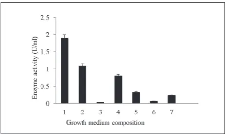
The effect of growth medium composition on chitinolytic activity of the M4 isolate was tested using 5-day-old bacterial cultures. The highest chitinase ac-tivity was achieved in a growth medium enriched with 1% of colloidal chitin or powdered crab shells (Fig. 1). It is interesting that the presence of 0.8% glucose in the culture medium resulted in reduced chitinolytic activ-ity of the bacteria. Enzymatic preparations obtained from 5-day-old bacteria cultures in growth medium with 1.0% colloidal chitin were subjected to a three-stage purification procedure, according to Table 1.
chitinases was checked by using zymograms. In the raw bacterial extract, four active chitinase forms were observed, whereas after hydrophobic chromatography, three forms were observed in fraction I and two in fraction II. After molecular sieve chromatography, only a single band of chitinase activity was observed in the zymogram (Fig. 3). The purified enzyme was subjected to proteomic identification using mass spec-trometry (MS). Proteomic analysis of the purified en-zymatic preparation demonstrated that the amino acid sequence of the studied chitinase is highly similar to the sequence of the chitinase A1 of Bacillus circulans Wl-12 (GenBank accession no P20533). The peptides derived from the studied chitinase overlap with 40% of the aa sequence of chitinase A1 by Bacillus circulans Wl-12. Chitinase A1 in strain Wl-12 is composed of 699 amino acid residues. Conserved domains typical for the family of 18 GHs were identified (Fig. 1S, Sup-plementary Material).
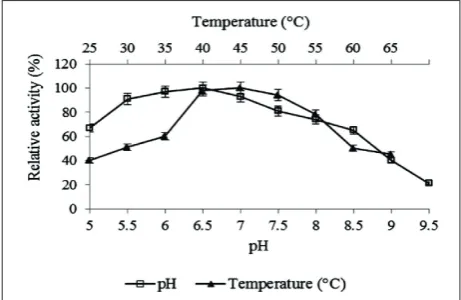
The purified enzyme exhibited activity in a broad range of pH values, ranging from 3.6 to 8.0, with maximum activity at pH 6.5 (Fig. 4). The tem-perature optimum was determined to be 45°C, al-though a broad range of temperatures in which the enzyme maintains activity could be observed (Fig. 4). The enzyme showed high thermal stability after 4 h of preincubation at 40°C. The studied chitinase demonstrated highest activity against 4-Nitrophenyl N,Nʹ-diacetyl-β-D-chitobioside as the substrate and Table 1. Purification of the chitinase type A1 produced by Pae-nibacillus sp. M4
Purification Step
To
ta
l ac
tiv
ity (U)
To
ta
l p
ro
te
in
(m
g)
Sp
ecif
ic ac
tiv
ity
(U/m
g)
Purif
ic
at
io
n f
ol
d
Re
co
ve
ry (%)
Cell free supernatant 430 70 6.1 1 100 Ammonium sulfate
precipitation (0-85%) 390 45 8.7 1.4 90 Hydrophobic chromatography
(samples 42-54) 230 5.5 41.8 6.9 53.5 Gel filtration 134 1.5 91.8 15 31.2
Fig. 4. Effect of pH and temperature on chitinase activity. In the case of optimum pH chitinolytic activity was studied in Britton-Robinson buffer in the 5.0-9.5 range. Optimum temperature was determined at pH 6.5 in the temperature range from 25 to 65°C.
Fig. 2. Elution profile of Paenibacillus sp. M4 chitinases after hy-drophobic chromatography with a decreasing ammonium sulphate gradient (1-0 M). On the chromatogram two protein fractions with chitinolytic activity, I and II, are indicated. Fraction II was taken for the subsequent step of protein purification, sieve chro-matography.
minimal activity against 4-Nitrophenyl β-D-N,Nʹ,Nʹʹ-triacetylchitotriose (Table 2).
The M4 isolate in dual cultures exhibited a vary-ing degree of antagonism towards such cereal patho-gens as F. oxysporum, F. solani, A. alternata, R. solani, R. cerealis and Ch. globosum. F. culmorum was found to be fully indifferent to the presence of the bacteria (Table 3).
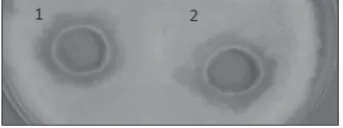
The fungistatic activity of A1-type chitinase and crude Paenibacillus sp. M4 chitinases was studied with regard to the same species of pathogens but using the agar well diffusion method, which has proven to be more effective in the case of liquid enzymatic prepa-rations. The species of pathogens susceptible to the action of these enzyme preparations are presented in Table 4. The purified chitinase type A1 showed a fungistatic effect on the growth of F. oxysporum and A. alternate. This enzyme did not have any effect on other pathogens. The fungistatic activity of the chiti-nase type A1 against F. oxysporum is shown in Fig. 5.
DISCUSSION
Interest in chitinolytic microorganisms is motivated by the possibility of their use in biological plant pro-tection. Chitinases synthesized by these organisms are an effective and precise biocontrol tool. From this point of view, Paenibacillus sp. M4 is an interest-ing study entity because it is among the chitinolytic strains of bacteria that are at the same time antago-nistic towards phytopathogenic molds. The chitinase we isolated showed fungistatic activity. This enzyme is called chitinase type A1 because of the similarity of its amino acid sequence to that of B. circulars chi-tinase A1. Zymogram analysis indicated that under the experimental conditions, the studied M4 isolate secreted at least four chitinases. As indicated by the literature data, bacteria of the Paenibacillus genus usu-ally synthesize from a few to over ten extracellular chitinases [24,25].
Chitinolytic activity was also detected in bacterial cultures on growth media that did not contain chitin. The obtained results indicate that chitinases are se-Table 2. Substrate specificity of the chitinase type A1 from
Pae-nibacillus sp. M4
Substrate Activity (U/mL) Catalytic type
4-Nitrophenyl
N-acetyl-β-D-glucosaminide 0 Exochitinase
Nitrophenyl
N,Nʹ-diacetyl-β-D-chitobioside 22 (chitobiosidase)Exochitinase 4-Nitrophenyl
β-D-N,Nʹ,Nʹʹ-triacetylchitotriose 2 Endochitinase
Table 3. Antagonism of Paenibacillus sp. M4 isolate towards fungal phytopathogens.
Phytopathogens Antifungal activity (%)
Fusarium solani 55
Fusarium oxysporum 60
Alternaria alternata 57
Rhizoctonia solani 40
Rhizoctonia cerealis 32
Chaetomium globosum 15
Fusarium culmorum 0
Table 4. Fungistatic activity of the chitinases from Paenibacillus
sp. M4.
Phytopathogens after salting out with Crude chitinases ammonium sulfate
Chitinase type A1, after molecular sieve
chromatography
F. solani ±
-F. oxysporum + +
A. alternata ++ ++
no inhibition, –, weak inhibition, ±; <2 mm, moderate inhibition, +; 2–8 mm, strong inhibition, ++; >8 mm (23).
Fig. 5. Fungistatic activity of crude chitinases (well 1) and purified chitinase (well 2) against
creted constitutively. Itoh et al. [24] also described the constitutive nature of chitinases in the FPU-7 strain. Furthermore, the presence of glucose in the growth medium caused a catabolite repression effect, which is consistent with the results described for chitinase synthesized by Paenibacillus sp. D1 [26]. However, most of the chitinases described so far, including B. circulars chitinase A1, are induced in the presence of a substrate, most of all by colloidal chitin [27].
Strain M4 synthesizes a chitinase similar to chi-tinase A1 from chichi-tinase group A, which belongs to family 18 GH. Chitinase A1 has been characterized in detail in Bacillus circulans, with 6 different chitinases described (A1, A2, B1, B2, C and D). Among them, chitinase A1 is synthesized in the largest amounts and exhibits strong affinity to insoluble forms of chitin. As with most chitinases from 18 GH, it comprises a C-end chitin-binding domain (ChBD, N-end large domain containing the enzyme catalytic site, and a third fibronectin domain type III [28].
The results of substrate specificity tests indicate that the studied chitinase belongs to exochitinases called chitobioses, capable of detaching two N-acetylglucosamine units from the non-reducing end of the molecule. However, no β-N-acetylglucosaminidase activity was observed. This substrate specificity con-firms that the studied enzyme has similar properties to chitinase A1 [28]. Similar results were reported by Itoh et al. [24] for chitinases of the FPU-7 strain. P. chiti-nolyticus does not exhibit β-N-acetylglucosaminidase activity either, but it has endochitinase activity [29] similar to P. illinoisensis [30].
The optimal temperature of action of this enzyme is 45°C, which is typical for most group A chitinases
[31, 32]. On the other hand, chitinases from bacteria of the genus Paenibacillus have different temperature op-tima, e.g. 37°C for the enzyme from P. pasadenensis [33] and 60°C for P. thermoaerophilus [34]. Similarly to the studied enzyme, chitinases produced by bacteria of the genus Paenibacillus are frequently thermally stable [35].
The tested enzyme showed an optimum pH close to neutral, similarly to chitinase A from Bacillus li-cheniformis [36]. The results presented in the literature
indicate that chitinases synthesized by Paenibacillus bacteria may show optimal activity in either acidic [35] or alkaline environments [33].
In our studies, fungistatic activity towards ubiq-uitous fungal pathogens was demonstrated by both the M4 isolate and the chitinase type A1. In dual cul-tures, the M4 isolate strongly inhibited the growth of R. solani,F. oxysporum and A. alternata. The stud-ied chitinase type A1 showed less fungistatic activ-ity compared to the M4 isolate. Furthermore, there was no negative influence on the growth of R. solani and R. cerealis. Our results bring us to conclude that chitinases are an important biocontrol tool of these bacteria, but not the only one. Literature data point to the dependence of the antagonism of bacteria against phytopathogenic fungi on different enzymatic activi-ties. The results of Aktuganov et al. [37] indicate a synergism of antifungal mechanisms of fungal cell-wall lytic enzymes, in which β-1,3-glucanases are ini-tiators of cell-wall hydrolysis, which is amplified by chitinase activity. Nevertheless, it appears there is no single rule applicable to the role of bacterial lytic en-zymes in the antagonistic interactions of microorgan-ism. It should also be kept in mind that the final effect of bacterial antagonism towards fungal pathogens is also affected by the activity of other metabolites, such as siderophores and antibiotics. The fungistatic prop-erties of group A chitinases were described earlier for bacteria belonging to the genera Stenotrophomonas [32]. Similarly, there are numerous scientific studies that describe antifungal activity for many bacteria of the genus Paenibacillus. P. peoriae inhibits the growth of Fusarium moniliforme, Diplodia macrospora, Ce-phalosporium, Penicillium corylophilum and Colle-totrichumgraminicola [38]. In a similar plate test, P. ehimensis exhibited fungistatic activity, among others, towards Phytophthora capsici, Rhizoctonia cerealis and Rhizoctonia solani [39].
application in plant protection. These results are very useful because the damage to and diseases of farm crops caused by fungal pathogens are not only an eco-nomic problem for food producers but also a health problem for consumers. The need to produce healthy food requires the elaboration of more effective plant protection methods, which at the same time are safer for the environment. For this reason, it seems worth-while continuing these studies with the use of plants in field conditions.
Acknowledgments: This work was financially supported by the Department of Biochemistry, Warsaw University of Life Sciences – SGGW
Authors’ contributions: U. Jankiewicz participated in the design of the study, performed the experiments and data analysis, and wrote the manuscript. M. Swiontek Brzezinska participated in the research and helped to draft the manuscript. Both authors read and approved the final manuscript.
Conflict of interest disclosure: The authors declare no conflict of interest
REFERENCES
1. Goodrich JD, Winter WT. α-Chitin nanocrystals prepared from shrimp shells and their specific surface area measure-ment. Biomacromolecules, 2007;8(1):252-7.
2. Pillai CKS, Willi P, Chandra PS. Chitin and chitosan poly-mers: Chemistry, solubility and fiber formation. Prog Polym Sci. 2009;34(7):641–78.
3. Bhattacharya D, Nagpure A, Gupta RK. Bacterial chitinases: properties and potential. Crit Rev Biotech. 2007;27(1):21-8. 4. Henrissat B. A classification of glycosyl hydrolases based on
amino-acid sequence similarities. Biochem J. 1991;280(1): 309-316.
5. Cohen-Kupiec R, Chet I. The molecular biology of chitin digestion. Curr Opin Biotechnol. 1998;9(3):270–7.
6. Swiontek Brzezinska M, Jankiewicz U, Burkowska A, Wal-czak M. Chitinolytic microorganisms and their possible application in environmental protection. Current Microbiol. 2014;68(1):71-81.
7. Watanabe T, Kobori K, Miyashita K, Fujii T, Sakai H, Uchida M, Tanaka H. Identification of glutamic acid 204 and aspar-tic acid 200 in chitinase A1 of Bacillus circulans WL-12 as essential residues for chitinase activity. J Biol Chem. 1993;268(25):18567-72.
8. Frederiksen RF, Paspaliari DK, Larsen T, Storgaard BG, Larsen MH, Ingmer H Leisner J, Palcic M, Leisner J. Bacte-rial chitinases and chitin-binding proteins as virulence fac-tors. Microbiology, 2013;159(5):833-47.
9. Roberts WK, Selitrennikoff CP. Plant and bacterial chitinases differ in antifungal activity. J Gen Microbiol. 1988;134(1):169-76.
10. Gohel V, Singh A, Vimal M, Ashwini P, Chhatpar H. S. Review-Bioprospecting and antifungal potential of chitino-lytic microorganisms. Afr J Biotechnol. 2006;5(2):54-72. 11. Lal S, Tabacchioni S. Ecology and biotechnological potential
of Paenibacillus polymyxa: a minireview. Indian J. Microbiol. 2009;49(1):2-10.
12. Budi SW, van Tuinen D, Arnould C, Dumas-Gaudot E, Gianinazzi-Pearson V, Gianinazzi S. Hydrolytic enzyme activity of Paenibacillus sp. strain B2 and effects of the antagonistic bacterium on cell integrity of two soil-borne pathogenic fungi. Appl Soil Ecol. 2000;15 (2):191-9. 13. Shida O, Takagi H; Kadowaki K, Nakamura LK, Komagata
K. Emended description of Paenibacillus amylolyticus and description of Paenibacillus illinoisensis sp nov and Paeniba-cillus chibensis sp nov. Int J Syst Bacteriol. 1997;47(2):299– 306.
14. Buyer JS. A Soil and Rhizosphere Microorganism Isolation and Enumeration Medium That Inhibits Bacillus mycoides. Appl Environ Microbiol. 1995;61(5):1839-42.
15. Holt J, Krieg GNR, Sneath PHA, Staley JT, Williams ST. Ber-gey’s Manual of Determinative Bacteriology. Baltimore: Wil-liams and Wilkins; 1994.
16. Watanabe K, Kodama Y, Harayama S. Design and evaluation of PCR primers to amplify bacterial 16 S ribosomal DNA fragments used for community fingerprinting. J Microbiol Meth. 2001;44(3):253–62.
17. Miller GL. Use of dinitrosalicylic acid reagent for determina-tion of reducing sugar. Anal Chem. 1959;31(3):426–8. 18. Lee HS, Han DS, Choi SJ, Choi SW, Kim DS, Bai DH, Yu
JH. Purification, characterization, and primary structure of a chitinase from Pseudomonas sp. YHS-A2. Appl Microbiol Biotechnol. 2000;54(3):397-405.
19. Bradford MM. A rapid and sensitive method for the quan-tization of microgram quantities of protein utilizing the principle of protein-dye binding. Anal Biochem. 1976;72(1-2):248–54.
20. Laemmli UK. Cleavage of structural proteins during the assembly of the head of bacteriophage T4. Nature 1970;227 (5259) : 680–5.
21. Trudel J, Asselin A Detection of chitinase activity after polyacrylamide gel electrophoresis. Anal Biochem. 1989;178(2):362-6.
22. Narayana KJ, Vijayalakshmi M. Chitinase production by S trep-tomyces sp ANU 6277. Brazilian J Microbiol. 2009;40(4):725-33.
23. Ghasemi S, Ahmadian G, Jelodar N, B Rahimian H, Ghandili S, Dehestani A, Shariati P. Antifungal chitinases from Bacil-lus pumiBacil-lus SG2: preliminary report. World J Microb Biot. 2010;26(8):1437-43.
and cell surface-expressed chitinases from Paenibacillus sp strain FPU-7. Appl Environ Microbiol. 2013;79(23):7482-90. 25. Song YS, Seo DJ, Kim KY, Park RD, Jung WJ. Expression
patterns of chitinase produced from Paenibacillus chitino-lyticus with different two culture media. Carbohyd Polym. 2012;90(2):1187-92.
26. Singh A K. Optimization of culture conditions for thermo-stable chitinase production by Paenibacillus sp. D1. African J Microbiol Res. 2010;4(21):2291-8.
27. Watanabe T, Oyanagi W, Suzuki K, Tanaka H. Chitinase sys-tem of Bacillus circulans WL-12 and importance of chitinase A1 in chitin degradation. J Bacteriol. 1990;172(7):4017-22. 28. Watanabe T, Ito Y, Yamada T, Hashimoto M, Sekine S,
Tanaka H. The roles of the C-terminal domain and type III domains of chitinase A1 from Bacillus circulans WL-12 in chitin degradation. J Bacteriol, 1994;176(15):4465-72. 29. Ahmadi K, Yazdi MT, Najafi MF, Shahverdi AR, Faramarzi
MA, Zanini G, Behrava J. Isolation and characterization of a chitionolytic enzyme producing microorganism, Pae-nibacillus chitinolyticus JK2 from Iran. Res J Microbiol. 2008;3(6):395-404.
30. Jung WJ, An KN, Jin YL, Park RD, Lim KT, Kim KY, Kim TH. Biological control of damping-off caused by Rhizoctonia solani using chitinase-producing Paenibacillus illinoisensis
KJA-424. Soil Biol Biochem. 2003;35(9):1261-4.
31. Wu Y J, Cheng CY, Li YK. Cloning and Expression of Chi-tinase A from Serratia marcescens for Large‐Scale Prepa-ration of N, N‐Diacetyl Chitobiose. J Chin Chem Soc. 2009;56(4):688-95.
32. Suma K, Podile AR. Chitinase A from Stenotrophomonas maltophilia shows transglycosylation and antifungal activi-ties. Bioresource Technol, 2013;133(1):213-20.
33. Loni PP, Patil JU, Phugare SS, Bajekal SS. Purification and characterization of alkaline chitinase from Paenibacillus pasa-denensis NCIM 5434. J Basic Microbiol. 2014;54(10):1080-9. 34. Ueda J, Kurosawa N. Characterization of an extracellular
thermophilic chitinase from Paenibacillus thermoaerophilus
strain TC22-2b isolated from compost. World J Microbiol Biotechnol. 2015;31(1)135-43.
35. Meena S, Gothwal RK, Saxena J, Mohan MK, Ghosh P. Chi-tinase production by a newly isolated thermotolerant Paeni-bacillus sp. BISR-047. Annals Microbiol. 2014;64 (2):787-97. 36. Songsiriritthigul C, Lapboonrueng S, Pechsrichuang P, Pesat-cha P, Yamabhai M. Expression and Pesat-characterization of Bacil-lus licheniformis chitinase (ChiA) suitable for bioconversion of chitin waste. Bioresource Technol. 2010;101(11):4096-103. 37. Aktuganov G, Melentjev A, Galimzianova N, Khalikova E, Korpela T, Susi P. Wide-range antifungal antagonism of
Paenibacillus ehimensis IB-X-b and its dependence on chi-tinase and β-1,3- glucanase production. Can J Microbiol. 2008;54(7):577–87.
38. Weid I, Alviano DS, Santos ALS, Soares RMA, Alviano CS, Seldin L. Antimicrobial activity of Paenibacillus peoriae
strain NRRL BD‐62 against a broad spectrum of phytopatho-genic bacteria and fungi. J Appl Microbiol. 2003;95(5):1143-51.
39. Naing KW, Anees M, Kim SJ, Nam Y, Kim YC, Kim KY. Characterization of antifungal activity of Paenibacillus ehi-mensis KWN38 against soilborne phytopathogenic fungi belonging to various taxonomic groups. Annals Microbiol. 2014;64(1):55-63.
Supplementary data
Fig.1S. Sequence alignment for precursor protein of chitinase A1 from Bacillus circulans (GenBank, accession number P20533) and studied chitinase type A1 from Paenibacil-lus sp. M4. For proteomic analysis of the purified enzyme mass spectrometry was employed; amino acids were identi-fied following digestion of the protein with trypsin. Iden-tical amino acid sequences in both proteins are marked in red. In the presented amino acid sequence the main conserved domains were identified in the regions: 1-41 – Signal sequence, 42-460 – ChiA1, 46-438 – GH18 type II, 465-549 – Fibronectin type 3, 455-697 aa – Chitin-binding domain of Chi A1-like proteins. The lead amino acid of each domain is indicated by an arrow.