757
© 2018 by the Serbian Biological Society How to cite this article: Yu T, Li M, Dong H, Xie P, Sun W, Meng B, Mu Y. The effect of knockdown of insulin receptor substrates1 and 2 on glucose and lipid metabolism in human hepatoblastoma cells. Arch Biol Sci. 2018;70(4):757-64.
The effect of knockdown of insulin receptor substrates 1 and 2 on glucose and lipid
metabolism in human hepatoblastoma cells
Tianfei Yu1,2, Ming Li3, Huiying Dong2, Pengyu Xie2, Wanzhu Sun2, Bo Meng2 and Yanshuang Mu1,*
1Key Laboratory of Animal Cellular and Genetic Engineering of Heilongjiang Province, Northeast Agricultural University,
Harbin, China 150030
2College of Life Science and Agriculture Forestry, Qiqihar University, Qiqihar, China 161006 3College of Computer and Control Engineering, Qiqihar University, Qiqihar, China 161006
*Corresponding author: [email protected]
Received: July 5, 2018; Revised: September 15, 2018; Accepted: September 17, 2018; Published online: September 20, 2018
Abstract: Hepatic insulin signaling mediated by insulin receptor substrates 1 and 2 (IRS-1 and IRS-2, respectively) plays a central role in the development of type 2 diabetes mellitus. Although the functions of individual components in the sig-naling network have been extensively studied, our knowledge is still limited with regard to how the signals are integrated and coordinated in the complex network to render their functional roles. To investigate the specific functions of IRS-1 and IRS-2 in regulating liver function in humans, we developed a vector-mediated RNA interference (RNAi) technique in which short hairpin RNAs were used to knock down IRS-1, IRS-2, or both, by 50-60%, in cultured human hepatoblastoma (HepG2) cells. The knockdown of IRS-1 and IRS-2 resulted in upregulation of the gluconeogenic enzyme phosphoenol-pyruvate carboxykinase (PEPCK). Decreased IRS-1 was also associated with a decrease in glucokinase (GCK) expression, whereas the knockdown of IRS-2 resulted in the upregulation of lipogenic enzymes, sterol regulatory element-binding protein 1c (SREBP-1c) and cholesterol 7 alpha-hydroxylase (Cyp7a1). Dual-knockdown of IRS-1 and IRS-2 in HepG2 cells was associated with defective GTPase α serine/threonine-protein kinase (Akt) activation. Taken together, our results demonstrate that hepatic IRS-1 and IRS-2 play divergent roles in gene-signaling pathways and have complementary roles in the control of hepatic metabolism.
Key words: IRS-1; IRS-2; RNA interference (RNAi); hepatoblastoma cells; glucolipid metabolism
INTRODUCTION
Insulin resistance, defined as reduced responsiveness of tissues to normal insulin concentrations, is a principal feature of type 2 diabetes, which leads to compensatory hyperinsulinemia [1-3]. The liver plays an essential role in the metabolism of glucose, lipid and energy. In liver cells, impaired insulin sensitivity (insulin resist-ance) or a dysregulated insulin response, contribute greatly to the development of type 2 diabetes [4,5]. De-creased hepatic insulin sensitivity leads to postprandial hyperglycemia and increased hepatic glucose produc-tion, which exacerbates an already deleterious situa-tion of hyperglycemia and chronic hyperinsulinemia in diabetics [4]. In addition to affecting glucose levels, hepatic insulin resistance may also lead to dysregulated
lipid synthesis, which can lead to hepatic steatosis and further systemic insulin resistance [6].
phosphorylation of IRS in liver cells are the key mo-lecular events in the development of insulin resistance and type 2 diabetes [15,16].
The IRS proteins have a high level of homology in their N-termini, which contain two highly con-served domains that contribute to their recruitment to activated upstream receptors. Essential downstream kinase cascades activated by IRS involve the phosphati-dylinositol 3-kinase (PI3K)-Akt pathway [17] and the phosphorylation of transcription factor fork head box O1 (FOXO1) [18]. Although IRS-1 and IRS-2 are high-ly homologous and mediate insulin signaling through the PI3K-Akt signaling pathway, it was suggested that IRS-1 or IRS-2 play a unique role in hepatic insulin action [19,20]. Short hairpin RNA (shRNA) was used to specifically knockdown IRS-1 and IRS-2 in mouse liver and it was shown that hepatic IRS-1 and IRS-2 have complementary roles in the control of PI3K and FoxO1 activity. Single knockdown of IRS-1 or IRS-2 showed that IRS-1 was more closely linked to glucose homeostasis and IRS-2 was more closely linked to lipid metabolism [21]. Only dual-knockdown of IRS-1 and IRS-2 resulted in systemic insulin resistance, glucose intolerance and hepatic steatosis [21]. Individual glo-bal knockout of IRS-1 and IRS-2 did not result in an obvious hepatic phenotype, and IRS-1/IRS-2 knockout mice die in utero [8,22,23]. IRS-1 knockout mice have no defects in hepatic phosphotyrosine (PY)-associated PI3K activity as the constitutive increase in IRS-2 pro-tein levels is capable of fully compensating for defects in IRS-1 expression [24]. However, IRS-2 knockout mice exhibit a 50% decrease in PY-associated PI3K activity in the liver, with no increase in IRS-1 levels [25]. Mice with liver-specific IRS-1 knockout devel-oped insulin resistance only after refeeding but not during fasting, while IRS-2 knockout mice exhibited insulin resistance in the fasted state [26]. These experi-ments suggest that insulin signaling adapts to differ-ent physiological functions through IRS-1 and IRS-2 signaling with different responses.
In order to further determine the roles of IRS-1 and IRS-2 in human hepatic insulin action, we used a vector-mediated RNAi technique that utilizes shR-NAs to substantially and stably knockdown IRS-1 and IRS-2 expression specifically in human HepG2 cells. By knocking down IRS-1 and IRS-2 separately and together in human HepG2 cells, we showed that
IRS-1 and IRS-2 work together via mutual compensation to activate the PI3K-Akt signaling pathway, and that they have unique roles in gene regulation.
MATERIALS AND METHODS
shRNA preparation and plasmid construction
Silencer-validated small interfering RNA (siRNA) targeting human IRS-1 and IRS-2 mRNAs were syn-thesized by Invitrogen (Shanghai, China) [27]. The sequence of shIRS1 is: 5΄-CCC AAG AGC ATG CAC AAA CTT GTG TGC TGT CCG TTT GTG CAT GCT CTT GGG-3΄; the sequence of shIRS2 is: 5΄-GCG AGT ACA TCA ACA TCG ATT GTG TGC TGT CCT CGA TGT TGA TGT ACT CGC-3΄; the sequence of scram-bled shRNA is: 5΄-GGC GCA GTA GTA AGC TCT TGT GTG TGC TGT CCA AGA GCT TAC TAC TGC GCC-3΄. The scrambled nontargeting siRNA was used as a negative control. The shRNA sense oligonucle-otides contained a 4-nucleotide overhang to create a restriction site plus G, followed by a 21-nucleotide sense siRNA sequence, a 11-nucleotide loop (GTGT-GCTGTCC), a 21-nucleotide reverse complementary antisense siRNA sequence, and a polymerase III termi-nator (TTTTTA). The complementary antisense oli-gonucleotides contained a four-nucleotide overhang at the 5’ terminus to create another restriction site. DNA sequences containing a small hairpin structure were synthesized, annealed, and inserted into the digested pGeneSil-1 vector with BamHI and HindIII. Both shIRS1 and shIRS2 were then inserted into digested pGeneSil-2 vector with BamHI, HindIII site and Nhe|,
Xho|site. Successful construction of IRS1U6, IRS2U6
and IRS1/2U6 was confirmed by DNA sequencing. All restriction endonucleases were purchased from TaKaRa (Dalian, China).
Cell culture and reagents
Human hepatoblastoma cells (HepG2) were cultured in Dulbecco’s modified Eagle’s medium (DMEM; Invitrogen, Shanghai, China) with 10% fetal bovine serum (FBS; Gibco, Shanghai, China) and penicillin-streptomycin (penicillin: 10000 U/mL, penicillin-streptomycin: 10000 ng/mL; Invitrogen, Shanghai, China). Freshly trypsinized HepG2 cells were suspended at 5-105 cells/
at a density of 106 cells per well in standard six-well
tissue-culture plates. After seeding, the cells were incu-bated at 37°C in a 90% air/10% CO2 atmosphere, and 2 mL of fresh medium was supplied every other day to the cultures after removal of the supernatant. The HepG2 cells were cultured in standard medium for 5-6 days to achieve 90% confluence before treatment. Human insulin was purchased from Sigma-Aldrich (Shenyang, China) and was stocked in HEPES buffer. We treated the cells with insulin at a concentration of 1 nM to mimic physiological concentrations. Cells were deprived of serum for 16 h before insulin treatment.
For the transfection experiment, cells were plated in a 3.5-cm plate to achieve 80-90% confluence within 24 h in HepG2 culture medium without penicillin/strep-tomycin. The cells were transfected with lipofectamine 2000 (Invitrogen, Shanghai, China) according the man-ufacturer’s instructions at a ratio of 3:1 transfection rea-gent (mL): DNA (mg). Cells were passaged 24 h after transfection. Selection medium DMEM (high glucose supplemented with 10% FBS and 400 ng/mL G418) replaced the standard HepG2 culture medium after cell adhesion. Cells for stable selection were obtained by G418 selection for 10 days, when the non-transfected cells died completely, and cells for transient transfection were harvested 24 h after transfection.
Real-time quantitative PCR analysis
Total RNA was isolated from cultured cells using Trizol reagent (Invitrogen, Shanghai, China) accord-ing to the manufacturer’s protocol. RNA quality and quantity were evaluated by UV spectrophotometry at 260 and 280 nm. The RNA was reverse transcribed to cDNA using the Revert Aid H Minus First Strand cDNA Synthesis Kit (Fermentas, Beijing, China) ac-cording to the manufacturer’s instructions. The cDNA was either analyzed immediately or stored at -20°C until use. Quantitative PCR amplification was per-formed using the ABI 7500HT fast real-time PCR System (ABI Company, Beijing, China), and the SYBR Green I fluorescent dye method was used to quantify the cDNA (TaKaRa, Dalian, China). The primers used for quantitative RT-PCR analyses of hu-man IRS-1 (5΄-TCCACCTCGGATTGTCTCTT-3΄ and 5΄-AGGGACTGGAGCCATACTCA-3΄), human IRS-2 (5΄-CCACTCGGACAGCTTCTTCT-3΄ and
5΄-AGGATGGTCTCGTGGATGTT-3΄), and human GAPDH (5΄-AACTTTGGTATCGTGGAAGGA-3΄ and 5΄-CAGTAGAGGCAGGGATGATGT-3΄) were synthesized by Invitrogen. RT-PCR was performed as described previously and normalized to GAPDH expression levels [28].
Western blot analysis and immunoprecipitation
HepG2 cells were lysed as described previously [28]. Total protein levels were quantified using the bicin-choninic acid (BCA) assay kit from Pierce Biotech-nology (Rockford, USA). Twenty to 40 µg of total protein were resolved on sodium dodecyl sulfate (SDS)-polyacrylamide gel electrophoresis (PAGE) gels (Bio-Rad, Hercules, USA), transferred to PVDF membranes, and probed with primary and secondary antibodies. Biotinylated protein ladders (Invitrogen, Shanghai, China) were loaded into one well of each SDS-PAGE gel. Antibody detection was performed us-ing the enhanced chemiluminescence kit from Pierce Biotechnology (Rockford, USA). The Western blots were quantified using Quantity One software (Bio-Rad, Shanghai, China).
Anti-IRS1, anti-IRS2 and anti-β-actin antibodies were from Sigma-Aldrich (Shenyang, China). Anti-PEPCK, anti-F-1,6-BP and anti-GCK were purchased from Abcam Technology Inc (Cambridge, UK). Rabbit polyclonal Akt, phospho Akt (S473), anti-SREBP-1, anti-LXRA, anti-Abcg8 and anti-Cyp7a1 were purchased from Cell Signaling Technology (Bos-ton, USA). Secondary anti-rabbit and anti-mouse an-tibodies were purchased from Pierce Biotechnology (Rockford, USA). For Western blotting, anti-IRS1, anti-IRS2, anti-β-actin, anti-PEPCK, anti-F-1,6-BP, and anti-GCK, anti-Akt, anti-LXRA, anti-Abcg8 and anti-Cyp7a1 antibodies were diluted 1:1000. Anti-phospho Akt (S473) and anti-SREBP-1 antibodies were diluted 1: 500. Secondary rabbit and anti-mouse antibodies were diluted 1: 2000.
Statistical analysis
with Student’s t test was used to evaluate statistical significances between the different treatment groups. P<0.05 indicates significant differences.
RESULTS
RNAi caused specific knockdown of IRS-1 and IRS-2 in HepG2 cells
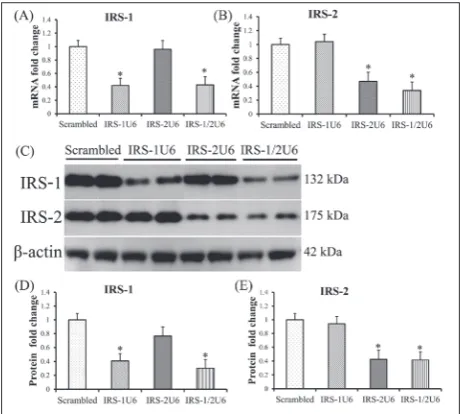
RNA interference can regulate gene expression by in-hibition of translation or by degradation of mRNA. To confirm that mRNA levels were decreased, the levels of expression of IRS-1 and IRS-2 mRNAs were measured by quantitative RT-PCR after G418 selec-tion. The results showed that IRS-1 mRNA was de-creased by 58% when an IRS1U6 or IRS1/2U6 vector was used. Similarly, the IRS2U6 vector caused a 54% drop in IRS-2 mRNA levels when used alone, and a 67% decrease when the IRS1/2U6 vector was used (Fig. 1A and B).
Following transfection with a vector expressing shRNAs against IRS-1 (IRS1U6), we observed an av-erage 60% decrease in IRS-1 protein levels in HepG2 cells as compared with HepG2 cells transfected with a control vector that expressed scrambled shRNAs (Fig. 1C). No associated decreases in actin and IRS-2 protein expression were observed. Similarly, a recom-binant RNAi vector against IRS-2 (IRS2U6) specifical-ly reduced IRS-2 protein expression by approximatespecifical-ly 60% without altering IRS-1 levels or actin controls (Fig. 1C, D and E).
The use of vector shRNAs to knockdown mul-tiple genes simultaneously is feasible. However, we were concerned that dual knockdown could result in greater nonspecific effects than the single vector treatment. We found that treatment with IRS1/2U6 vector caused substantial knockdown of both IRS-1 and IRS-2, with decreases of 70% and 60%, respec-tively, without inducing decreases in the levels of actin protein (Fig, 1C, D and E).
Downregulated IRS protein expression in HepG2 cells results in changes in the expression of glycometabolism genes
Using Western blot analysis, we measured the protein levels of two key gluconeogenic enzymes, PEPCK and fructose-1,6-bisphosphatase (F-1,6-BP) (Fig. 2A, B, and C). PEPCK was upregulated in double-knock-down HepG2 cells 1.7-fold. F-1,6-BP was upregulated in double-knockdown HepG2 cells 1.2-fold, which was not statistically significant.
Interestingly, treatment with IRS1U6 or IRS2U6 alone did not increase the protein levels of PEPCK and F-1,6-BP. Glucokinase (GCK) plays a key role in regu-lating glucose homeostasis, and GCK gene mutations in humans cause maturity onset diabetes of the young, type 2 (MODY-2). The MODY phenotype can arise partly from dysregulated hepatic glucose disposal, as mice with liver-specific ablation of glucokinase exhibit severe hyperglycemia [29]. Thus, we measured GCK expression in HepG2 cells treated with RNAi vector and found that GCK expression was closely related to IRS-1 expression (Fig. 2A and D). Treatment with IRS1U6 caused a 50% drop in GCK expression, while dual treatment resulted in a 46% decrease in GCK levels. Treatment with IRS2U6 resulted in a modest
23% downregulation of GCK, which was not statistically significant (Fig. 2A and D).
Downregulated IRS protein expression in HepG2 cells results in changes in the expression of lipid metabolism genes
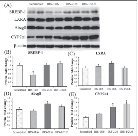
The inappropriate activation of glu-coneogenesis in insulin-resistant states is often accompanied by de-fects in hepatic lipid regulation. We measured the expression of key lipogenic genes such as Srebp1c in response to IRS-1 and IRS-2 knock-down by vector shRNAs. IRS-2 knockdown alone increased hepatic Srebp1 expression 1.7-fold, whereas decreased hepatic IRS-2 expression and dual knockdown also increased hepatic Srebp1 expression (Fig. 3A and B).
Insulin can also regulate lipogenic gene transcrip-tion through pathways independent of the IRS proteins, most notably through liver X receptor A (LXRA) activa-tion [30]. Using Western blot analysis, we found that LXRA protein levels were unchanged after treatment with IRS1U6 or IRS1/2U6 (Fig. 3A and C, respectively), while treatment with IRS2U6 alone resulted in a modest 24% upregulation of LXRA, which was not statistically significant (Fig. 3A and C). However, these results do not preclude the possibility that LXRA activity may be increased. As a proxy for LXR activation, we measured the expression of downstream LXRA genes, such as Abcg8 (Fig. 3A and D), and cytochrome P450, isoform 7A1 (Cyp7a1; Fig. 3A and E). Abcg8 protein levels were unchanged after treatment with IRS1U6 or IRS1/2U6 (Fig. 3A and D, respectively), while treatment with IRS2U6 alone resulted in a modest 26% upregulation of Abcg8, which was not statistically significant (Fig. 3A and D). IRS-1 knockdown alone did not have any effect on Cyp7a1 expression and decreased hepatic IRS-2 expression alone was sufficient to induce a 1.9-fold upregulation of Cyp7a1. Moreover, dual knockdown of IRS-1 and IRS-2 resulted in a 2.2-fold increase in Cyp7a1 genes when compared with the control shRNA treatment (Fig. 3A and E).
Fig. 2. Downregulated IRS protein expression in HepG2 cells resulted in changes in the expression of glycol-metabolism genes. A – Western blots of PEPCK, F-1,6-BP, and GCK in HepG2 cells treated with shRNA vector. B – Corresponding densitometric analyses of the protein bands of PEPCK/actin. C – Corresponding densitometric analyses of the protein bands of F-1,6-BP/actin. D – Corresponding densitometric analyses of the protein bands of GCK/actin. Statistical analysis was performed on 1U6, 2U6 and IRS-1/2U2 vs scrambled, respectively. *P<0.05 indicates significant differences.
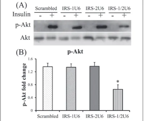
Significant defects in Akt activation in HepG2 cells treated with IRS1U6 and IRS2U6
To better understand the molecular mechanisms of these physiological defects, we measured Akt phos-phorylation in HepG2 cells. Akt phosphos-phorylation was decreased only in HepG2 cells with dual defects in IRS-1 and IRS-2 expression (Fig. 4A). The administra-tion of either IRS1U6 or IRS2U6 alone did not result in any detectable changes in Akt activation when com-pared with the control shRNA treatment (Fig. 4B). However, concomitant treatment with IRS1U6 and IRS2U6 caused a 50% decrease in serine 473 phos-phorylation of Akt compared with the control shRNA treatment (Fig. 4B).
DISCUSSION
In the present study, we utilized an in vitro RNAi tech-nique that included a vector to deliver shRNAs against IRS-1, IRS-2, or both in human hepatoblastoma cells. This vector-mediated RNAi technique allowed us to stably knockdown the expression of IRS-1 and IRS-2 in human hepatoblastoma cells, and to define their individual roles in hepatic insulin signaling and he-patic metabolism.
Previous reports have suggested that IRS-1 and IRS-2 have unique individual roles in regulating key genes in glucose and lipid homeostasis, although they work in a complementary fashion to maintain PI3K signaling [11,13]. In mouse liver, single knockdown of IRS-1 caused a significant increases in mRNAs for the essential gluconeogenic enzymes, PEPCK and F-1,6-BP [21]. However, in this study, the increased expres-sion of the PEPCK gene was a consequence of the marked downregulation of both IRS-1 and IRS-2. In addition, the expression of F-1,6-BP was not changed significantly following knockdown of IRS-1 or IRS-2. This difference may be the result of a dosage effect as in the current study IRS-1 and IRS-2 were knocked down by about 60-70%, while in a previous study they were knocked down by about 70-80%. It was shown that IRS-1 can regulate GCK mRNA levels in cultured hepatocytes by activating PI3K [12]. Our data also indicate that IRS-1 is more prominent than IRS-2 in the regulation of GCK expression, as IRS1U6, either alone or in combination with IRS2U6, caused about a 50% decrease in GCK expression.
It is known that decreased expression of IRS-2 in the liver alone is sufficient to elevate the levels of Srebf1 and can occur independently of the known positive regulatory effects of insulin on Srebf1 expres-sion [31]. In IRS-2 knockout mice, the expresexpres-sion of SREBP-1 was upregulated, and in primary hepato-cytes from SREBP-1 knockout mice, the expression of IRS-2 was upregulated [6,32,33]. Our results showed that decreased IRS-2 increased Srebf1 expression in HepG2 cells. The LXR signaling pathway is a distinct pathway that contributes to dysregulated lipogenesis and hepatic steatosis. Our data suggest that the ex-pression of LXRA was not statistically significantly changed, while the expression of Cyp7a1, which is a downstream LXRA gene, was increased. These results preclude the possibility that LXRA activity may be in-creased. As insulin-stimulated LXR activity increased SREBP-1c transcription via a signaling-independent mechanism [30], increased Srebf1 expression may be the result of increased LXR activity.
PI3K/Akt signaling is incorporated in the main signaling pathway for insulin-dependent regulation of glucose metabolism [34]. After insulin activates its re-ceptors, PI3K must recruit an insulin substrate protein to promote Akt Ser473 phosphorylation [35]. IRS-1
and IRS-2 play complementary roles in the liver at the level of intermediate signaling of PI3K activation. Akt is the main downstream gene of PI3K, and Akt phosphorylation and insulin-stimulated Akt activity can correspond to PI3K activity [36]. Compensatory mechanisms occur in the livers of IRS-1 knockout mice, where IRS-2 expression significantly increases to maintain total PI3K activity and Akt activation [24]. IRS-2 knockout mice do not exhibit this increase in IRS-1-associated PI3K activity in the liver, whereas IRS-2 knockdown results in about a 50% decrease in hepatic Akt activation [37]. The reduction in 60-70% in IRS-1 or IRS-2 individually does not perturb total Akt activity as a reciprocal increase in PI3K activity through the unaltered IRS antipode helps to maintain full PI3K signaling. Reductions in IRS-1 and IRS-2 si-multaneously disturb the balance of IRS, whereas Akt phosphorylation and insulin-stimulated Akt activity were decreased in human hepatoblastoma cells, with dual defects in IRS-1 and IRS-2 expression.
CONCLUSION
The present study shows that dual downregulation of IRS-1 and IRS-2 increased the expression of PEPCK gene, and that a reduction in IRS-1 caused an ap-proximately 50% decrease in GCK expression. The decrease in IRS-2 increased the expression of Srebf1 and Cyp7a1 in human hepatoblastoma cells. Insulin-stimulated Akt activity was decreased in human hepa-toblastoma cells with dual defects in IRS-1 and IRS-2 expression. Thus, the differential modulation of he-patic IRS expression and signaling may represent an important component of the molecular pathophysiol-ogy that underlies both type 2 diabetes mellitus and the metabolic syndrome.
Acknowledgments: This study was supported by the Open
Projects of Key Laboratory of Animal Cellular and Genetic En-gineering of Heilongjiang Province (KF201708).
Author contributions: Tianfei Yu and Ming Li collected the data and contributed equally to the paper. Huiying Dong, Pengyu Xie and Wanzhu Sun collected the data and performed the statisti-cal analysis of the data. Tianfei Yu, Bo Meng and Yanshuang Mu prepared the manuscript. Yanshuang Mu conceived the study. All authors contributed to and approved the final manuscript. Conflict of interest disclosure: The authors have no financial interests or potential conflicts of interests to declare.
REFERENCES
1. Fisher SJ and Kahn CR. Insulin signaling is required for insulin’s direct and indirect action on hepatic glucose pro-duction. J Clin Invest. 2003;111:463-8.
2. Kim SP, Ellmerer M, Van Citters GW,Bergman RN. Primacy of hepatic insulin resistance in the development of the meta-bolic syndrome induced by an isocaloric moderate-fat diet in the dog. Diabetes. 2003;52:2453-60.
3. Tripathy D, Eriksson KF, Orho-Melander M, Fredriksson J, Ahlqvist G, Groop L. Parallel manifestation of insulin resistance and beta cell decompensation is compatible with a common defect in Type 2 diabetes. Diabetologia. 2004;47:782-93.
4. Michael MD, Kulkarni RN, Postic C, Previs SF, Shulman GI, Magnuson MA, Kahn CR. Loss of insulin signaling in hepatocytes leads to severe insulin resistance and progressive hepatic dysfunction. Mol Cell. 2000;6:87-97.
5. Huang C, Wu M, Du J, Liu D, Chan C. Systematic modeling for the insulin signaling network mediated by IRS(1) and IRS(2). J Theor Biol. 2014;355:40-52.
6. Shimomura I, Matsuda M, Hammer RE, Bashmakov Y, Brown MS, Goldstein JL. Decreased IRS-2 and increased SREBP-1c lead to mixed insulin resistance and sensitivity in livers of lipodystrophic and ob/ob mice. Mol Cell. 2000;6:77-86.
7. Patti ME and Kahn CR. The insulin receptor-a critical link in glucose homeostasis and insulin action. J Basic Clin Physiol Pharmacol. 1998;9:89-109.
8. Withers DJ, Gutierrez JS, Towery H, Burks DJ, Ren JM, Pre-vis S, Zhang Y, Bernal D, Pons S, Shulman GI, Bonner-Weir S, White MF. Disruption of IRS-2 causes type 2 diabetes in mice. Nature. 1998;391:900-4.
9. Sun XJ, Rothenberg P, Kahn CR, Backer JM, Araki E, Wilden PA, Cahill DA, Goldstein BJ, White MF. Structure of the insulin receptor substrate IRS-1 defines a unique signal transduction protein. Nature. 1991;352:73-7.
10. Anai M, Funaki M, Ogihara T, Terasaki J, Inukai K, Kata-giri H, Fukushima Y, Yazaki Y, Kikuchi M, Oka Y, Asano T. Altered expression levels and impaired steps in the path-way to phosphatidylinositol 3-kinase activation via insulin receptor substrates 1 and 2 in Zucker fatty rats. Diabetes. 1998;47:13-23.
11. Aytug S, Reich D, Sapiro LE, Bernstein D, Begum N. Impaired IRS-1/PI3-kinase signaling in patients with HCV: a mechanism for increased prevalence of type 2 diabetes. Hepatology. 2003;38:1384-92.
12. Kerouz NJ, Horsch D, Pons S, Kahn CR. Differential regula-tion of insulin receptor substrates-1 and -2 (1 and IRS-2) and phosphatidylinositol 3-kinase isoforms in liver and muscle of the obese diabetic (ob/ob) mouse. J Clin Invest. 1997;100:3164-72.
13. Kim YB, Peroni OD, Franke TF, Kahn BB. Divergent regula-tion of Akt1 and Akt2 isoforms in insulin target tissues of obese Zucker rats. Diabetes. 2000;49:847-56.
15. Gual P, Le Marchand-Brustel Y, Tanti JF. Positive and nega-tive regulation of insulin signaling through IRS-1 phosphor-ylation. Biochimie. 2005;87:99-109.
16. Zick Y. Uncoupling insulin signalling by serine/threonine phosphorylation: a molecular basis for insulin resistance. Biochem Soc Trans. 2004;32:812-6.
17. Brunet A, Bonni A, Zigmond MJ, Lin MZ, Juo P, Hu LS, Anderson MJ, Arden KC, Blenis J, Greenberg ME. Akt promotes cell survival by phosphorylating and inhibiting a Forkhead transcription factor. Cell. 1999;96:857-68. 18. Puigserver P, Rhee J, Donovan J, Walkey CJ, Yoon JC,
Ori-ente F, Kitamura Y, Altomonte J, Dong H, Accili D, Spiegel-man BM. Insulin-regulated hepatic gluconeogenesis through FOXO1-PGC-1alpha interaction. Nature. 2003;423:550-5. 19. Fritsche L, Weigert C, Haring HU, Lehmann R. How insulin
receptor substrate proteins regulate the metabolic capacity of the liver--implications for health and disease. Curr Med Chem. 2008;15:1316-29.
20. Haeusler RA and Accili D. The double life of Irs. Cell Metab. 2008;8:7-9.
21. Taniguchi CM, Ueki K, Kahn R. Complementary roles of IRS-1 and IRS-2 in the hepatic regulation of metabolism. J Clin Invest. 2005;115:718-27.
22. Araki E, Lipes MA, Patti ME, Bruning JC, Haag B, Johnson RS, Kahn CR. Alternative pathway of insulin signalling in mice with targeted disruption of the IRS-1 gene. Nature. 1994;372:186-90.
23. Withers DJ, Burks DJ, Towery HH, Altamuro SL, Flint CL, White MF. Irs-2 coordinates Igf-1 receptor-mediated beta-cell development and peripheral insulin signalling. Nat Genet. 1999;23:32-40.
24. Yamauchi T, Tobe K, Tamemoto H, Ueki K, Kaburagi Y, Yamamoto-Honda R, Takahashi Y, Yoshizawa F, Aizawa S, Akanuma Y, Sonenberg N, Yazaki Y, Kadowaki T. Insulin signalling and insulin actions in the muscles and livers of insulin-resistant, insulin receptor substrate 1-deficient mice. Mol Cell Biol. 1996;16:3074-84.
25. Kubota N, Tobe K, Terauchi Y, Eto K, Yamauchi T, Suzuki R, Tsubamoto Y, Komeda K, Nakano R, Miki H, Satoh S, Sekihara H, Sciacchitano S, Lesniak M, Aizawa S, Nagai R, Kimura S, Akanuma Y, Taylor SI, Kadowaki T. Disruption of insulin receptor substrate 2 causes type 2 diabetes because of liver insulin resistance and lack of compensatory beta-cell hyperplasia. Diabetes. 2000;49:1880-9.
26. Kubota N, Kubota T, Itoh S, Kumagai H, Kozono H, Taka-moto I, Mineyama T, Ogata H, Tokuyama K, Ohsugi M, Sasako T, Moroi M, Sugi K, Kakuta S, Iwakura Y, Noda T, Ohnishi S, Nagai R, Tobe K, Terauchi Y, Ueki K, Kadowaki T. Dynamic functional relay between insulin receptor substrate
1 and 2 in hepatic insulin signaling during fasting and feed-ing. Cell Metab. 2008;8:49-64.
27. Yang X, Nath A, Opperman MJ, Chan C. The double-stranded RNA-dependent protein kinase differentially regu-lates insulin receptor substrates 1 and 2 in HepG2 cells. Mol Biol Cell. 2010; 21: 3449-58.
28. Yang X and Chan C. Repression of PKR mediates palmitate-induced apoptosis in HepG2 cells through regulation of Bcl-2. Cell Res. 2009;19:469-86.
29. Postic C, Shiota M, Niswender KD, Jetton TL, Chen Y, Moates JM, Shelton KD, Lindner J, Cherrington AD, Mag-nuson MA. Dual roles for glucokinase in glucose homeo-stasis as determined by liver and pancreatic beta cell-spe-cific gene knock-outs using Cre recombinase. J Biol Chem. 1999;274:305-15.
30. Chen G, Liang G, Ou J, Goldstein JL, Brown MS. Central role for liver X receptor in insulin-mediated activation of Srebp-1c transcription and stimulation of fatty acid synthesis in liver. Proc Natl Acad Sci U S A. 2004;101:11245-50. 31. Shimomura I, Bashmakov Y, Ikemoto S, Horton JD, Brown
MS, Goldstein JL. Insulin selectively increases SREBP-1c mRNA in the livers of rats with streptozotocin-induced diabetes. Proc Natl Acad Sci U S A. 1999;96:13656-61. 32. Ide T, Shimano H, Yahagi N, Matsuzaka T, Nakakuki M,
Yamamoto T, Nakagawa Y, Takahashi A, Suzuki H, Sone H, Toyoshima H, Fukamizu A, Yamada N. SREBPs suppress IRS-2-mediated insulin signalling in the liver. Nat Cell Biol. 2004;6:351-7.
33. Tobe K, Suzuki R, Aoyama M, Yamauchi T, Kamon J, Kubota N, Terauchi Y, Matsui J, Akanuma Y, Kimura S, Tanaka J, Abe M, Ohsumi J, Nagai R, Kadowaki T. Increased expres-sion of the sterol regulatory element-binding protein-1 gene in insulin receptor substrate-2(-/-) mouse liver. J Biol Chem. 2001;276:38337-40.
34. Hers I, Vincent EE, Tavare JM. Akt signalling in health and disease. Cell Signal. 2011;23:1515-27.
35. Stiles B, Wang Y, Stahl A, Bassilian S, Lee WP, Kim YJ, Sher-win R, Devaskar S, Lesche R, Magnuson MA, Wu H. Liver-specific deletion of negative regulator Pten results in fatty liver and insulin hypersensitivity [corrected]. Proc Natl Acad Sci U S A. 2004;101:2082-7.
36. Du K, Herzig S, Kulkarni RN, Montminy M. TRB3: a tribbles homolog that inhibits Akt/PKB activation by insulin in liver. Science. 2003;300:1574-7.