© 2019 by the Serbian Biological Society 703
Expression of PCNA, CD-31 and HER-2 in Serbian patients with oral squamous cell
carcinoma
Slobodan A. Lončarević1,2, Denis O. Brajković1,3, Milka F. Gardašević2, Olivera Lj. Lončarević4, Nebojša G.
Lađević5,6, Dejan M. Nešić6, Dušica M. Stamenković7,8, Ivana S. Likić-Lađević6,9, Nikola N. Lađević10 and
Nemanja K. Rančić8,11,12,*
1Department of Dentistry; Faculty of Medical Sciences University of Kragujevac, Kragujevac, Serbia 2Clinic for Maxillofacial Surgery, Military Medical Academy, Belgrade, Serbia
3Resident at Clinic for Maxillofacial Surgery, Military Medical Academy, Belgrade, Serbia 4Pulmology Clinic, Military Medical Academy, Belgrade, Serbia
5Center for Anesthesiology and Reanimatology, Clinical Center of Serbia, Belgrade, Serbia 6Faculty of Medicine, University of Belgrade, Belgrade, Serbia
7Department of Anesthesiology and Intensive Care, Military Medical Academy, Belgrade, Serbia 8Medical Faculty Military Medical Academy, University of Defense, Belgrade, Serbia
9Clinic of Gynecology and Obstetrics, Clinical Center of Serbia, Belgrade, Serbia 10Urology Clinic, Clinical Center of Serbia, Belgrade, Serbia
11Centre for Clinical Pharmacology, Military Medical Academy, Belgrade, Serbia 12Institute of Radiology, Military Medical Academy, Belgrade, Serbia
*Corresponding author: [email protected]
Received: July 5, 2019; Revised: August 4, 2019; Accepted: August 6, 2019; Published online: August 30, 2019
Abstract: Several studies have investigated the expression of tumor markers, including p53, HER-2, PCNA, EGFR, VEGFR CD-31 and Bcl-2 in patients with oral squamous carcinoma (OSC). This study aimed to determine the expression of pro-liferating cell nuclear antigen (PCNA), endothelial functions of platelet/endothelial cell adhesion molecule-1 (CD31) and human epidermal growth factor receptor-2 (HER-2) according to OSC stage. The prospective study included 62 patients diagnosed with OSC stages II and III. Surgical specimens were obtained from tumor and peritumoral tissues. We deter-mined the pathohistological degree of tumor differentiation and the immunohistochemical expression of PCNA, CD-31 and HER-2 for each specimen. Immunohistochemical analysis of the expression of PCNA in tumor cells demonstrated poor staining of immunoreactive tumor cells in 23 patients (10 in stage II, 7 in stage IIIa and 6 in stage IIIb). Moderately expressed PCNA-immunoreactivity in the tumor cells in 17 patients (7 in stage II, 6 in stage IIIa and 4 in stage III), and extremely strong PCNA-immunoreactive staining in tumor cells of 10 patients with IIIb stage, was observed. These results suggest that PCNA expression combined with pathohistological findings could possess a prognostic value in determining the survival rates for patients with oral squamous cell carcinoma.
Keywords: oral carcinoma; squamous cell carcinoma; tumor markers; PCNA, CD-31, HER-2
How to cite this article: Lončarević SA, Brajković DO, Gardašević MF, Lončarević OLj, Lađević NG, Nešić DM, Stamenković DM, Likić-Lađević IS, Lađević NN, Rančić NK. Expression of PCNA, CD-31 and HER-2 in Serbian patients with oral squamous cell carcinoma. Arch Biol Sci. 2019;71(4):703-10.
INTRODUCTION
Cancer is a multifactorial disease caused by a series of genetic alterations that lead to cellular prolifera-tion and differentiaprolifera-tion [1]. Cellular proliferaprolifera-tion is an important indicator of the biological aggressive-ness of cancer. Oral squamous cell carcinoma (OSC)
Prognosis and survival rate are low despite the ad-vance in OSC diagnosis and treatment recorded in recent years [4-6]. Evaluation of molecular changes in cancer and their extrapolation on clinical parameters might be beneficial in future diagnostic and treatment approaches [7-9]. The expression of different tumor markers in patients with OSC, including tumor pro-tein 53 (p53), human epidermal growth factor re-ceptor-2 (HER-2), proliferating cell nuclear antigen (PCNA), epidermal growth factor receptor (EGFR), vascular endothelial growth factor receptor (VEGFR), endothelial functions of platelet/endothelial cell ad-hesion molecule-1 (CD-31) and B-cell leukemia/ lymphoma 2 (Bcl-2), has been explored [10-13]. The presence of tumor cells in tissue at the tumor margins is a common finding in high-grade OSC and can be a direct or indirect indicator of disease progression during five-year survival rate monitoring [14].
Biological markers provide information about the differentiation and proliferation of tumor cells and carcinoma progression and therefore play a signifi-cant role in disease prognosis. The nuclear antigen of PCNA is an acidic nuclear protein that fluctuates dur-ing the cell cycle and is connected with cell prolifera-tion [15]. PCNA is important for DNA synthesis and repair, cell proliferation and cycle progression. The levels of PCNA are low and undetectable in quiescent cells, but the production of this protein occurs before DNA replication. PCNA increases at G1/S phases, de-creases at phase G2 of the cell cycle, and hence it is a sensitive index of proliferation [16,17]. Increased PCNA expression follows tissue progression from a normal epithelium to hyperplasia and dysplasia and premalignant and malignant lesion of the oral cavity [18,19].
Increased expression of different vascular markers such as VEGF, von Willebrand factor (VWF), CD-31, CD-34 and endoglin (CD105) in OSC was found to accompany the progression of histological abnormal-ity [20]. CD-31 and CD-34 were shown to be more sensitive than factor VWF for evaluating tumor blood vessels, but no significant relationship between CD-31 and OSC stages was detected [20].
The expression of HER-2 in OSC was seen to range between 2.5-88% [21]. HER-2 belongs to the HER gene family that regulates cell growth, survival,
differentiation and migration. HER-2 expression and gene amplification were rarely detectable in OSC and are apparently unrelated to tumor phenotype or sur-vival rates of patients with OSC [22].
Most of these studies aimed to clarify the molecu-lar origin of OSC and to determine the biomarkers for early detection, prognosis, prediction of outcome and therefore more efficient treatment regimens of patients with OSC [7,8,23,24]. The present study aimed to determine the expression of PCNA, CD-31 and HER-2 in patients with OSC according to histo-pathologic and clinical tumor stage.
MATERIALS AND METHODS Study design
The study was designed as a prospective study of a series of cases. The principles of ICH Good Clinical Practice were strictly followed, and approval from the Ethics Committee of the Military Medical Academy was obtained.
The study population included 62 patients with OSC stages II and III according to TNM (T-tumor, N-lymph node, M-metastasis) classification [25,26]. All patients underwent the same type of surgery at the Clinic for Maxillofacial Surgery of Military Medical Academy in the period between 1995 and 2015. Stage II (T1N0M0) includes tumors between 2 and 4 cm in size, with no metastasis detected in distant organs and lymph nodes. Stage IIIa (T3, N0, M0) includes tumors larger than 4 cm. In stage IIIb (T1-3, N1, M0) tumor size can range between 2 and more than 4 cm, with metastasis detected in the ipsilateral lymph node with a size up to 3 cm [25,26].
Histopathological analysis
For histopathological analysis, tumor tissue sec-tions were fixed in 5% buffered neutral formalin and processed in a V.I.P. Sakura apparatus (Sakura, Netherlands) for automatic fixation, dehydration and embedding tissue, and were then embedded in paraf-fin. Prepared tissue blocks were sliced (thickness 5-7 µm). The tissue sections were mounted on separate adherent chips (SuperFrost, Thermo Fisher Scientific Gerhard Menzel B.V. and Co., Germany) and dried at 56°C for 1 h.
The histological grade of the tumor, the degree of infiltration of the peritumoral tissue as well as perivascular, perineural and lymphatic infiltration were determined using an Olympus AX70 (Olympus Optical co. GMBH., Germany) microscope. The de-gree of infiltration of connective, adipose, muscle and bone tissues was scored as follows: 0 – no infiltration; 1 – infiltration is present at 0.5 cm from the edge of the tumor; 2 – presence of infiltration at 0.5-1 cm from the edge of the tumor; 3 – infiltration of tissue at >1cm from the edge of the tumor.
Immunohistochemical analysis
Marking of tumor cells in the peritumoral tissue was performed with monoclonal and polyclonal anti-human antibodies (Dako Cytomation, Denmark). Visualization of the marker was carried out using highly sensitive and specific methods, IHC and la-belling with the streptavidin-biotin complex method (Universal LSAB™+ Kit HRP). We determined the presence and number of tumor cells in the peritu-moral tissue by semiquantitative analysis of speci-mens using PCNA, HER-2 and CD-31 tumor markers (Supplementary Fig. S1). The findings were defined as the level of expression of a particular tumor marker (PCNA, HER-2 and CD-31) in the peritumoral tissue specimen. The level of expression was scored based on the intensity of staining and the number of immuno-reactive cells, as follows: 0 – no immunoimmuno-reactive cells; 1 – poor staining, rare positive cells; 2 – moderate staining, small number of positive cells; 3 – intense staining, numerous positive cells.
Statistical analysis
For statistical analysis, we used the commercial statisti-cal program SPSS for Windows, ver. 18.0. To test the normality of the distribution of the statistical charac-teristics, we used the Kolmogorov-Smirnov test. To test the intergroup differences, we used the Kruskal-Wallis test and analysis of variance (ANOVA), depending on the type of variables. All categorical variables were pre-sented as the percentage frequency of certain categories. For categorical variables, the statistical significance of differences was examined using the chi-square test.
RESULTS
Sociodemographic characteristics
The present study enrolled 62 patients, 28 patients with stage II and 34 with stage III (Fig. 1) cancer. The age of the patients was 56.19±1.41 years (range 39-85 years) (ANOVA, p>0.05). Tumor size less than 2 cm (T1 stage) was found in 38.71% of patients, while tumors larger than 4 cm (T3 stage) were found in 50% of patients.
Immunohistochemical analysis according to tumor stage
Tumor marker (PCNA, CD-31 and HER-2) expres-sion corresponding to tumor size was significant only for PCNA (chi-square test, p=0.002) (Table 1).
Immunohistochemical analysis of PCNA expression in tumor cells demonstrated poor staining of immu-noreactive tumor cells in 23 (37%) patients, of which 10 (36%) were in stage II, 7 (54%) in stage IIIa and 6 (29%) in the stage IIIb. Moderately expressed PCNA-immunoreactivity in tumor cells was detected in 17 patients (7 or 25% of the patients in stage II, 6 or 46% in stage IIIa, and 4 or 19% in stage IIIb); extremely strong PCNA-immunoreactive staining was noted in tumor cells of 10 or 48% of the patients with stage IIIb
stage of the disease. Differences in the level of PCNA expression in peritumoral tissue cells, depending on the stage of the disease, were statistically significant (Kruskal-Wallis test; p<0.001).
Differences in the degree of expression of PCNA, CD-31 and HER-2, according to clinical tumor stag-ing, was statistically significant for all investigated markers (Table 2). At stages IIIa and IIIb, 20 (59%) of the patients had a higher level of PCNA (expression levels 2 and 3) compared to patients in stage II. The expression of HER-2 and CD-31 were positive only in 10 patients with IIIb stage.
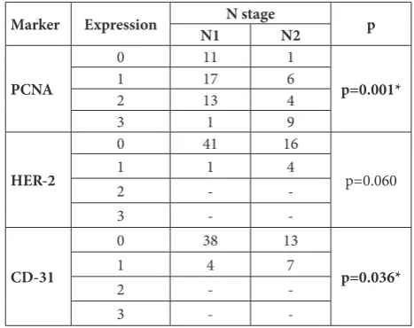
Significant differences were found in the level of expression of PCNA and CD-31 in metastatic lymph nodes (N1 and N2). PCNA had 1 and 2 levels of ex-pression (Table 3). On the other hand, CD-31 expres-sion level was 0 in most of the cases (Table 3).
DISCUSSION
We investigated the level of expression of certain markers in OSC specimens. We found that the levels of PCNA expression depended significantly on tumor size, TNM stage and metastatic lymph node presence.
OSC is the result of the final stage in the process of carcinogenesis, resulting from genetic and epigenetic
Table 3. Expression of PCNA, HER-2 and CD-31 in peritumoral tissue cell according to lymph node status.
Marker Expression N1N stageN2 p
PCNA
0 11 1
p=0.001*
1 17 6
2 13 4
3 1 9
HER-2
0 41 16
p=0.060
1 1 4
2 -
-3 -
-CD-31
0 38 13
p=0.036*
1 4 7
2 -
-3 -
-Level of expression: 0 – no immunoreactive cells; 1 – poor staining, rare positive cells; 2 – moderate staining, a small number of positive cells; 3 – intense staining, numerous positive cells.
*p<0.05 (Chi-square test). Table 1. Expression of PCNA, HER-2 and CD-31 in peritumoral
tissues according to tumor size.
Marker Expression Tumor size p
T1 T2 T3
PCNA
0 9 2 1
0.002*
1 10 - 13
2 5 4 8
3 - 1 9
HER-2
0 24 7 26
0.066
1 - - 5
2 - -
-3 - -
-CD-31
0 22 4 25
0.103
1 2 3 6
2 - -
-3 - 0
-Expression: 0 – no immunoreactive cells; 1 – poor staining, rare positive cells; 2 – moderate staining, a small number of positive cells; 3 – intense staining, a greater number of positive cells.
Tumor size: T1 – tumor <2 cm; T2 – tumor 2-4 cm; T3 – tumor >4 cm. *p<0.05 (chi-square test).
Table 2. Expression of PCNA, HER-2 and CD-31 in peritumoral tissue cell according to clinical tumor stage.
Marker Expression Stage
II IIIaStage IIIIIIb p
PCNA
0 11 - 1
0.001*
1 10 7 6
2 7 6 4
3 - - 10
HER-2
0 28 13 16
0.005*
1 - - 5
2 - -
-3 - -
-CD-31
0 24 13 14
0.038*
1 4 - 7
2 - -
-3 - -
-Level of expression: 0 – no immunoreactive cells; 1 – poor staining, rare positive cells; 2 – moderate staining, a small number of positive cells; 3 – intense staining, numerous positive cells.
defects, and potentiated by the action of cancerogenic substances. Two classes of genes have a key role in this process: proto-oncogenes and tumor suppressor genes, which are necessary for the control of prolifera-tion, differentiation and cell death [27-29].
A set of proteins, including p53, Bcl-2, HER-2, PCNA, epidermal growth factor (EGF) and vascu-lar endothelial growth factor (VEGF), were analyzed and correlated with histopathologic parameters in studies that aimed to analyze the metastatic poten-tial of tumors [12,30,31]. Numerous studies aimed to detect and identify the prognostic properties of the tumor markers for the growth of the OSC and to establish the correlation between the expression level of certain markers and disease prognosis [12,31-33]. Contemporary studies are investigating the role of early detection of premalignant and malignant le-sions, early detection of local recurrence and distant metastasis, and early prevention of secondary tumors, as well as a selection of the most effective treatment for the patient [7,17,34-36].
Basic morphological features for estimation of the malignancy level include the histological grade, nuclear grade, mitotic index and nucleocytoplasmic ratio [37-40]. These parameters are significantly cor-related with the degree of tumor infiltration of mar-ginal resection tissue [41,42]. Studies have shown differences in the survival rate of patients with OSC [4-6]. Additionally, the low survival rate of patients diagnosed with OSC was correlated with increased expression of PCNA [43,44]. Thus, it was suggested that PCNA might be used as a biomarker for the initial assessment of OSC aggressiveness [43,44].
No specific antigenic properties were found in OSC. To date, the most studied genes and markers are the genes for p53, cyclin D1, transforming growth fac-tor (TGF), EGF, VEGF, E-cadherins and collagen VII [10,45,46]. However, none of these markers has proved to be specific for the prognosis and diagnosis of OSC.
Previous studies found a positive correlation be-tween PCNA proteins with tumor grade and the clini-cal stage of OSC [43,44]. Our results indicate that the expression of PCNA in OSC is in a high correlation with the histological grade of the tumor and the degree of infiltration of peritumoral tissue, which are indi-cators of poor prognosis and reduced survival rates
of patients with OSC. The prognostic value of PCNA could be significant for patients with OSC. The synthe-sis of PCNA protein correlates with cell proliferation as it is expressed in the nuclei of cells during DNA synthesis [43]. Tumor proliferation was identified as an important predictor of biologic behavior for vari-ous malignancies [47]. PCNA is one of the proteins important in cell proliferation, and antibodies to this protein help to assess the tumor proliferation activity of oral lesions. Varying degrees of atypia in OSC were analyzed employing PCNA immunoreactivity. Focal immunoreactivity of PCNA was noted in highly dif-ferentiated OSC and elevated cell proliferation, and aneuploidy was observed in the entire tumor in poorly differentiated OSC [48]. The intensity of PCNA im-munoreactivity is in correlation with tumor cell dif-ferentiation, nuclear atypia and the patterns of inva-sive margins in the underlying connective tissue[44]. Increased expression of PCNA was indicative of poor differentiation, higher nuclear atypia and more inva-sive growth of tumor cells [44]. The presence of PCNA proteins in chronic and premalignant lesions, such as oral lichen planus and epithelial dysplasia. could be helpful in the evaluation of malignant potential [49].
Based on the significant difference in the PCNA index between preinvasive and invasive squamous epithelial lesions of the cervix, Pahuja et al. [50] sug-gested that the PCNA index might have a predictive value for the prognosis of patients with squamous cell carcinoma. The PCNA index could be helpful in the identification of patients whose carcinoma’s in situ
lesion will progress and require treatment, and those whose lesion will remain static or regress [51]. PCNA in tissue sections can be used as an adjunct to diag-nose preneoplastic and neoplastic lesions [52]. The use of a specific biomarker of dysplasia in conjunction with histological procedures could improve the accu-racy, precision and sensitivity of potential screening program. Accordingly, PCNA could play an important role in the pathogenesis, progression and metastasis in patients with OSC [53,54].
invasion, and enhancement of the malignant potential in epithelial tissues [55]. HER-2 has been reported to be overexpressed in many cancers and associated with poor prognosis. [56]. Immunohistochemical staining has been the most common method used to detect overexpression of ErbB receptors. Bernardes et al.[55] showed that 97.8% of tumor specimens were negative for HER-2 in OSC patients. Thus, HER-2 was not con-firmed as a prognostic factor, with which our results are in agreement.
Neoangiogenesis is one of the essential events in neoplastic progression. Vascular marker CD-31 is an important marker used for quantifying microvessels and correlating with tumor growth and prognosis [57]. CD-31 expression in tumor blood vessels in OSC showed no significant relationship with OSC stage [20]. However, overexpression of CD-31 correlated with the progression of histological abnormality [20]. In our study, we found overexpression of CD-31 in 11 tumor specimens. Tumor angiogenesis and the density of newly formed vessels have a potential prognostic relevance in the assessment of OSC, with CD-34 pos-sessing a higher correlation in the assessment of tumor vascularization than CD-31 [58,59].
The present study evaluated the immunohisto-chemical expression of PCNA, HER-a and CD-31 in tumor samples after OSC resections. Expression of positively stained PCNA cells increased with more advanced stages of the disease, i.e. from II to the IIIb TNM stage, suggesting an increase in the proliferative activity of tumor cells. Therefore, PCNA expression along with clinical characteristics might be useful in predicting the aggressiveness and recurrence rate of OSC. Large-scale multicentric studies are needed to define the economic feasibility for implementing this type of testing in all patients diagnosed with OSC be-cause of the high costs of immunohistochemical anal-ysis, especially in developing countries. The present study has some limitations as this was a single-center study that included a small sample of the patients and the expression of only three tumor markers, PCNA, HER-2 and CD-31.
CONCLUSIONS
Immunohistochemical analysis of PCNA expression in tumor cells demonstrated poor staining of
immu-noreactive tumor cells in 23 patients (10 in stage II, 7 in stage IIIa and 6 in stage IIIb), and moderate ex-pression of PCNA-immunoreactivity in 17 patients (7 in stage II, 6 in stage IIIa and 4 in stage IIIb). Very strong PCNA-immunoreactive staining was observed in tumor cells in 10 patients with stage IIIb of the dis-ease. Differences in the level of expression of PCNA in peritumoral tissue cells were statistically signifi-cant depending on the tumor stage. Further work is needed to fully elucidate the role and use of PCNA in early detection of premalignant and malignant lesions, local recurrence and distant metastasis, prevention of secondary tumors and choice of an appropriate therapeutic protocol. The presented results suggest that PCNA, in combination with histopathological parameters, could be an important prognostic factor.
Funding: This study was supported by the Clinic for Maxillofacial Surgery, Military Medical Academy, Belgrade, Serbia.
Author contributions: S.L. initiated the study, collected and in-terpreted the data and wrote the manuscript. D.B. collected and interpreted the data and wrote the manuscript. M.G. performed the statistical data analysis, literature search and collected the data. O.L. performed the literature search and wrote the manuscript. N.L. performed the literature search and revised the manuscript. D.N. initiated the study, performed the literature search and re-vised the manuscript. D.S. performed the literature search, col-lected the data, performed the anesthesia and revised the manu-script. I.L.L. interpreted the data and revised the manumanu-script. N.L. performed the literature search and wrote the manuscript. N.R. performed the statistical data analysis, interpreted the data and revised the manuscript. All authors read and approved the final draft of the manuscript.
Conflict of interest disclosure: The authors declare no conflict of interest.
REFERENCES
1. Martínez-Lara I, González-Moles MA, Ruiz-Avila I, Bravo M, Ramos MC, Fernández-Martínez JA. Proliferating cell nuclear antigen (PCNA) as a marker of dysplasia in oral mucosa. Acta Stomatol Belg. 1996;93(1):29-32.
2. Parkin DM, Bray F, Ferlay J, Pisani P. Estimating the world cancer burden: Globocan 2000. Int J Cancer. 2001;94(2):153-6. 3. World Health Organization. International Agency for
Research on Cancer. Lip, oral cavity. In: The Global Cancer Observatory. Cancer Today, Database. [last updated 2018 Mar; cited 2019 Jul 25]. Available from: https://gco.iarc.fr/ 4. Wang B, Zhang S, Yue K, Wang XD. The recurrence and
5. Tonchev K, Vladimirov B. Survival rates in oral cancer patients - a 10-year retrospective study. J IMAB. 2016;22(4):1385-8. 6. Listl S, Jansen L, Stenzinger A, Freier K, Emrich K, Holleczek
B, Katalinic A, Gondos A, Brenner H; GEKID Cancer Sur-vival Working Group. SurSur-vival of patients with oral cavity cancer in Germany. PLoS One. 2013;8(1):e53415.
7. Loncarević S, Vignjević S, Jović N, Aćimović L, Gardasević M, Todorović V, Dimitrijević J. Significance of pathohisto-logical findings and the expression of Bcl-2 in diagnosis and treatment of oral planocellular carcinoma. Vojnosanit Pregl. 2012;69(4):314-9.
8. Fuchs A, Hartmann S, Ernestus K, Mutzbauer G, Linz C, Brands RC, Kübler AC, Müller-Richter UD. Mandibular intraosseous pseudocarcinomatous hyperplasia: a case report. J Med Case Rep. 2016;10(1):268.
9. Dimitrijević J. Clinical pathology. Srp Arh Celok Lek. 2006;134(Suppl 1):78-83.
10. Wilson GD, Saunders MI, Dische S, Richman PI, Daley FM, Bentzen SM. Bcl-2 expression in head and neck cancer: an enigmatic prognostic marker. Int J Radiat Oncol Biol Phys. 2001;49(2):435-41.
11. Rodrigo JP, Coto E, González MV, Maldonado M, Suárez C. P53 allelic loss and outcome in patients with squamous cell carcinoma of the head and neck. Eur Arch Otorhinolaryn-gol. 2001;258(5):255-8.
12. Monteiro LS, Diniz-Freitas M, Garcia-Caballero T, Forteza J, Fraga M. EGFR, and Ki-67 expression in oral squamous cell carcinoma using tissue microarray technology. J Oral Pathol Med. 2010;39(7):571-8.
13. Choontharu MM, Binda A, Bhat S, Sharma SM. Role of tumor markers in oral squamous cell carcinoma: Review of literature and future consideration. SRM J Res Dental Sci. 2012;3(4):251-6.
14. Scully C, Sudbø J, Speight PM. Progress in determining the malignant potential of oral lesions. J Oral Pathol Med. 2003;32(5):251-6.
15. Kurki P, Ogata K, Tan EM. Monoclonal antibodies to pro-liferating cell nuclear antigen (PCNA)/cyclin as probes for proliferating cells by immunofluorescence microscopy and flow cytometry. J Immunol Methods. 1988;109(1):49-59. 16. Chiang CP, Lang MJ, Liu BY, Wang JT, Leu JS, Hahn LJ,
Kuo MY. Expression of proliferating cell nuclear antigen (PCNA) in oral submucous fibrosis, oral epithelial hyperker-atosis, and oral epithelial dysplasia in Taiwan. Oral Oncol. 2000;36(4):353-9.
17. Srinivasan M, Jewell SD. Quantitative estimation of PCNA, c-myc, EGFR and TGF-alpha in oral submucous fibrosis - an immunohistochemical study. Oral Oncol. 2001;37(5):461-7. 18. Shahela T, Aesha S, Ranganathan K, T R, Roa K UD, Joshua
E, Ahmed AS, Chittamsetty H. Immunohistochemical Expression of PCNA in Epithelial Linings of Selected Odon-togenic Lesions. J Clin Diagn Res. 2013;7(11):2615-8. 19. Keshav R, Narayanappa U. Expression of Proliferating Cell
Nuclear Antigen (PCNA) in Oral Submucous Fibrosis: An Immunohistochemical Study. J Clin Diagn Res. 2015;9(5): ZC20-3.
20. Jaafari Ashkavandi Z, Moshref M, Mashhadi-Abbas F, Sar-golzaie S, Taghavi N. Evaluation of CD31 Expression and
Mast Cell Count in Dysplastic Lesions and Squamous Cell Carcinoma of the Oral Cavity. IRCMJ. 2010;12(3):272-6. 21. Papavasileiou D, Tosios K, Christopoulos P, Goutas N,
Vla-chodimitropoulos D. Her-2 immunohistochemical expres-sion in oral squamous cell carcinomas is associated with polysomy of chromosome 17, not Her-2 amplification. Head Neck Pathol. 2009;3(4):263-70.
22. Hanken H, Gaudin R, Gröbe A, Fraederich M, Eichhorn W, Smeets R, Simon R, Sauter G, Grupp K, Izbicki JR, Sehner S, Heiland M, Blessmann M. Her2 expression and gene ampli-fication is rarely detectable in patients with oral squamous cell carcinomas. J Oral Pathol Med. 2014;43(4):304-8. 23. Lo Muzio L, Santarelli A, Panzarella V, Campisi G, Carella
M, Ciavarella D, Di Cosola M, Giannone N, Bascones A. Oral squamous cell carcinoma and biological markers: an update on the molecules mainly involved in oral carcino-genesis. Minerva Stomatol. 2007;56(6):341-7.
24. Smilek P, Dusek L, Veselý K, Rottenberg J, Kostrica R. Prognostic significance of mitotic and apoptotic index and the DNA cytometry in head and neck cancer. Neoplasma. 2005;52(3):199-207.
25. Amin MB, Greene FL, Edge SB, Compton CC, Gershen-wald JE, Brookland RK, Meyer L, Gress DM, Byrd DR, Winchester DP. The Eighth Edition AJCC Cancer Staging Manual: Continuing to build a bridge from a population-based to a more “personalized” approach to cancer staging. CA Cancer J Clin. 2017;67(2): 93-9.
26. Amin MB, Edge S, Greene F, Byrd DR, Brookland RK, Washington MK, Gershenwald JE, Compton CC, Hess KR, Sullivan DC, Jessup JM, Brierley JD, Gaspar LE, Schilsky RL, Balch CM, Winchester DP, Asare EA, Madera M, Gress DM, Meyer LR, editors. AJCC Cancer Staging Manual. 8th ed. New York, NY: Springer International Publishing; 2017. 27. Wang LH, Wu CF, Rajasekaran N, Shin YK. Loss of Tumor
Suppressor Gene Function in Human Cancer: An Overview. Cell Physiol Biochem. 2018;51(6):2647-93.
28. Ali J, Sabiha B, Jan HU, Haider SA, Khan AA, Ali SS. Genetic etiology of oral cancer. Oral Oncol. 2017;70:23-8.
29. Sugerman PB, Joseph BK, Savage NW. The role of onco-genes, tumor suppressor onco-genes, and growth factors in oral squamous cell carcinoma: a case of apoptosis versus prolif-eration. Oral Dis. 1995;1(3):172-88.
30. Papavasileiou D, Tosios K, Christopoulos P, Goutas N, Vla-chodimitropoulos D. Her-2 immunohistochemical expres-sion in oral squamous cell carcinomas is associated with polysomy of chromosome 17, not Her-2 amplification. Head Neck Pathol. 2009;3(4):263-70.
31. Lopes MA, Nikitakis NG, Reynolds MA, Ord RA, Sauk J Jr. Biomarkers predictive of lymph node metastases in oral squamous cell carcinoma. J Oral Maxillofac Surg. 2002;60(2):142-147.
32. Ribeiro-Silva LROA. Prognostic significance of immunohis-tochemical biomarkers in oral squamous cell carcinoma. Int J Oral Maxillofac Surg. 2011;40(3):298-307.
34. Lothaire P, de Azambuja E, Dequanter D, Lalami Y, Sotiriou C, Andry G, Castro G Jr, Awada A. Molecular markers of head and neck squamous cell carcinoma: promising signs in need of prospective evaluation. Head Neck. 2006;28(3):256-69. 35. Lallemant B, Evrard A, Chambon G, Sabra O, Kacha S,
Lal-lemant JG, Lumbroso S, Brouillet JP. Gene expression pro-filing in head and neck squamous cell carcinoma: Clinical Perspectives. Head Neck. 2010;32(12):1712-9.
36. Yuen AP, Lam KY, Choy JT, Ho WK, Wong LY, Wei WI. Clinicopathologic significance of Bcl-2 expression in the surgical treatment of oral tongue carcinoma. Eur J Surg Oncol. 2002;28(6):667-72.
37. Baba AI, Câtoi C. Comparative Oncology. Bucharest: The Publishing House of the Romanian Academy; 2007. 38. Fischer AH, Zhao C, Li QK, Gustafson KS, Eltoum IE,
Tam-bouret R, Benstein B, Savaloja LC, Kulesza P. The cytologic criteria of malignancy. J Cell Biochem. 2010;110(4):795-811. 39. Chen J, Wu M, Liu R, Li S, Gao R, Song B. Preoperative
evaluation of the histological grade of hepatocellular car-cinoma with diffusion-weighted imaging: a meta-analysis. PLoS One. 2015;10(2):e0117661.
40. Sung WW, Lin YM, Wu PR, Yen HH, Lai HW, Su TC, Huang RH, Wen CK, Chen CY, Chen CJ1, Yeh KT. High nuclear/ cytoplasmic ratio of Cdk1 expression predicts poor progno-sis in colorectal cancer patients. BMC Cancer. 2014;14:951. 41. Golubović M, Asanin B, Jelovac D, Petrović M, Antunović
M. Correlation between disease progression and histopatho-logic criterions of the lip squamous cell carcinoma. Vojno-sanit Pregl. 2010;67(1):19-24.
42. Brusevold IJ, Søland TM, Khuu C, Christoffersen T, Bryne M. Nuclear and cytoplasmic expression of Met in oral squa-mous cell carcinoma and an organotypic oral cancer model. Eur J Oral Sci. 2010;118(4):342-9.
43. Poosarla C, Ramesh M, Ramesh K, Gudiseva S, Bala S, Sundar M. Proliferating Cell Nuclear Antigen in Premalig-nancy and Oral Squamous Cell Carcinoma. J Clin Diagn Res. 2015;9(6):ZC39-41.
44. Lan HA, Zain RB, Saitoh M, Muramatsu Y, Shrestha P, Mori M. Proliferating cell nuclear antigen (PCNA) and p53 in epithelial dysplasia and squamous cell carcinoma of oral mucosa--a marker for poor tumor differentiation, increasing nuclear atypia and invasiveness? Anticancer Res. 1996;16(5B):3059-65.
45. Broćić M, Kozomara R, Cerović S, Jović N, Vukelić-Marković S, Stosić S. Clinical significance of vascular endothelial growth factor expression in patients with car-cinoma of the mouth floor and tongue. Vojnosanit Pregl. 2009;66(6):440-8.
46. Motta Rda R, Zettler CG, Cambruzzi E, Jotz GP, Berni RB. Ki-67 and p53 correlation prognostic value in squamous cell carcinomas of the oral cavity and tongue. Braz J Otorhino-laryngol. 2009;75(4):544-9.
47. Tsuji T, Sasaki K, Kimura Y, Yamada K, Mori M, Shinozaki F. Measurement of proliferating cell nuclear antigen (PCNA)
and its clinical application in oral cancers. Int J Oral Maxil-lofac Surg. 1992;21(6):369-72.
48. Steinbeck RG, Moege J, Heselmeyer KM, Klebe W, Neuge-bauer W, Borg B, Auer GU. DNA content and PCNA immu-noreactivity in oral precancerous and cancerous lesions. Eur J Cancer B Oral Oncol. 1993;29B(4):279-84.
49. Al-Azzawi Lehadh M. Immunohistochemical Analysis of PCNA and P53 Proteins in Oral Lichen Planus, Oral Dyspla-sia and Normal Oral Mucosa. Diyala J Med. 2014;6(1):41-7. 50. Pahuja S, Choudhury M, Gupta U. Ki-67 immunostaining
in Pap smears of cervix: assessment of proliferation in pre-invasive and pre-invasive squamous epithelial lesions. Indian J Pathol Microbiol. 2004;47(1):1-3.
51. Heatley MK. What is the value of proliferation markers in the normal and neoplastic cervix? Histol Histopathol. 1998;13(1):249-54.
52. Srivastava S. P16INK4A and MIB-1: An immunohistochem-ical expression in preneoplasia and neoplasia of the cervix. Indian J Pathol Microbiol. 2010;533(3):518-24.
53. Goel MM, Mehrotra A. Immunohistochemical expression of MIB-1 and PCNA in precancerous and cancerous lesions of uterine cervix. Indian J Cancer. 2013;50(3):200-5.
54. Wan Y, Wu X. Expressions of PCNA and EMMPRIN in esophageal squamous cell carcinoma and their clinical significance. Tumor. 2011;31(2):141-4.
55. Bernardes VF, Gleber-Netto FO, Sousa SF, Silva TA, Aguiar MC. Clinical significance of EGFR, Her-2 and EGF in oral squamous cell carcinoma: a case control study. J Exp Clin Cancer Res. 2010;29:40.
56. Chen IH, Chang JT, Liao CT, Wang HM, Hsieh LL, Cheng AJ. Prognostic significance of EGFR and Her-2 in oral cav-ity cancer in betel quid prevalent area cancer prognosis. Br J Cancer. 2003;89(4):681-6.
57. Fukumitsu R, Takagi Y, Yoshida K, Miyamoto S. Endog-lin (CD105) is a more appropriate marker than CD31 for detecting microvessels in carotid artery plaques. Surg Neurol Int. 2013;4:132.
58. Ingaleshwar PS, Pandit S, Desai D, Redder CP, Shetty AS, Mithun KM. Immunohistochemical analysis of angiogenesis by CD34 and mast cells by toluidine blue in different grades of oral squamous cell carcinoma. J Oral Maxillofac Pathol. 2016;20(3):467-73.
59. Li SH, Hung PH, Chou KC, Hsieh SH, Shieh YS. Tumor angiogenesis in oral squamous cell carcinomas: The signifi-cance of endothelial markers and hotspot selection. J Med Sci. 2009;29:67-74.
Supplementary Material