Journal of Global Pharma Technology
Available Online at:
www.jgpt.co.in
RESEARCH ARTICLE
Evaluation of Crestal Bone Resorption around Dental Implants In
Flapped and Flapless Surgical Techniques Depending on Cone
Beam CT Scan (Comparative Study)
Mustafa Abdul Hussein Al Najjar
,Sahar Shakir Al- Adili
Iraq/University of Baghdad/College of Dentistry.
Abstract
Background: The long term survival of dental implants is evaluated by the amount of crestal bone loss around the implants. Some initial loss of bone around dental implants is generally expected. There is reason to believe that reflecting a mucoperiosteal flap promotes crestal bone loss in the initial phase after an implant has been inserted. The surgical placement of a dental implant fixture is constantly changing and in recent years, there has been some interest in developing techniques that minimize the invasive nature of the procedure, with flapless implant surgery being advocated. The purpose of this study was to compare the radiographic level of the peri- implant bone after implant placement between traditional flapped surgery and flapless approach depending on CBCT during 24 weeks healing period. Materials and Methods: A total of 25 Iraqi patients with 46 implants aged 20-60 years .Were randomized into two groups: control group which involved 27 implants inserted by conventional flapped surgical approach and study group which involved 19 implants inserted by flapless surgical approach. The bone level was measured by CBCT for each implant at buccal and palata/ lingual sides at two times, immediately after implant placement (base line data), and after 24 weeks healing period. Results: There was no significant difference between study (flapless) and control (flapped) groups in the mean of total crestal bone resorption for buccal and palatal sides. Conclusions: Bone resoption around dental implants placed with conventional flap surgery compared to flapless surgery does not seem to be influenced during the healing period before implant loading.
Introduction
Osseo integrated dental implants are usually placed with a flap approach, which is based on soft tissue flap reflection and repositioning with suture after implant placement. This traditional approach has several drawbacks: decreased supraperiosteal blood supply due to flap elevation procedures, which can theoretically lead to bone loss; patient discomfort, which includes pain, bleeding, edema, and a longer surgical time [1, 2]. The objective of modern implant treatment involves not only the successful osseointegration of the dental implant but furthermore an esthetic and functional restoration.
This implant required to be surrounded by stable peri-implant tissue levels that are in harmony with the existing dentition. Stable bone levels at or close to the implant margin (shoulder) are among the factors used to consider implant treatment successful [3, 4].
Successful prosthetic reconstruction by dental implant mainly depends on the preservation of peri-implant bone {5, 6]. In recent years, flapless surgery is increasingly becoming a routine clinical procedure, and has been reported to have a predictable outcome with high success rate. This is facilitated by modern radiographic technologies and dental implant treatment planning software to perform three-dimensional evaluation of bone volume at anticipated implant sites [7]. Flapless surgery has several theoretical advantages. From a patient point of view, it shortens the surgical time, decreases discomfort by reducing swelling and pain and accelerates post-surgical healing.
Nevertheless, flapless surgery harbors some risks due to the limitations of exposure in the surgical site. A reported risk is buccal or lingual cortical plate perforation. Therefore, the technique requires advanced clinical experience, and surgical judgement for appropriate case selection (patients with sufficient alveolar three dimensional bones and adequate keratinized gingiva) [9].
Several studies report on bone resorption and ridge alterations after implant placement in humans [10] and animals [11, 12]. Many studies in animals assess that leaving the periosteum in place with flapless implant surgery clearly decreases the bony resorption rate [13, 14], and according to some authors, the flapless procedure is superior to flap implant procedures for maintaining original mucosal shape surrounding implants [15].
Therefore, it can be speculated that similar events may occur in humans after implant surgery. Imaging may aid in evaluating implant therapy outcomes, such as peri implant bone defects and level and bone to implant interface [16, 17]. Radiographic evaluations of the peri implant tissues only at mesial and distal aspects of the implant. This may lead to an underestimation of bone due to poor radiographic visibility of the marginal bone crest on the buccal and lingual aspects of the implants.
Recently, cone beam computed tomography (CBCT) has been heavily marketed for implant-based oral rehabilitation procedures, mainly for treatment planning [18]. Nevertheless, some studies have used the method for assessment of the marginal peri-implant bone level and thickness, primarily of the buccal bone, and outcome of regenerative procedures [19, 20, 21].
Materials and Methods
This clinical prospective study was conducted from December 2017 to November 2018; a total of 25 Iraqi patients with 47 implants aged 20-60 years. (One implant in the control group failed so it is excluded from the statistical analysis except in the analysis of survival and failure rates).
Were randomized into two groups: control group which involved 27 implants inserted by conventional flapped surgical approach and study group which involved 19 implants inserted by flapless surgical approach.
Preoperative clinical and radiographic assessments were done for all patients and estimation of alveolar bone was done for study group by bone (ridge) mapping procedure. One implant system was utilized in the study (Nucleoss Co., Turkey).
Inclusion Criteria
Only those patients who did not need any soft or hard tissue augmentation.
A minimum distance of 2 mm to adjacent anatomical structures (mandibular canal, maxillary sinus, mental foramen, and adjacent teeth).
Implants to be placed at least 6 months after teeth extraction (healed sites).
The Presence of attached non-mobile soft tissue at least 1.5 mm in thickness above the crest of bone in the area receiving the implant, as measured by reamer and stopper.
The presence of adequate bone width at least 5 mm, 3 mm apical to crest measured by bone caliper after measurement of soft tissue above the crest, precluding the need for bone augmentation procedures.
Patients had to be at least 18 years of age.
Patients with good oral hygiene.
Exclusion Criteria
Insufficient keratinized tissue above the implant site (less than 2 mm above the ridge crest).
Insufficient bone width (less than 5 mm, 3 mm apical to crest).
Presence of any pathological condition adjacent to proposed implant site or at the implant zone.
Any medical conditions that could not withstand implant surgery or conditions that would interfere with normal healing mechanism including uncontrolled diabetes, osteoporosis, psychosis, current pregnancy at the time of surgical procedures.
Heavy smokers (more than 20 cigarette per day)
History or clinical evidence of para-functional habits such as bruxism and clenching.
Patients with periodontitis.
Any dehiscence or fenestration of alveolar bone happened during the operation of implant placement.
Surgical Procedures
Flap Procedure (Control Group)
Anaesthetization of the planned surgical field with Lidocaine 2% by infiltration technique then three sided flap (extensive flap design) or two sided flap was made initiated via paracrestal incision with palatal bias for better visibility, preserving a wider band of keratinized attached gingiva for more solid wound closure and avoiding wound dehiscense. Full thickness mucoperiosteal flap reflection to expose crestal and buccal alveolar bone using Haworth periosteal elevator .The implant bed was prepared by the conventional drilling procedure .The predetermined DI size installed in its position, followed by subjoining the cover screw into the fixture. Bone closure achieved with 3/0 black silk non absorbable suture (simple interrupted technique).
Flapless Procedure (Study Group)
Anaesthetization of the planned surgical field with Lidocaine 2% by infiltration technique, the surgical stent was rubbed by antiseptic alcohol and was placed in the mouth, the hole in it help in determining the point in which the punch cut by localizing it with pilot drill before using the punch. This step was followed by using the tissue punch with speed 400-600 rpm to remove the soft tissue and expose the bone in which the implant to be driven in. The drilling procedure was started by pilot drill, and then the parallelism and angulation of drilling holes checked with the aid of parallel pins .Followed by sequential stepped drilling until reaching the appropriate final drill size according to manufacturer instructions.
The thickness of mucosa that was measured at the beginning of procedure should be added to the length of the drill to avoid merging of the margin of the implant above the bone level. The predetermined DI size installed in its position and the margin of implant checked by periodontal probe to ensure that it was with the level of the
surrounding bone followed by subjoining the cover screw into the fixture. Then single 3/0 black silk suture was used for just to preserve blood clot above the implant area. After six months the patients re-attended for second stage surgery and placement of healing abutments for about (10-14) days, then the patients were referred for prosthesis fabrication.
Data Collection and Post-operative Radiological Assessment
All patients in both surgical groups were informed to taking CBCT (time 1) in the same day or at the second day of surgery to assess the position of the implants within the bone (especially for implants placed with flapless procedure as this technique is blind), the relation of the implant/s to the other dentition, vital structures and the relation of the implant to the crestal bone level in 3 planes. Data were collected first at the seventh day after surgery (first follow up) when patients were attended for suture removal and checkup they brought CBCT.
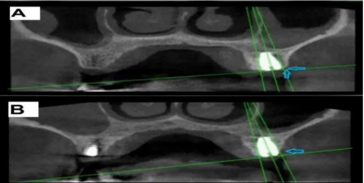
Determination of bone level immediately that represents as base line data was done for each implant in both group on two sides, buccal side and palata/lingual side, in first a vertical line was drawn at the center with the long axis of the dental implant passing through the notch of the cover screw which represent a reference point, then another horizontal line was drawn at the top of implant to determine its level with the crestal bone, also another two vertical lines along the buccal and palatal/lingual sides of implant were drawn where from these lines about 1mm the bone level was measured on each side in implants placed in posterior area when the bone thickness was enough, or about 0.5 mm in implants placed in anterior region, Fig. (1 A).
relations (the first one immediately after implant placement and the second one after six months) will be compared and the difference will be calculated which represents bone loss that showed as a shadow in the
area, Fig. (1 B).In order to view the same section in the CBCT & to avoid any errors that may affect the result in reading the section, the notch of the cover screw is used as a reference point.
Figure 1: CBCT (oblique view) for the same posterior DI placed with flapped procedure. (A) Immediate post-operative CBCT illustrating the buccal side bone level of DI (arrows) in relation to the horizontal line. (B) 24 weeks post-operative CBCT illustrating the difference in bone level (arrow) that presented as shadow
Table 1: Comparison according to total mean bone loss in buccal and palatal sides between two surgical groups
Sides
Total Bone Loss in Surgical Groups
P- Value Flapped Group
Mean ± SD Flapless Group Mean ± SD
Buccal Side 0.5 ± 0.43 0.39 ± 0.35 0.393
Palatal Side 0.13 ± 0.18 0.06 ± 0.14 0.214
Values are expressed in mean ± SD
Table 2: Comparison between Total Resorption of Buccal and Palatal sides
Group Buccal side Total Bone loss of Buccal and Palatal Sides P- Value
Mean ± SD Palatal side Mean ± SD
Flapped group 0.5 ± 0.43 0.13 ± 0.18 0.001
Flapless group 0.39± 0.35 0.06 ± 0.14 0.001
Values are expressed in mean ± SD
Results
There was no significant difference between study (flapless) and control (flapped) groups in the mean of total crestal bone resorption for buccal and palatal sides after 24 weeks from implant placement (P= 0.393 for buccal side and P= 0.214 for palatal side), Table (1). There was highly significant differences between buccal and palatal sides regarding crestal bone loss around implants measured by CBCT after 24 weeks from implants placement for both flapped and flapless surgical techniques (P = 0.001), Table (2).
Discussion
Based on the results obtained from this study, the choice of type of surgical approach does not affect peri-implant bone resoption. According to this study the measurement and evaluation were taken place before implants loading, so we can expect that significant difference between the two surgical techniques may occurs after implants loading and function. Our data on bone resoption are in line with the majority of data in the literature Lin et al [23].That reported no significant reduction of marginal bone resoption with flapless technique.
Interestingly, also studies showed that crestal bone loss was comparable among implants placed either using flapped or flapless surgical technique [24, 25].The findings of this study disagree with Tsoukaki et al [8].Who reported that no bone resoption around flapless implants and [26] who reported a significantly lower resoption around implants in flapless group. According to this study findings, there was significant difference between total mean of buccal and palatal aspects (P = 0.001) for both flapped and flapless group. There was obvious that bone resoption in buccal side much more than palatal / lingual side in both groups.
Also bone resoption in buccal side was a little higher in maxilla than mandible and anteriorly more than posteriorly for the flapped group, while for flapless group was nearly the same?
The data from this study can be accounted for the following reasons:
According to anterior maxilla the bone loss in buccal/facial side was higher than palatal side in both flapped and flapless groups, this may be related to the little thickness of the facial plate that leading to more bone resoption after implant placement. This was supported by EL Nahass & Naiem [27]. They reported that in the incisor region, the buccal bone plate around a tooth was thinner than 1 mm in 86% of the cases as demonstrated by Computerized tomography.
According to posterior maxilla and mandible in flapless group also the bone loss was higher in buccal side than palatal/lingual side, there was no clear interpretation for this result but after evaluation of the buccal plates for implants placed with flapless procedures, we noticed that most of them were thinner than palatal/lingual plates, this may be either they were initially thin or implant were deviated slightly towards buccal side during placement made them more thin. So it is, however noteworthy that thin buccal plates lost more bone than thick buccal plates. The deviation of implants supported by a study done by Van de Velde et al [28].
Performed an in vitro model study to analyze deviations in the position and inclination of implants placed with flapless surgery compared with the ideal, virtual planned position and they concluded that location of implants installed with a flapless approach differed significantly from the ideal position. The above two causes (1&2) were supported or in line with a hypothesis reported by [29, 30], stated that a thin buccal plate is less resistant to the different types of trauma an implant can endure and would therefore be more prone to resoption and buccal implant exposition.
According to posterior maxilla and mandible in flapped group the buccal bone loss was higher than palatal/lingual bone; this may be related to the flap elevation during surgical procedure on the buccal side and subsequent trauma that occurred more buccally rather than palatal/ lingual sides where there was no flap elevation. This was supported and in accordance with the fundamental studies reported by Merheb et al [30]. Stated that flap elevation lead to a bone resoption from the surgical trauma of up to 0.4 mm. Conclusions: Bone resoption around dental implants placed with conventional flap surgery compared to flapless surgery does not seem to be influenced during the healing period before implant loading. Bone resoption in buccal side facing dental implants compared to palatal side seems to be influenced in conventional flap and flapless surgery.
References
1. Kim JI, Choi BH, Li J, Xuan F, Jeong SM
(2009) Blood vessels of the peri-implant mucosa: a comparison between flap and flapless procedures. Oral Surgery, Oral
Medicine, Oral Pathology, Oral Radiology, and Endodontology, 1: 107(4):508-12.
2. Lei Q, Chen J, Jiang J, Fu X, Lin H, Cai Z
around implants in beagle dogs: flap surgery versus flapless surgery. Oral surgery, oral medicine, oral pathology and oral radiology, 1: 115(3):e21-7.
3. Albrektsson T, Zarb G, Worthington P,
Eriksson AR (1986) The long-term efficacy of currently used dental implants: a review and proposed criteria of success. Int. J. oral. maxillofac implants, 1(1):11-25.
4. Weber HP, Crohin CC, Fiorellini JP (2000)
A 5‐year prospective clinical and
radiographic study of non‐submerged
dental implants. Clinical Oral Implants Research, 11(2):144-53.
5. Guirado JL, Yuguero MR, Zamora GP,
Barrio EM (2007) Immediate
provisionalization on a new implant design for esthetic restoration and preserving crestal bone. Implant Dentistry, 1: 16(2):155-64.
6. Hürzeler M, Fickl S, Zuhr O, Wachtel HC (2007) Peri-implant bone level around implants with platform-switched abutments: Preliminary data from a prospective study. J Oral Maxillofac Surg., 65 (7): 1:33-9.
7. Sclar AG (2007) Guidelines for flapless
surgery. Journal of oral and maxillofacial surgery, 1: 65(7):20-32.
8. Tsoukaki M, Kalpidis CD, Sakellari D,
Tsalikis L, Mikrogiorgis G, Konstantinidis
A (2013) Clinical, radiographic,
microbiological, and immunological
outcomes of flapped vs. flapless dental implants: a prospective randomized controlled clinical trial. Clinical oral implants research, 24(9):969-76.
9. Campelo LD, Camara JR (2002) Flapless
implant surgery: a 10-year clinical
retrospective analysis. International
Journal of Oral & Maxillofacial Implants, 1: 17(2).
10.Matarasso S, Salvi GE, Iorio Siciliano V,
Cafiero C, Blasi A, Lang NP (2009) Dimensional ridge alterations following immediate implant placement in molar
extraction sites: a six‐month prospective
cohort study with surgical re‐entry.
Clinical oral implants research,
20(10):1092-8.
11.Blanco J, Liñares A, Pérez J, Muñoz F
(2011) Ridge alterations following flapless immediate implant placement with or without immediate loading. Part II: a
histometric study in the Beagle dog. Journal of clinical period ontology, 38(8):762-70.
12.Calvo‐Guirado JL, Gomez Moreno G,
Aguilar‐Salvatierra A, Mate Sanchez de
Val JE, Abboud M, Nemcovsky CE (2015) Bone remodeling at implants with
different configurations and placed
immediately at different depth into extraction sockets. Experimental study in dogs. Clinical oral implants research, 26(5):507-15.
13.Jeong SM, Choi BH, Li J, Kim HS, Ko CY,
Jung JH, Lee HJ, Lee SH, Engelke W (2007) Flapless implant surgery: an experimental study. Oral Surgery, Oral Medicine, Oral Pathology, Oral Radiology, and Endodontology, 1: 104(1):24-8.
14.You TM, Choi BH, Li J, Xuan F, Jeong
SM, Jang SO (2009) Morphogenesis of the
peri-implant mucosa: a comparison
between flap and flapless procedures in the canine mandible. Oral Surgery, Oral Medicine, Oral Pathology, Oral Radiology, and Endodontology, 1:107(1):66-70.
15.Lee DH, Choi BH, Jeong SM, Xuan F, Kim
HR (2011) Effects of flapless implant surgery on soft tissue profiles: a prospective clinical study. Clinical implant dentistry and related research, 13(4):324-9.
16.Reddy MS, Wang IC (1999) Radiographic
determinants of implant performance. Advances in dental research, 13(1):136-45.
17.Tyndall DA, Price JB, Tetradis S, Ganz
SD, Hildebolt C, Scarfe WC (2012) Position statement of the American Academy of Oral and Maxillofacial Radiology on selection criteria for the use of radiology in dental implantology with emphasis on cone beam computed tomography. Oral surgery, oral medicine, oral pathology and oral radiology, 1: 113(6):817-26.
18.Benavides E, Rios HF, Ganz SD, An CH, Resnik R, Reardon GT, Feldman SJ, Mah JK, Hatcher D, Kim MJ, Sohn DS, Palti A, Perel ML, Judy KW, Misch CE, Wang HL (2012) Use of cone beam computed tomography in implant dentistry: the International Congress of Oral Implantologists consensus report. Implant Dentistry, 21: 78-86.
19.Fienitz T, Schwarz F, Ritter L, Dreiseidler
of cone beam computed tomography in
assessing peri‐implant bone defect
regeneration: a histologically controlled study in dogs. Clinical oral implants research, 23(7):882-7.
20.Vera C, De Kok IJ, Chen W, Reside G,
Tyndall D, Cooper LF (2012) Evaluation of post-implant buccal bone resorption using cone beam computed tomography: a clinical pilot study. International Journal of Oral & Maxillofacial Implants, 1 (27): 5.
21.Schropp L, Wenzel A, Spin‐Neto R,
Stavropoulos A (2015) Fate of the buccal bone at implants placed early, delayed, or late after tooth extraction analyzed by
cone beam CT: 10‐year results from a
randomized, controlled, clinical study. Clinical oral implants research, 26(5):492-500.
22.Nickenig H-J, Wichmann M, Schlegel KA, Nkenke E, Eitner S (2010) Radiographic evaluation of marginal bone levels during healing period, adjacent to parallel-screw cylinder implants inserted in the posterior zone of the jaws, placed with flapless surgery. Clin. Oral. Impl. Res, 21 (I): 386-I393.
23.Lin GH, Chan HL, Bashutski JD, Oh TJ,
Wang HL (2014) The effect of flapless surgery on implant survival and marginal bone level: a systematic review and
meta‐analysis. Journal of periodontology,
85(5):e91-103.
24.Froum SJ, Cho SC, Elian N, et al (2011) Survival rate of one-piece dental implants placed with a flapless or flap protocol, a randomized, controlled study: 12-month
results. Int J Periodontics Restorative Dent, 31: 591-601.
25.De Bruyn H, Atashkadeh M, Cosyn J, et al (2011) Clinical outcome and bone preservation of single TiUnite implants installed with flapless or flap surgery. Clin Implant Dent Relat. Res, 13: 175-183.
26.Job S, Bhat V, Naidu EM (2008) In vivo
evaluation of crestal bone heights following implant placement with 'flapless' and' with-flap' techniques in sites of immediately loaded implants. Indian Journal of Dental Research, 1: 19(4):320.
27.El Nahass H, N Naiem S (2015) Analysis
of the dimensions of the labial bone wall in
the anterior maxilla: a cone‐beam
computed tomography study. Clinical oral implants research, 26(4):e57-61.
28.Van de Velde T, Glor F, De Bruyn H (2008)
A model study on flapless implant placement by clinicians with a different experience level in implant surgery. Clinical oral implants research, 19(1):66-72.
29.Teughels W, Merheb J, Quirynen M (2009)
Critical horizontal dimensions of
interproximal and buccal bone around implants for optimal aesthetic outcomes: a systematic review. Clinical oral implants research, 20:134-45.