Shape-Specific Hydrogel Nanoparticles with Defined Composition and Surface Properties for Gene Silencing
Stuart Scott Dunn
A dissertation submitted to the faculty of the University of North Carolina at Chapel Hill in partial fulfillment of the requirements for the degree of Doctor of Philosophy in the
Department of Chemistry
Chapel Hill 2012
Approved by
Joseph M. DeSimone
Wei You
Maurice S. Brookhart
Sergei S. Sheiko
ii Abstract
STUART SCOTT DUNN: Shape-Specific Hydrogel Nanoparticles with Defined Composition and Surface Properties for Gene Silencing
(under the direction of Prof. Joseph M. DeSimone)
acid-iii
labile system capable of targeting the transferrin receptor, endosomal escape, and delivery of siRNA.
iv
ACKNOWLEDGEMENTS
The author acknowledges those who contributed to and assisted with the work herein: Dr. Shaomin Tian for providing essential input into data analysis and planning siRNA experiments such that the entire project could have not been pursued without her skilled expertise in conducting all in vitro and in vivo work; Dr. Warefta Hasan for conversing about the siRNA projects and research directions in general; Dr. J. Christopher Luft for his insightful contributions to research discussions, in vitro work, and collaboration on additional endeavors; Dr. Steve Blake for his great contribution to the project and for synthesizing acid-labile PEGs; other group members who have been part of the siRNA team, including Dr. Jing Xu, Dr. Jin Wang, Luke Roode, Dr. Dorian Canelas, Xin Gao, and Dr. Charlie Bowerman; Liquidia siRNA team members aided significantly in the development of PRINT nanoparticles described herein for RNAi therapies.
v
science. Prof. T. Daniel Crawford delightfully taught Physical Chemistry and provided excellent mentorship on computational quantum chemistry research.
Professors at UNC who supplemented knowledge in organic and polymer chemistry as well as advances in drug delivery are much appreciated. Facilities and training for chemical and particle analysis were provided by Dr. Sohrab Habibi (mass spectrometry), Dr. Marc ter Horst (NMR), and Drs. Carrie Donley, Amar Kumbhar, and Wallace Ambrose (CHANL).
I highly value the extracurricular times at Chapel Hill, playing basketball, Frisbee, softball, and flag football with peers. It has been a privilege to sit next to Tammy Shen who made the past years such a pleasurable time while providing essential energy sources at the snack shack. Cathy Fromen continually made lab entertaining and had a pumpkin always filled with lovely goodies. Vicki Haithcock and Crista Farrell were very helpful, resourceful, and pleasant in enabling group operations. I would like to especially thank everyone in the Chemistry Department and DeSimone Lab for their kindness and hospitality in providing an enjoyable place to conduct research over the years.
vi
thanks to Prof. Mary Napier as well for always being open, understanding, and extremely insightful in discussing research and directions. Prof. Mary Napier was essential in pushing research forward, exploring new ideas, and staying focused on attainable paths.
vii
Table of Contents
Chapter 1: Introduction to Gene Therapy and RNA Interference………..1
1.1 Introduction to gene therapy and RNA interference……….... 1
1.1.1 Gene therapy and background on nucleic acids……….………... 1
1.1.2 Nucleic acids for gene therapy……….. 3
1.1.3 RNAi for silencing expression of genes in cells………... 8
1.2 RNAi for therapeutic applications and targeting diseases………... 9
1.2.1 Design of siRNA for effective gene knockdown……….. 9
1.2.2 Targeted diseases in the clinic for RNAi……… 11
1.3 Platforms for RNAi therapies………...13
1.3.1 Route of administration for delivery of RNAi therapeutics……… 13
1.3.2 In vivo barriers to systemic delivery of siRNA………... 14
1.3.3 Viral vectors for gene therapy in vivo………. 18
1.3.4 Non-viral, synthetic vectors for RNAi………... 19
1.4 PRINT technology and application to RNAi………. 24
1.4.1 Background to PRINT technology and applications in materials and life sciences……… 24
1.4.2 Incorporation of biologically-relevant cargos into PRINT particles for drug delivery……….... 30
1.4.3 Design of particles for gene silencing………. 32
References……… .………...34
viii
2.1 Introduction to hydrogels for delivery of siRNA………... 41
2.2 Cationic poly(vinyl pyrrolidone)-based, degradable hydrogel nanoparticles for delivery of siRNA……….. 43
2.2.1 Experimental………... 46
2.2.1.1 Materials………...46
2.2.1.2 Synthesis of bis(ethylene acrylate) disulfide………....46
2.2.1.3 Fabrication of PVP-based hydrogel nanoparticles………... 47
2.2.1.4 Characterization of hydrogels………. .47
2.2.1.5 Cell culture and assays………. 48
2.2.2 Activity of siRNA after exposure to UV light with photoinitiator: compatibility with particle fabrication conditions………... 49
2.2.3 Physicochemical characteristics of PVP-based hydrogels……….. 50
2.2.4 Dosing PVP hydrogels on HeLa cells for cell uptake, cytocompatibility, and gene silencing……… 51
2.3 Poly(ethylene glycol)-based cationic hydrogel nanoparticles for delivery of siRNA……….. 52
2.3.1 Experimental………... 53
2.3.1.1 Materials……….. 53
2.3.1.2 Fabrication of PEG-based hydrogels containing siRNA………. 54
2.3.1.3 Functionalization of PEG particles……….. 55
2.3.1.4 Characterization of hydrogel nanoparticles………. 55
2.3.1.5 Cell culture and confocal laser scanning microscopy………..…… 56
2.3.2 Fabrication and characterization of PEG-based nanoparticles………... 56
2.3.3 Delivery of siRNA via hydrogels PEGylated with imines………. 58
ix
2.3.5 Conclusions………. 63
2.4 Targeting cancer cells with ligand-conjugated hydrogels for gene silencing……… 63
2.4.1 Experimental………... 65
2.4.1.1 Materials……….. 65
2.4.1.2 Synthesis of carboxylic acid-functionalized 2,3-dimethyl maleic anhydride (CDM)………..………….. .65
2.4.1.3 Synthesis of CDM-PEG-biotin……….... 66
2.4.1.4 Synthesis of carboxylic acid-functionalized tetrahydrophthalic anhydride (CTA) precursor……….. 67
2.4.1.5 Synthesis of carboxylated tetrahydrophthalic anhydride-derivatized mPEG12……… 68
2.4.1.6 Fabrication and functionalization of hydrogel nanoparticles………... 69
2.5.1.7 Characterization of hydrogel nanoparticles………. 69
2.4.2 Functionalization of hydrogels for targeting transferrin receptor………... 70
2.4.3 Targeted delivery of siRNA to cancer cells via hydrogels conjugated with targeting and stealthing ligands through labile bonds………. 72
2.4.4 Conjugation of PEGylated ligands and stealthing groups to nanoparticles for targeted delivery of siRNA……….. 79
2.5 Optimization of rice-shaped hydrogel nanoparticles composition for gene silencing in hepatocytes………...85
2.5.1 Experimental………... 87
2.5.1.1 Materials……….. 87
2.5.1.2 Fabrication of cationic rice-shaped nanoparticles……… 87
2.5.1.3 Particle characterization, cell culture, and cell assay………... 88
x
2.6 Reversibly coating rice-shaped particles with terpolymer ligands
through polyelectrolyte attraction for targeting hepatocytes………... 91
2.6.1 Experimental………... 93
2.6.1.1 Materials……….. 93
2.6.1.2 Synthesis of ligands for stealthing particles and targeting hepatocytes………... 93
2.6.1.3 Characterization of ligands, cell assays, and coating cationic hydrogels with poly(acrylic acid)-based ligands……….. 95
2.6.1.4 Intravenous injection of nanoparticles and analysis of tissues……….... 97
2.6.2 Behavior of cationic nanoparticles coating with terpolymer ligands……….. 98
2.6.3 Evaluation of in vitro targeting hepatocytes with coated nanoparticles………....100
2.6.4 Factor VII gene knockdown in vitro by rice-shaped hydrogel nanoparticles………... 104
2.6.5 Injection of rice-shaped hydrogel nanoparticles into mice………..………. 108
2.7 Future directions for delivery of physically-entrapped siRNA in PRINT particles……….. 109
2.7.1 Range of particle matrices for encapsulation of siRNA and delivery to cells……….. 110
2.7.2 Cationic hydrogels……… 113
2.7.3 Ligands for reversible covalent conjugation………. 115
2.7.4 Ligands for electrostatic complexation to nanoparticles……….. 116
References……….. 117
Chapter 3: Reductively-Responsive siRNA-Conjugated Hydrogel Nanoparticles for Gene Silencing in Liver Hepatocytes………122
3.1 Introduction to siRNA conjugates for gene silencing……….. 122
3.2 Preparation of polymerizable siRNA macromers for covalent incorporation into hydrogel nanoparticles……… 123
3.2.1 Experimental………. 124
xi
3.2.1.2 Synthesis of siRNA macromers………. 125
3.2.1.3 Characterization of siRNA macromers and analysis of bioactivity………... 126
3.2.1.4 Particle characterization, analysis of siRNA by gel electrophoresis, and cell studies………... 129
3.2.2 Fabrication of hydrogels via PRINT process for covalent incorporation of siRNA……… 129
3.2.3 pro-siRNA hydrogels for gene silencing………..……… 131
3.2.3.1 Screening the amine content of hydrogel nanoparticles……….…………... 131
3.2.3.2 Implementing additional siRNA cargos into hydrogel nanoparticles to investigate gene silencing specificity……… 133
3.3 Targeting hepatocytes with PRINT particles………... 138
3.3.1 Experimental………. 139
3.3.1.1 Materials……… 139
3.3.1.2 Particle fabrication and characterization as well as cell studies……… 139
3.3.1.3 Functionalization of particles with lactoferrin and p17-derived peptide…………... 139
3.3.2 Lactoferrin conjugation strategies for enabling receptor-mediated endocytosis of 200 nm cylindrical hydrogels……… 140
3.3.3 Screening lactoferrin density on 80x180 nm hydrogels for targeting hepatocytes………. 143
3.3.4 Evaluating human lactoferrin-conjugated nanoparticles targeted to mouse hepatocytes in vitro……… 145
3.3.5 Targeting human and mouse hepatocytes with lactoferrin and peptide-conjugated nanoparticles compared to macrophages……… 147
3.3.6 Screening peptide density on hydrogel nanoparticles for targeting hepatocytes in vitro………..….. 150
3.4 Targeted PRINT particles for in vivo delivery to hepatocytes………. 153
xii
3.4.1.1 Materials……… 153
3.4.1.2 Fabrication, functionalization, and analysis of particles as well as in vitro cell studies……….. 153
3.4.1.3 In vivo studies……….153
3.4.2 Investigating the effect of covalent quenching on biodistribution of functionalized hydrogel nanoparticles………... 153
3.4.3 Electrostatic quenching of ligand-functionalized hydrogel nanoparticles for targeting hepatocytes………. 157
3.4.3.1 Evaluating noncovalent quenching of functionalized cationic hydrogels for targeting hepatocytes in vitro………... 157
3.4.3.2 Administration of hydrogel nanoparticles with different surface properties to mice to observe bioaccumulation in organs……….. 159
3.4.3.3 siRNA-conjugated hydrogel nanoparticles for in vivo gene silencing………...162
3.4.3.4 Targeting hepatocytes with ligand-functionalized hydrogel nanoparticles prepared with different reaction conditions………. 164
3.5 Recovery of siRNA throughout particle fabrication and pursuing scalability…………. 166
3.5.1 Experimental………. 167
3.5.1.1 Materials………...167
3.5.1.2 Particle fabrication and siRNA recovery methods……….………167
3.5.1.3 Fabrication of particles through scalable protocols………... 168
3.5.1.4 Particle characterization and cell studies………... 168
3.5.2 Recovery of siRNA in PRINT process………. 168
3.5.3 Scaling up pro-siRNA hydrogels of various dimensions for gene knockdown………. 170
3.5.3.1 Exploring fabrication conditions for open face curing particles……… 170
xiii
3.6 Future work for siRNA-conjugated, functionalized hydrogels………175
3.6.1 siRNA macromers with different degradable linkages………. 175
3.6.2 Additional matrices for siRNA-conjugated hydrogels………. 176
3.6.3 Endosmolytic coatings on nanoparticles for gene delivery ………. 177
3.6.4 Conjugation of siRNA post-particle fabrication……….……….. 178
3.6.5 Alternative delivery targets for RNAi………...179
References……….. 181
Appendix: Biodegradable Poly(ester)-Based, Rice-Shaped Nanoparticles For Gene Silencing in Cancer Cells………... 184
A.1 Introduction to biodegradable poly(ester)s for delivery of siRNA………. 184
A.2 PLGA and PCL particle matrices for gene silencing in cancer cells……….. 185
A.2.1 Experimental……… 186
A.2.1.1 Materials………186
A.2.1.2 Fabrication of siRNA-containing, lipid-coated PLGA and PCL nanoparticles……….. 186
A.2.2 PLGA particles………. 186
A.2.3 PCL particles……… 188
A.2.4 Future Work………..……... 190
xiv List of Tables
Table 1.1 Clinical trials for RNAi therapies………... 12
Table 1.2 Different routes of administration for RNAi therapies………... 13
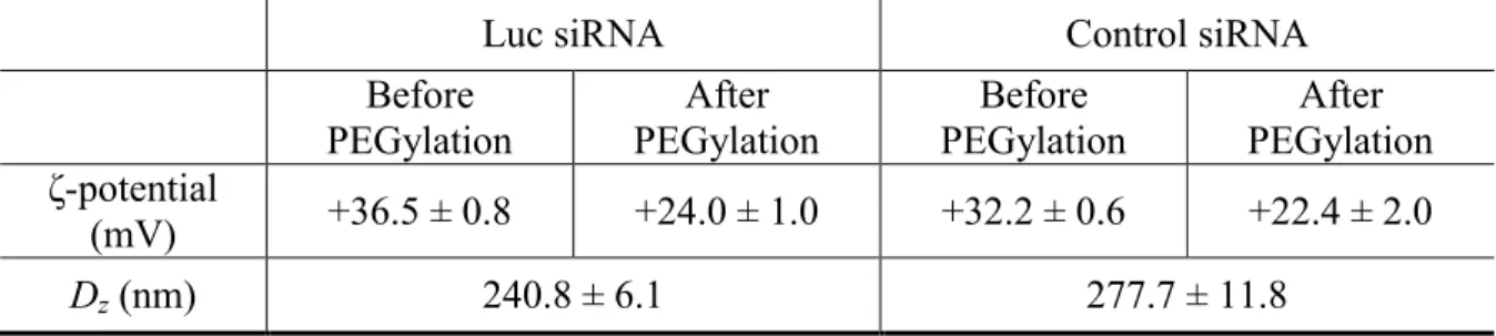
Table 2.1 ζ-potential and dynamic light scattering analysis of siRNA-charged, PEG-based particles before and after PEGylation………... 41
Table 2.2 Zetasizer analysis of siRNA-charged hydrogels before and after PEGylation………... 58
Table 2.3 ζ-potential and DLS of protein-functionalized nanoparticles………. 61
Table 2.4 ζ-potential of antibody-conjugated particles quenched with different maleic anhydrides……… 71
Table 2.5 ζ-potential analysis of functionalized particles after exposure to physiological and endosomal conditions………. 75
Table 2.6 Characterization of antibody-conjugated, TPA/NHS-mPEG12- quenched hydrogels………. 83
Table 2.7 Compositions of pre-particle solution for screening siRNA delivery efficiency………..88
Table 2.8 ζ-potentials of cationic hydrogel nanoparticles prepared with different amine-containing monomers……… 90
Table 2.9 Molecular weight (MW) analysis by MALDI-MS of galactose-PEG- NH2 with predicted and observed mass based on repeat units (n) of PEG……….. 96
Table 2.10 Zetasizer analysis of hydrogels dosed on hepatocytes……… 102
Table 2.11 ζ-potential of bare and ligand-coated cationic hydrogels……… 103
Table 2.12 Composition of cationic hydrogels for delivery of Factor VII………... 105
Table 2.13. Zetasizer analysis of hydrogels dosed on AML12 cells……….105
Table 3.1 Composition of pre-particle solution for fabrication of pro-siRNA hydrogels…..……….. 132
Table 3.2 Zetasizer analysis of pro-siRNA hydrogels with variable amine content………. 132
xv
Table 3.4 Zetasizer analysis of ligand-conjugated nanoparticles……….. 141
Table 3.5 Zetasizer analysis of functionalized 80x180 nm hydrogel nanoparticles………. 144
Table 3.6 Zetasizer analysis of functionalized hydrogels………. 147
Table 3.7 Zetasizer analysis of peptide and control ligand-conjugated particles………….. 151
Table 3.8 Zetasizer analysis of functionalized hydrogel nanoparticles……… 154
Table 3.9 Zetasizer analysis of functionalized 200 nm hydrogels……… 158
Table 3.10 Zetasizer and BCA analysis of nanoparticles injected into mice……… 159
Table 3.11 Zetasizer and BCA analysis of pro-siRNA nanoparticles for in vivo
gene silencing……….162
Table 3.12 Zetasizer and BCA analysis of functionalized nanoparticles……….. 164
Table 3.13 Compositions of pre-particle solution for fabrication of pro-siRNA
hydrogels……… 170
Table 3.14 Compositions of LED-cured pro-siRNA hydrogels……… 172
Table 3.15 Zetasizer analysis of LED-cured pro-siRNA hydrogels………. 172
Table 3.16 Zetasizer analysis of LED-cured 80x320 nm hydrogel nanoparticles………… 174
Table A.1 Zetasizer characterization of lipid-coated, PLGA rice………. 187
xvi List of Figures
Figure 1.1 Biological processes for the synthesis of proteins from DNA……… 2
Figure 1.2 Gene level triple-helix formation to impede transcription through
introduction of TFO, which binds to a Watson-Crick paired duplex……….…... 4
Figure 1.3 Illustration of intracellular gene silencing mechanism mediated by exogenously introduced synthetic siRNAs incorporated into the RNA-Induced
Silencing Complex (RISC)……… 8
Figure 1.4 Structures of oligonucleotide derivatives for enhanced stability,
mRNA binding, and intracellular delivery………...10
Figure 1.5In vivo barriers to systemic delivery of siRNA-containing
nanoparticles……… 15
Figure 1.6 Hepatic liver sinusoids with ca. 150 nm fenestrations……….. 17
Figure 1.7 Structure of adenovirus with knobs as cell receptor binding agents,
spheres as proteins, and duplexes as nucleic acids inside……… 18
Figure 1.8 Chemical structures of cationic polymers used for siRNA delivery………. 20
Figure 1.9 Non-viral polycation systems for effective in vivo RNAi………. 21
Figure 1.10 Structures of DOPE and DOTAP lipids as well as an illustration of a
multilamellar lipoplex with nucleic acid sandwiched between bilayers……….. 22
Figure 1.11 Lipid-based delivery vectors for effective RNAi in vivo………. 24
Figure 1.12 Illustration of PRINT process……….. 25
Figure 1.13 Range of particle shapes, sizes, and compositions accessible with
PRINT……….. 26
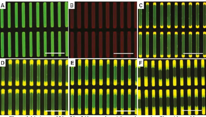
Figure 1.14. Array of 20 x 20 x 240 μm rod particles on harvesting film with
tunable dimensions in ABA triblock structures………... 27
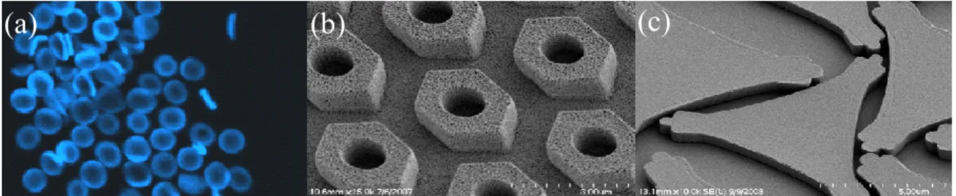
Figure 1.15 (a) Representative SEM image showing 96 wt % PEGDA
80 × 5000 nm particles………. 29
Figure 1.16 Controlled release of drugs from silyl ether prodrug-conjugated
nanoparticles……… 31
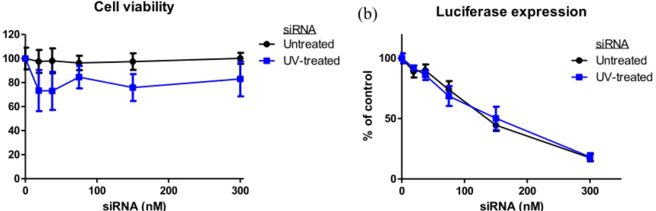
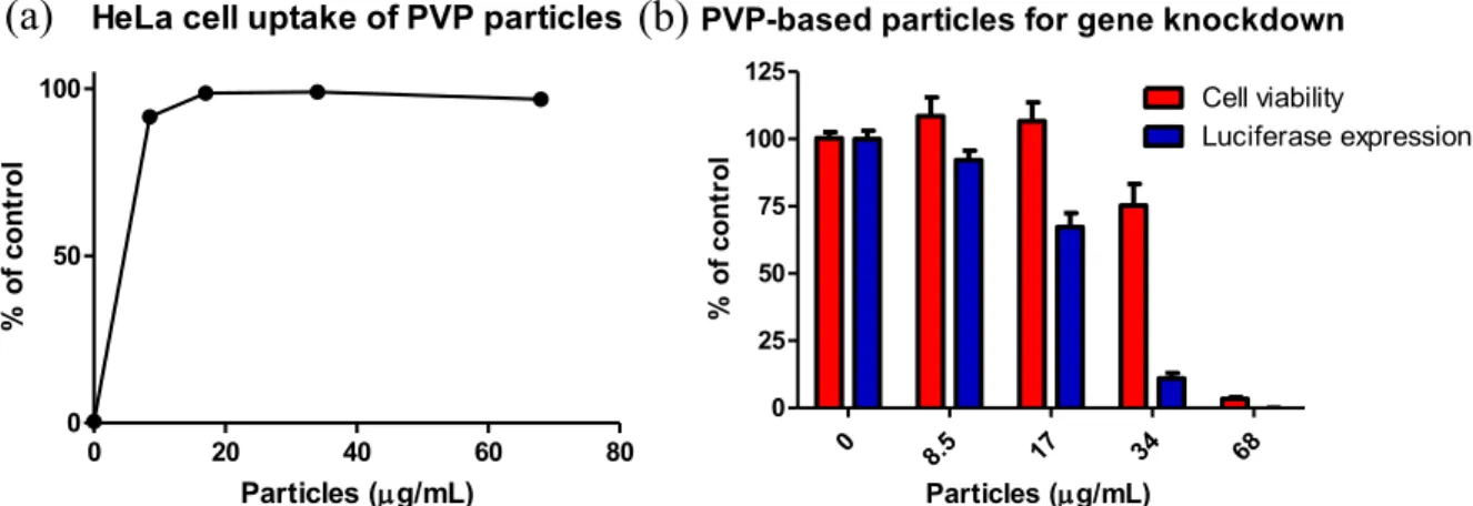
Figure 2.1 (a) Viability and (b) luciferase expression of HeLa cells dosed with
xvii
Figure 2.2 (a)Chemical structures of monomers implemented in PVP-based cationic hydrogel nanoparticles. (b) SEM micrographs of PVP-based particles
containing siRNA illustrate cylindrical morphology and 200 x 200 nm dimensions………..50
Figure 2.3 (a) Uptake of PVP-based particles by HeLa cells and (b) viability and luciferase expression of HeLa cells dosed with PVP particles for 4 h followed
by 72 h incubation at 37 ºC………... 51
Figure 2.4 (a) Chemical structures of monomers and macromonomers
implemented in cationic PRINT particles. (b) Reaction scheme for PEGylation of primary amine-containing hydrogels with aldehyde-terminated mPEG5K through imine formation. (c) SEM micrographs of cationic PEG-based hydrogels
illustrate cylindrical dimensions and soft qualities……….. 57
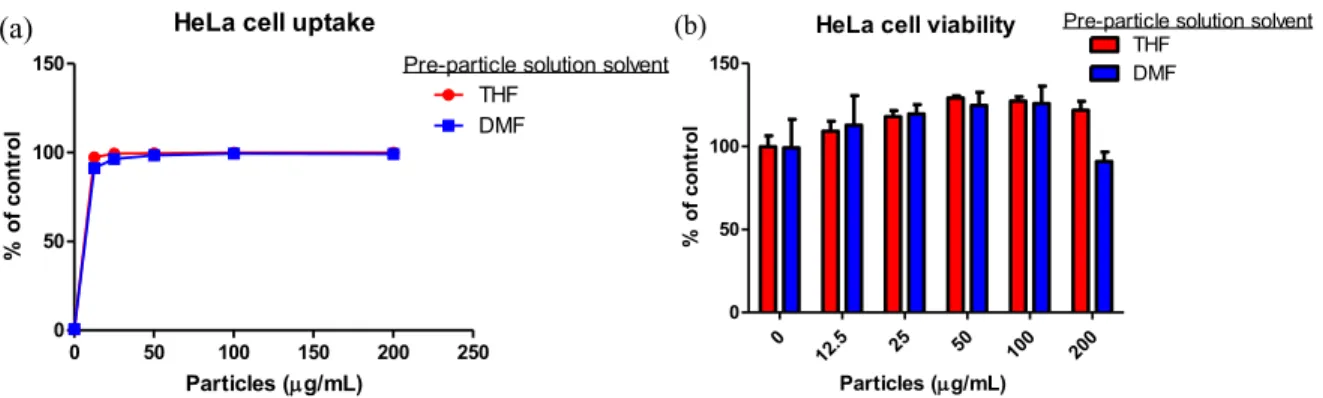
Figure 2.5 (a) HeLa cell uptake of PEG-based particles prepared from THF and DMF pre-particle solutions. (b) Viability of HeLa cells dosed with PEG-based
particles prepared from THF and DMF pre-particle solutions……… 57
Figure 2.6 SEM micrographs of PEG-based hydrogel nanoparticles confirms
cylindrical dimensions………. 58
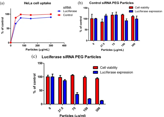
Figure 2.7 (a) HeLa cell uptake of PEG-based hydrogel nanoparticles after 4 h dosing time. Viability and luciferase expression of HeLa cells dosed with (b) control and (c) luciferase siRNA-containing nanoparticles for 4 h followed by
72 h incubation at 37 ºC………... 59
Figure 2.8 (a) Reaction scheme for the PEGylation of hydrogels with succinimidyl succinate monomethoxy PEG2K through amidation. (b) SEM micrographs of PEG-based hydrogel nanoparticles demonstrates cylindrical features, monodispersity, and 200 x 200 nm dimensions. (c) Gel electrophoresis analysis of the time-dependent release of siRNA from hydrogels incubated in
PBS at 2 mg/mL and 37 ºC……….. 60
Figure 2.9 (a) HeLa cell uptake of PEGylated, siRNA-containing hydrogel nanoparticles after 4 h dosing at 37 ºC in cell media. (b) Luciferase expression of HeLa cells dosed with luciferase and control siRNA-containing particles for 4 h followed by 72 h incubation at 37 ºC. (c) Viability of HeLa cell dosed with PEGylated, siRNA-containing hydrogels. Cells were dosed with particles for 4 h
and incubated for 72 h in media………... 62
Figure 2.10. Confocal micrographs of HeLa/luc cells dosed with 50 µg/mL particles containing (a) luciferase or (b) control siRNA cargos for 4 h. Cellular actin cytoskeleton was stained with phalloidin (red) and nuclei with DAPI (blue), while particles (green) were labeled with the fluorescent monomer fluorescein
xviii
Figure 2.11 Reaction scheme for multi-step preparation of targeted particles through conjugation of biotinylated proteins to amine-quenched, avidinated
particles……….... 70
Figure 2.12 HeLa cell uptake of (a) OKT9- and (b) IgG-conjugated hydrogel nanoparticles at multiple antibody:particle ratios and particle dosing
concentrations. HeLa cells were dosed with particles for 4 h at 37 ºC in media. Antibody:particle ratio symbolizes milliequivalents of protein per mg particle
(wt/wt)……….. 71
Figure 2.13 Reaction scheme for reversibly quenching amines with maleic anhydride derivatives, whose chemical structures are illustrated. Chemical structure of CDM-activated, biotin-terminated PEG2K is shown for the
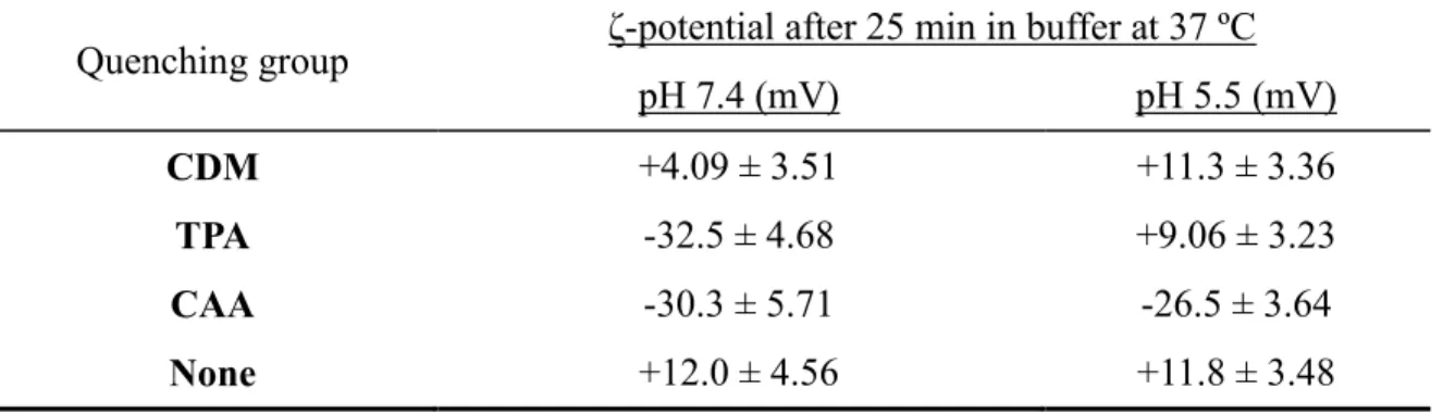
preparation of particles functionalized with acid-labile ligands.………. 73
Figure 2.14 HeLa cell uptake of OKT9- and IgG-conjugated particles quenched
with different maleic anhydrides………. 74
Figure 2.15 ζ-potential of CDM- and Ac2O-quenched particles as a function of
pH over 25 min……… 76
Figure 2.16 Viability and luciferase expression of HeLa cells dosed with (a) TPA- and (b) CDM-quenched, antibody-conjugated particles. (c) Gel electrophoresis of siRNA released from (A) PEGylated nanoparticles and
(B) transferrin receptor-targeted nanoparticles……… 77
Figure 2.17 (a) Gel electrophoresis of siRNA released from particles exposed to different reaction conditions after incubation in PBS at 37 ºC for 22 h. (b) Gel electrophoresis of siRNA remaining from particles loaded into wells after
incubation in water or KCl solutions of different ionic strength………. 78
Figure 2.18 Gel electrophoresis of siRNA released from particles functionalized
with different ligands after incubation in PBS at 2 mg/mL and 37 ºC……… 79
Figure 2.19 (a) HeLa cell uptake of OKT9- and IgG-conjugated particles quenched with CDM-mPEG12 after 4 h dosing time. (b) Viability and luciferase expression of HeLa cells dosed with CDM-masked, OKT9-conjugated particles
for 4 h followed by 48 h incubation in media at 37 ºC……… 80
Figure 2.20 (a) Establishment of wholly acid-labile system: functionalization of hydrogel nanoparticles with biotinylated targeting ligand and different amine masking agents. (b) Gel electrophoresis comparison of siRNA released after 48 h incubation from (A) cationic NP-mPEG2K, (B) CTA-mPEG12- and
xix
Figure 2.21 HeLa cell uptake of CTA-mPEG12-quenched particles after 4 h dosing expressed as (a) % control and (b) mean fluorescence intensity. (c) HeLa
cell uptake of NHS-mPEG12-quenched particles after 4 h dosing………... 83
Figure 2.22 Viability and luciferase expression of HeLa cells dosed with CTA-mPEG12-quenched particles containing control or luciferase siRNA conjugated with OKT9 or IgG antibodies for 4 h followed by 48 h incubation
in media……… 84
Figure 2.23 Viability and luciferase expression of HeLa cells dosed with NHS-mPEG12-quenched particles containing luciferase or control siRNA conjugated with OKT9 or IgG1 antibodies when dosed at 200 µg/mL for 4 h
followed by 48 h incubation in media………..85
Figure 2.24 Time-dependent release of siRNA from hydrogels after post- fabrication functionalization with targeting ligands when incubated at 2 mg/mL and 37 °C in PBS demonstrates loss of physically entrapped cargo (0.7 wt% encapsulated compared to 1.4 wt% originally encapsulated). Post-fabrication functionalization involved step-wise biotinyation, PEGylation, avidination, and conjugation of biotinylated protein to particles accompanied by multiple
washes of particles………... 85
Figure 2.25 Chemical structures of (a) primary amine and quaternary ammonium (meth)acrylate and (meth)acrylamide monomers and (b) new monomers
implemented in cationic hydrogel matrices. (c) SEM micrographs of select rice-
shaped hydrogel nanoparticles (30% AEM, 5 wt% siRNA)……….... 89
Figure 2.26 Viability and luciferase expression of HeLa cells dosed with AEM- based cationic hydrogel nanoparticles containing siRNA. Half-maximal effective concentration (EC50) of siRNA (nM) required for gene knockdown is based on
5 wt% siRNA charged into the pre-particle solution………... 90
Figure 2.27 Half maximal effective concentration (EC50) of siRNA (nM) required to reduce gene expression by 50%. For each particle sample, EC50s were based on
5 or 10 wt% siRNA loading into hydrogel nanoparticles……….... 91
Figure 2.28 (a) Structural breakdown of galactose-PEG-NH2 for predicting molecular weight as would be observed by MALDI-MS in Table 2.9.
(b) MALDI-MS spectrum of galactose-PEG-NH2……….. 96
Figure 2.29 Two-step reaction scheme for preparation of PAA-based terpolymers
with graphical illustrations………... 98
Figure 2.30 Illustration of coating cationic nanoparticles with PAA-based terpolymer ligands under physiological conditions followed by dissociation
xx
Figure 2.31 (a) ζ-potential of cationic nanoparticles as a function of ligand concentration, demonstrating ligand density-dependent surface properties. (b) ζ-potential of coated nanoparticles subjected to buffers of decreasing pH,
mimicking the progression of the endosome………. 100
Figure 2.32 (a) Time-dependent AML12 cell uptake of bare 80x320 nm particles and ligand-coated particles at 100 µg/mL dosing concentration. (b) Viability of AML12 cells dosed with cationic and ligand-coated particles at 48 h incubation
at 37 ºC………... 101
Figure 2.33 Time-dependent AML12 cell uptake of cationic particles coated with terpolymer ligands bearing different degrees of ligand substitution when
dosed at 100 µg/mL………... 103
Figure 2.34 HepG2 cell uptake of bare 80x320 nm hydrogel nanoparticles and ligand-coated hydrogels bearing different mol% of ligand after (a) 1 h and
(b) 24 h dosing times………..103
Figure 2.35 Gel electrophoresis analysis siRNA released from particles incubated
in 10x PBS………. 105
Figure 2.36 (a) AML12 cell uptake of bare and ligand-coated hydrogel
nanoparticles. (b) Viability of AML12 cells dosed with bare and ligand-coated, siRNA-containing nanoparticles. *Denotes that particles were washed in buffer
prior to dosing on cells………... 106
Figure 2.37 Confocal microscopy overlay images of AML12 cells incubated with cationic and coated hydrogels (tagged green with DyLight 488 maleimide). Nucleus was stained with DAPI (blue) and actin was stained with Phalloidin
(red)……… 107
Figure 2.38 qRT-PCR for relative FVII mRNA level in AML12 cells dosed with hydrogels containing FVII siRNA and LipofectamineTM 2000 mixed with either ApoB or FVII siRNA. Cationic rice particles or hydrogels coated with PAA- mPEG or -gal were dosed on cells and incubated for 48 h. *Denotes that particles
were washed in buffer prior to dosing on cells……….. 107
Figure 2.39 Confocal microscopy of tissues from mice injected with different nanoparticles: (a) PAA-glu and (b) PAA-gal with 30 mol% substitution of
saccharides, (c) cationic nanoparticles, and (d) no nanoparticles (PBS)………... 109
Figure 2.40 Structures of lipidated chlorosilanes, hydrophobic bischlorosilanes,
and dichlorosilanes where R may consist of different alkyl substituents……….. 111
xxi
Figure 2.42 Structures of amine-containing acrylic monomers that may be
purchased commercially or specially synthesized………. 114
Figure 2.43 Reaction schemes for the synthesis of amine-containing monomers……….... 114
Figure 2.44 Structures of amine-containing small molecules that may be used to synthesize acrylamide-based or thiol-presenting cationic monomers using acryloyl
chloride or thiirane. R = H or CH3………. 115
Figure 3.1 Analysis of native siRNA and siRNA macromers by reversed phase
HPLC………. 127
Figure 3.2 (a) Luciferase expression and (b) viability of HeLa/luc cells dosed with luciferase and control sequences of native (siRNA-NH2) and degradable siRNA prodrug (PD) complexed to Lipofectamine TM 2000 and incubated for 48 h. Retention of siRNA activity after macromonomer synthesis was confirmed
by evaluating transfection efficiency before and after siRNA derivatization……… 128
Figure 3.3 (a) Structures of degradable and non-degradable siRNA macromers as well as native siRNA, (b) Illustration of pro-siRNA hydrogel behavior under physiological and intracellular conditions, and (c) SEM micrograph of pro-
siRNA, 200 x 200 nm cylindrical nanoparticles (scale bar = 2 µm)………. 129
Figure 3.4 Release profiles and stability of siRNA in 30% AEM-based hydrogels. All hydrogels were washed with 10x PBS buffer to remove the sol fraction
containing unconjugated siRNA before release studies were performed……….. 130
Figure 3.5 Gel electrophoresis of siRNA released from hydrogels prepared with different amine monomer contents. Hydrogels were incubated in 10x PBS
containing 5 mM glutathione at 1.7 mg/mL for 4 h at 37 °C……… 132
Figure 3.6 (a) Luciferase expression and (b) viability of HeLa/luc cells dosed with cationic pro-siRNA hydrogels fabricated with different amine (AEM)
contents.………... 133
Figure 3.7 Gel electrophoresis of native siRNA-NH2 released from 30 wt% AEM-based hydrogels over time in PBS at 1 mg/mL and 37 ºC shows rapid
release of cargo……….. 134
Figure 3.8 Characterization of particle work-up and behavior after harvesting by gel electrophoresis. Particles prepared from 30 wt% AEM-based composition without dye were charged with native siRNA (NH2), disulfide pro-drug macromer (PD), and acrylamide macromer (AA) in addition to blank particles without
xxii
Figure 3.9 Gel electrophoresis analysis of siRNAs (abbreviation below) released from hydrogels incubated at 2.5 mg/mL for control knockdown studies under reducing conditions (5 mM glutathione, GSH) in 10x PBS for 4 h at 37 °C. lucAA1: acrylamide non-degradable luciferase siRNA; NH22: native amine luciferase siRNA; lucPD3: degradable luciferase siRNA prodrug; crtlPD4:
degradable control siRNA prodrug……… 135
Figure 3.10 Viability of HeLa/luc cells dosed with cationic hydrogels charged with different siRNA cargos. Cells were dosed with particles for 4 h followed by
removal of particles and 48 h incubation in media……… 136
Figure 3.11 30% AEM-based hydrogel particles charged with different siRNA cargos for transfection of HeLa cells. (a) Cellular uptake. HeLa/luc cells were dosed with particles for 4 h followed by trypan blue treatment and flow cytometry analysis. (b) Luciferase expression. HeLa/luc cells were dosed with particles for 4 h followed by removal of particles and 48 h incubation in media. Data in (a) and (b) represent one of two independent experiments. The error bars represent
standard deviation from triplicate wells in the same experiment. Note that all hydrogels were thoroughly washed after fabrication to remove non-conjugated siRNA in the sol fraction. (c)-(f) Confocal micrographs. HeLa/luc cells were dosed with 50 µg/mL hydrogels containing (c) luc PD (d) luc siRNA-NH2, (e) luc acrylamide, and (f) control PD siRNA cargos for 4 h. Cellular actin cytoskeleton was stained with phalloidin (red) and nuclei with DAPI (blue) while particles
(green) were labeled with the fluorescent monomer, DyLight 488 maleimide………. 137
Figure 3.12 Reaction scheme for preparation of ligand-conjugated hydrogels……… 141
Figure 3.13 AML12 cell uptake of ligand-conjugated particles expressed as
(a) % of control cells and (b) mean fluorescence intensity……… 142
Figure 3.14. AML12 cell uptake after dosing with 200 nm ligand-conjugated
particles expressed as (a) % of control cells and (b) mean fluorescence intensity………… 143
Figure 3.15 AML12 cell uptake of targeted particles incubated in
(a) OPTI-MEM or (b) complete medium………...145
Figure 3.16 (a) AML12 cell uptake (%) of hydrogel nanoparticles functionalized with hLTF or EtOHNH2. (b) Mean fluorescence intensity of AML12 cells (right)
dosed with functionalized particles……….... 146
Figure 3.17 SEM micrograph of 80x180 nm hydrogel nanoparticles………... 147
Figure 3.18 Cell uptake of ligand-conjugated particles. (a) AML12, (b) HepG2, and (c) RAW 264.7 cells were dosed with particles to investigate internalization
xxiii
Figure 3.19 (a)-(d) Confocal micrographs of AML12 cells dosed with ligand- conjugated particles. (a) Cells were not dosed with particles. (b)-(d) AML12 cells were dosed with (b) control particles, (c) human lactoferrin-conjugated particles, (d) peptide-conjugated particles. Actin was stained with Phalloidin (Alexa Fluor 555, red), nuclei were stained with DAPI (blue), and particles
were labeled with DyLight 488 maleimide……….... 149
Figure 3.20 Confocal micrographs of (a)-(c) RAW cells and (d) HepG2 cells dosed with ligand-conjugated particles. (a)-(c) RAW cells were dosed with (a) control particles, (b) human lactoferrin-conjugated particles, and(c) peptide- conjugated particles. (d) HepG2 cells were dosed with lactoferrin-conjugated particles. Actin was stained with Phalloidin (Alexa Fluor 555, red), nuclei were stained with DAPI (blue), and particles were labeled with DyLight 488
maleimide………... 150
Figure 3.21 (a) AML12 cell uptake of peptide-functionalized cylindrical nanoparticles. (b) Mean fluorescence intensity of AML12 cells dosed with
functionalized nanoparticles……….. 152
Figure 3.22 Images of liver sections from different mice dosed with 80x180 nm (a,b) unquenched, PEGylated particles, or (c,d) quenched, PEGylated particles. Nuclei were stained with DAPI (blue), actin was stained with phalloidin (Alexa Fluor 555, gray), macrophages were marked with MARCO (AlexaFluor 488,
green), and particles were labeled with DyLight 680 (red)………... 155
Figure 3.23 Images of liver sections from different mice dosed with (a,b) unquenched, lactoferrin-conjugated particles, or (c,d) quenched, lactoferrin- conjugated particles. Nuclei were stained with DAPI (blue), actin was stained with phalloidin (Alexa Fluor 555, gray), macrophages were marked with MARCO
(AlexaFluor 488, green), and particles were labeled with DyLight 680 (red)………... 156
Figure 3.24 AML12 cell uptake of functionalized hydrogels coated with PAA
(a) before and (b) after ligand conjugation……… 158
Figure 3.25 (a) Organ imaging of untreated mice and those injected with
nanoparticles for biodistribution analysis. (b) Fluorescence intensity of untreated
mice and those injected with nanoparticles………... 160
Figure 3.26 Liver histology from mice injected with cationic, PAA-mPEG- coated, and Lactoferrin-conjugated, PAA-coated nanoparticles (two images per particle sample). Particles were labeled with DyLight 488 maleimide (green), cell nuclei were stained blue with DAPI, and actin was stained red with
xxiv
Figure 3.27 Gel electrophoresis analysis of siRNA-containing nanoparticles. Particles were incubated in 5 mM glutathione-containing 10x PBS (0.05% PVA) at 2 mg/mL for 4 h followed by isolation of supernatants for loading into the gel. Encapsulation efficiency was calculated to be 28-33% for the samples listed.
Corresponding sample labels are provided in the Zetasizer table………..163
Figure 3.28 Relative FVII gene expression of mice without treatment or after administration of different hydrogel nanoparticles. n.s. not significant;
** P < 0.01; t-test, double-tailed, n = 3………. 163
Figure 3.29 Uptake of functionalized hydrogel nanoparticles by AML12 cells
shown as (a) percentage of cells with particles and (b) MFI of cells……….165
Figure 3.30 Gel electrophoresis of siRNA recovered from delivery sheet and washed from particles. (1-3) siRNA standard of 50, 100, and 200 ng; (4-5) delivery sheet contents after filling the mold; (6) sol fraction of siRNA macromer
recovered in 10x PBS……….168
Figure 3.31 siRNA released from hydrogels charged with recovered siRNA that
were incubated in GSH-containing 10x PBS for examining cargo release………... 170
Figure 3.32 Release of siRNA from hydrogels cured under different conditions, which were incubated at 0.5 mg/mL in 10x PBS containing glutathione for 4 h at
37 °C……….. 171
Figure 3.33 Gel analysis of particles incubated in 10x PBS (5 mM) at 37 °C for 4 h illustrates that that encapsulation efficiency appeared to be high for all
samples (ca. 75% or greater except ca. 55% for 5 wt% siRNA-charged particles)………... 172
Figure 3.34 (a) HeLa cell uptake and (b) viability, and (c) luciferase expression
after dosing with particles containing different siRNA loadings………..173
Figure 3.35 (a) Viability and (b) luciferase expression of HeLa cells dosed with rice-shaped, pro-siRNA hydrogel nanoparticles fabricated through scalable
procedure………174
Figure 3.36 Synthetic routes to siRNA pro-prodrug macromers containing degradable linkages. (a) 2’-hydroxyl groups of the backbone or the (b) 5’ terminal alcohol of the oligonucleotide may be reacted with acrylate-functionalized chlorosilanes. (c) Amine-terminated siRNA may be reacted with an NHS- activated chlorosilane to yield a pendant silyl ether prodrug. (d) siRNA may be end-functionalized with a NHS-activated, methacrylated Cathepsin-B cleavable
peptide for use as a prodrug………... 175
xxv
Figure 3.38 Structures of monomers that may be used to conjugate siRNA post-
particle fabrication and corresponding bonds formed with siRNA………... 179
Figure A.1 Structures of poly(glycolic acid), poly(lactic acid), and
poly(ε-caprolactone)……….. 184
Figure A.2 SEM micrographs of (a) irrelevant and (b) anti-luciferase siRNA-
loaded lipidated PLGA rice………... 187
Figure A.3 Viability and luciferase expression of HeLa cells dosed with lipidated PLGA rice containing (a) control or (b) anti-luciferase siRNA for
4 h followed by 72 h incubation at 37 ºC in media……… 187
Figure A.4 SEM micrographs of (a) control and (b) anti-luciferase siRNA-
containing lipidated PCL rice……… 188
Figure A.5 (a) Luciferase expression and (b) viability of HeLa cells dosed with particles coated with increasing amounts of DOTAP:DOPE and at increasing particle concentration for 4 h followed by 72 h incubation at 37 ºC
in media. EC50s were calculated based on 100% encapsulation efficiency………... 189
Figure A.6 Retrosynthetic schemes toward siRNA-PCL pro-drug conjugates
xxvi
List of abbreviations ACN – acetonitrile
AEM – 2-aminoethylmethacrylate hydrochloride AON – antisense oligonucleotide
DCM – dichloromethane
DMF – N,N-dimethylformamide DLS – dynamic light scattering DMSO – dimethylsulfoxide DNA – deoxyribonucleic acid
DOPE – 1,2-dioleoyl-sn-Glycero-3-Phosphoethanolamine
DOTAP – N-[1-(2,3-Dioleoyloxy)propyl]-N,N,N-trimethylammonium chloride Dz – Z-average diameter
EC50 – half maximal effective concentration
EDC – 1-ethyl-3-(3-dimethylaminopropyl) carbodiimide EtOAc – ethyl acetate
HeLa – human cervical adenocarcinoma cell line HPLC – high performance liquid chromatography kDa – kilodaltons (1,000 g/mol)
NHS – N-hydroxysuccinimide NIPAM – N-isopropylacrylamide PBS – phosphate buffered saline PEG – poly(ethylene glycol) PFPE – perfluoropolyether
PLGA – poly(lactic-co-glycolic acid)
xxvii PVA – poly(vinyl alcohol)
RES – reticuloendothelial system
CHAPTER 1
INTRODUCTION TO GENE THERAPY AND RNA INTEFERENCE
1.1Introduction to gene therapy and RNA interference
1.1.1 Gene therapy and background on nucleic acids
Diseases and cancers may be treated with gene therapy through the introduction of nucleic acids into target cells to modulate gene expression and remedy cellular malfunctioning. When errors arise in the production or processing of nucleic acids, propagation of genetic mutations can lead to diseases that pose serious threats to health. Cancer has received the most attention in clinical trials worldwide, comprising 64.7% of all trials while the next most commonly treated diseases are monogenic (8.5%) and cardiovascular (8.4%).1 Examples of monogenic diseases include cystic fibrosis, haemophilia, and severe combined immunodeficiency disorder. In addition to monogenic and polygenic (e.g. cancer) diseases, gene therapy efforts have been focused on infectious diseases (HIV/AIDS), Alzheimer’s disease, and Parkinson’s disease in clinical trials.
2
acids are polynucleotides (linear polymers of nucleotides) linked 3’ to 5’ via phosphodiester bridges (Figure 1.1). There are five bases present in nucleic acids, where one is unique to DNA (thymine) and the other to RNA (uracil). The bases provide inter-chain hydrogen bonding that dictate structure, e.g. the double helix. DNA and RNA are the two natural forms of nucleic acids that are responsible for information storage (due to greater stability) and expression of the code (due to catalytic capabilities), respectively. An exception to this statement can be noted in some viruses that store their genetic information as RNA.
DNA has one critical biological role: carry and protect genetic code in chromosomes to provide information for generating all biofunctional macromolecules. Conversely, RNA can be seen in multiple copies, forms, and functions with one crucial role being the transfer of DNA to protein. Nucleic acid processing involves intricate pathways (Figure 1.1),1 which play a role in essentially all cellular metabolic processes. DNA replication may yield an identical molecule, e.g. during cell division, while transcription is carried out by RNA polymerase to create complementary
3
base sequences to DNA in a single-stranded messenger RNA (mRNA). Three-base codons on mRNA define a particular amino acid and the sequence of a protein through translation. Transfer RNAs carry amino acids and recognize the mRNA codons for synthesis of proteins in the ribosome. Biological processes proceed through the transfer of information, starting from DNA, moving to mRNA, and then to protein, that governs biological behaviors and functions.
1.1.2 Nucleic acids for gene therapy
To fix malfunctions in genetic processing, nucleic acids may be delivered to restore normal cellular behavior that alleviates the disease. Currently, some of the most commonly delivered nucleic acids are plasmid DNA (pDNA), antisense oligonucleotides (AONs), and small interfering RNA (siRNA), each with unique repairing mechanisms. First, pDNA is an extrachromosomal form of DNA that generally exists as double stranded and ranges from 1-200 kilobase pairs, adopting open circular, linear, or supercoiled conformations. Once inside the cytoplasm, pDNA must travel to the nucleus by crossing the nuclear pore membrane for its expression. In therapeutic applications, pDNA must efficiently insert the critical genes to be expressed at specific chromosome locations within the genome to ultimately produce desired proteins. Several gene therapies based on plasmids have been approved and are in clinical trials, some designed to cure severe combined immunodeficiency, neuroblastoma, or cystic fibrosis by replacing or repairing defective genes while cancer may be treated through expression of tumor suppressing genes.2
4
area with the discovery of the natural role of antisense RNA in gene regulation and modification, thereby promoting its potential to be harnessed for therapeutics.4 Antisense therapies can be envisioned to take action at the gene or mRNA level. Single stranded triple-helix-forming oligodeoxynucleotides (TFOs) are an example of gene level regulators: they halt transcription by binding to the major groove of duplex DNA through Hoogsteen bonds (Figure 1.2) in purine- or pyrimidine-rich sequences, ultimately preventing the unwinding of DNA. TFOs can also induce mutations that repair an inherited or acquired defective gene by activating the cellular nucleotide excision repair system.5,6 A requirement for successful TFO targeting and hybridization is that 10-30 nucleotide stretches of pyridines on one strand with pyrimidines on the other must be present, which makes TFO methods less appealing.
Figure 1.2 Gene level triple-helix formation to impede transcription through introduction of TFO, which binds to a Watson-Crick paired duplex.7
5
factors or proteins11 involved in diseases. However, aptamers are sometimes limited by the requirement for local administration: systemic in vivo delivery would impair proteins in untargeted organs; still, there is hope with new delivery technologies.
Now, therapeutics working at the mRNA level will be addressed: stoichiometrically, it seems less effective than attacking the genetic “source” (DNA); however, mRNA is more susceptible to attack since it is single-stranded and unprotected in transcription, transport from nucleus to cytoplasm, and translation. Once again, decoys (RNA now) may be employed to compete with protein binding sites that serve as translational activators or mRNA stabilizers.12,13 Consequently, decoys can prevent translation or induce mRNA instability, leading to its degradation. More commonly, antisense approaches are taken to interfere with gene expression by providing a nucleic acid with a complementary sequence to the mRNA of interest for hybridization and subsequent treatment. Direct sequence-specific cleavage of transcripts may be achieved with ribozymes or DNAzymes that bind to target RNA through Watson-Crick base pairing and contain a catalytic moiety that cleaves the hybrid.14,15
6
less toxic and show high targeting specificity.18 Nevertheless, degradation of mRNA through RNAse H cannot be triggered and translation of mRNA is generally blocked by duplex formation; therefore, silencing efficiency is relatively low. Not directly binding mRNA, antisense 2’-O-methylribooligonucleotides can be targeted against specific mutated sequences in pre-mRNAs to restore correct splicing of RNAs in vitro; this has been seen in targeting β-globin pre-mRNA to treat β-thalassemia and prevent anemia.19
micro RNA have only limited complementarity (in the seed region) to the target mRNA, resulting in repression of translation and non-sequence-specific mRNA degradation.20 Design of AONs is constantly improving as can be seen with recent developments of peptide nucleic acids21 and morpholino deoxynucleotides,22 which have significant modifications to the backbone and nucleotide structures (bases are maintained). These highly derivatized AONs exhibit complete nuclease resistance and a translation-blocking mechanism of the target instead of activating RNAse H for target degradation.
7
Regardless of the therapeutic nucleic acid identity and function, the cargo must be delivered to the cells of interest, which often proves to be a challenge. To facilitate delivery of therapeutic payloads into target tissues, cells, and intracellular compartments, delivery vehicles or external devices are often implemented.
External devices that utilize electric currents include electroporation, which involves applying an electric field to permeabilize cell membranes, and iontophoresis, which uses a small electric current to deliver charged molecules through skin and tissue. Ultrasound therapy has been used along with microbubbles in sonoporation to permeabilize membranes for gene delivery. The gene gun was invented to transfect DNA-coated nanoparticles into cells to express genes, which has been used in vaccine and plasmid delivery.
8
1.1.3 RNAi for silencing expression of genes in cells
After covering different therapeutic nucleic acids and their functions for regulating gene expression, notable highlights for siRNA include simple delivery to the cytoplasm (instead of the nucleus), harnessing the natural pathway of RNA interference (RNAi), high potency with catalytic activity, minimization of adverse side effects (that could be caused with drugs), and the ability to theoretically target any gene of interest. Avoiding delivery to specific intracellular compartments (e.g. vesicles or the nucleus) enables rather facile intracellular delivery of siRNA. The post-transcriptional gene regulating phenomenon known as RNAi provides innate defense against invading viruses and transposable elements.24 In 1998, Fire and Mello discovered RNAi in the nematode Caenorhabditis elegans25 for which they received the Nobel Prize in Physiology or Medicine in 2006. Next, Tuschl and coworkers demonstrated that synthetic, 21- to 22-nucleotide long double-stranded RNAs are sequence-specific mediators for RNAi.26
Figure 1.3 Illustration of intracellular gene silencing mechanism mediated by exogenously introduced synthetic siRNAs incorporated into the RNA-Induced Silencing Complex (RISC).
Adapted from Alnylam, Inc.
Synthetic siRNAs can be designed to target a specific gene with minimal off-targeting effects. After introduction into the cytoplasm of a cell, siRNA becomes incorporated into the
9
RNA Induced Silencing Complex (RISC), which unwinds the double-stranded siRNA such that the antisense strands then targets its complementary mRNA sequence (Figure 1.3). Once the antisense strand of siRNA binds to mRNA in the RISC, the mRNA is degraded in the cell, preventing its translation and the expression of a target protein. siRNA acts catalytically by incorporating into the RISC, whose endonuclease activity is directed by the siRNA, and should minimally shutdown off-target genes due to the extensive complementarity required between the mRNA and siRNA.27 Multiple siRNA libraries exist in literature and industry for addressing different gene families and functions.
1.2 RNAi for therapeutic applications and targeting diseases
1.2.1 Design of siRNA for effective gene knockdown
10
Modifications to siRNA and antisense oligonucleotides have been explored to alter bioavailability, serum stability, and transfection. Chemical structures of different oligonucleotide chemistries focus on backbone and substituent modifications (Figure 1.4). For example, phosphorothioate (PS) backbones and 2’-O-methyl substituents increase resistance to degradation by nucleases and enhance binding to target RNAs.30 Locked nucleic acids (LNA) also increase binding to target mRNA and phosphorodiamidate morpholino oligomers (PMO) yield neutral oligonucleotide analogs that are extremely stable in the presence of nucleases. Positively charged piperazine, amino, and arginine-containing PMOs offer enhanced cellular internalization and transfection. Modifications to siRNA that enhance resistance to nuclease stability may not increase the duration of gene silencing, but rather protect siRNA from degradation in the extracellular environment.
Figure 1.4 Structures of oligonucleotide derivatives for enhanced stability, mRNA binding, and intracellular delivery.30
11
really depend on cell division: unmodified siRNA achieved gene silencing that lasted roughly a week in rapidly dividing cells while knockdown persisted for a month in slowly dividing fibroblast cells.32 To confirm gene silencing kinetic mechanisms, cells with low proliferation were investigated: prolonged RNAi was observed in primary macrophages33 and mammalian cells.34 After optimizing sequence specificity and stability of siRNAs, in vivo efficacy mainly depends on effective delivery to the target tissue and cell. Design of delivery vectors and devices plays a crucial role in influencing biodistribution, cell uptake, and delivery to the cytoplasm. First, one must begin with the disease at hand to develop delivery systems before determining the best route of administration.
1.2.2 Targeted diseases in the clinic for RNAi
12
efficient in gene transmission into human hematopoietic cells, resulting in inhibited cell growth and increased sensitivity to imatinib, a tyrosine kinase inhibitor.35
For the injury of kidney acute renal failure, Quark Pharmaceuticals intravenously injected naked siRNA (I5NP) to inhibit the expression of the pro-apoptotic protein, p53. Dose-dependent attenuation of apoptotic signal was observed where analysis of renal histology and apoptosis revealed improved injury scores in both cortical and corticomedullary regions. Furthermore, this siRNA to p53 was also effective in a model of cisplatin-induced kidney injury.37
Table 1.1 Clinical trials for RNAi therapies.35
Disease Sponsor Drug Target Status
Macular degeneration
Opko Health,
Inc. Bevasiranib VEGF Phase II
Glaucoma Sylentis SYL040012 β2 adrenergic receptor Phase II
Solid tumor Calando Pharm. CALAA-01 RRM2 Phase I
Metastatic
melanoma Duke University NCT00672542
LMP2, LMP7,
and MECL1 Phase I
Liver cancer NCI TKM-080301 PLK1 gene
product Phase I Hepatitis B
virus
Nucleonics,
Alnylam Pharm. NUC B1000 HBV mRNA Phase I Hepatitis C
virus Santaris Pharma SPC3649 miR-122 Phase II
Respiratory syncytial
virus
Alnylam Pharm. ALN-RSV01 RSV N-gene Phase II Kidney injury Quark Pharm, Inc. 1002/I5NP QPI- p53 Phase I
13
not attained. Baseline treatment with VEGF may be required first to take care of pre-existing VEGF to realize therapeutic value for Cand5.38 To attain RNAi efficacy in clinical trials, platform technologies are under development to enhance delivery of siRNA.
1.3 Platforms for RNAi therapies
1.3.1 Route of administration for delivery of RNAi therapeutics
Accessibility to diseased tissue by siRNA determines the route of administration, which broadly includes local, topical, and systemic. Localized delivery of siRNA involves direct application to the accessible tissue of interest. Administration of siRNA to the skin comprises topical modes of delivery. In tissues which are not accessible directly or through topical application of RNAi therapies, systemic administration becomes necessary. Systemic delivery concerns the intravenous injection of therapeutics, which take on the intravascular journey, encountering in vivo barriers en route to the target tissue and cell. Select in vivo studies implementing local, topical, and systemic delivery approaches are outlined as therapeutic interventions in Table 1.2.
Table 1.2 Different routes of administration for RNAi therapies.
Route of
administration Disease Organ siRNA target
Electroporation39 Cancer Prostate VEGF
Iontophoresis40 Atopic dermatitis Epidermis IL-10
Ultrasound41 Yolk sac carcinoma Tumor MDR1
Topical42 Inflammatory bowel Rectum CCR5
Inhalation43 Cancer Lung PI 3-kinase, NF-κB
Topical44 Angiogensis Eye TLR3
Intracranial45 Alzheimer Brain BACE1
14
As a form of local delivery, electroporation involves application of controlled electric fields to permeabilize cells for the introduction of siRNA. Implementing “plate and fork” electrodes, efficient delivery of siRNA in vivo through electroporation was achieved to suppress growth of prostate cancer tumors.39 Application of ultrasound to gas-encapsulated microbubbles enables sonoporation (permeabilization of cells with sound), which can be harnessed for gene therapy. Perfluoropropane gas-encapsulated, PEGylated bubble liposomes were combined with siRNA and ultrasound was applied to enable gene silencing in vitro and in vivo.47 Inhalation of RNAi therapies offers a unique way to reach previously untreatable respiratory conditions. Inflammatory and immune conditions, cystic fibrosis, infectious diseases, and cancer may be tackled with RNAi therapies through inhalation.48
Alnylam has completed Phase 2b clinical trials on a novel RNAi antiviral therapeutic directed against respiratory syncytial virus. Developing systemically-administrable siRNA delivery platforms that enable tissue- and cell-targeting capabilities without immunogenicity or toxicity should offer translation to other routes of administration. In search of robust RNAi delivery platforms, nanoparticles present opportunities to enhance stability, increase bioavailability, target tissues and cells, and optimize transfection through combinatorial material screening.
1.3.2 In vivo barriers to systemic delivery of siRNA
15
filtration, phagocytosis, and degradation in the bloodstream, (B) extracellular matrix diffusion, (C) cellular receptor targeting and uptake, (D) endosomal escape, (E) siRNA release or dissociation from the carrier, and (F) intracellular trafficking to the RISC to degrade mRNA as illustrated in Figure 1.5. Delivery of naked siRNA may be performed; yet, it is highly susceptible to rapid clearance and nuclease degradation, showing low cell uptake in systemic delivery.
Figure 1.5In vivo barriers to systemic delivery of siRNA-containing nanoparticles. Administration of nanoparticles, which must: A, avoid clearance and degradation in serum; B, travel across the vascular endothelium; C, diffuse in the extracellular matrix to target cells
16
Hydrodynamic injection of naked nucleic acids may overcome some barriers by using a large volume of high pressure solution; however, potential side effects like high blood pressure, low heart rate, and death tarnish the appeal. Encapsulation of siRNA in delivery vehicles may allow for improved efficiency, stability, and cell specificity while avoiding immune responses. Methods to obtain proper packaging of siRNAs, stability, resistance to nuclease degradation, and efficient transfections are constantly being developed. Ideal nanoparticle features for RNAi in liver or tumor targets include small size (< 150 nm), effective siRNA loading and stability, surface decorations like PEG and targeting ligands, dispersion stability in serum, endosomolytic (endosome lysing) behavior, and effective unpackaging of siRNA cargo in the intracellular environment.
17
upon nanoparticles, which must be below the size of fenestrations to effectively reach liver parenchymal cells (hepatocytes).
After diffusing through extracellular space to the target cell, ligand-conjugated nanoparticles may bind to receptors to undergo receptor-mediated endocytosis whereby the cell internalizes the vector into an endocytic vesicle. Depending on the cell line and individual species, approximately a thirty minute window exists for the nanoparticle to escape the endocytic vesicle before lysosomal fusion occurs in which degradation of contents by proteases and nucleases will occur. Often amine-containing materials are implemented in nanoparticle formulations to escape the endosome by harnessing the so-called “proton sponge effect”. Progression of the endosomal vesicle over time is accompanied by decreasing pH, proceeding from physiological values to ca. pH 5.5 at the late-stage of the endosome’s lifetime. Ionizable groups that have pKa values ranging from physiological to endosomal values are capable of buffering the decreasing pH of the endosome, resulting in an influx of protons and chloride ions to maintain the pH of the endosome. Resulting, osmotic pressure may increase such that the endosome becomes unstable and ruptures, releasing its contents into the cytoplasm. Alternatives to exploiting the “proton sponge effect” include use of membrane-lysing or -fusing species as well as pore-forming peptides. In the process of or after endosomal escape, nanoparticles may release siRNA into the cytoplasm where it incorporates into the RISC to silence gene expression by binding to and triggering degradation of target mRNA.
18
1.3.3 Viral vectors for gene therapy in vivo
Produced by nature, viruses are highly evolved nanoparticles of uniform size that are coated by a protein shell and contain RNA- or DNA-based genes, which are responsible for diseases ranging from minor afflictions such as chickenpox or cold sores to major ones like acquired immune deficiency syndrome or severe acute respiratory syndrome. The distinct shapes (e.g. icosahedral, helical, or enveloped) and sizes of viruses provide superior abilities to infect organisms. Noting their efficiency for inducing disease, viruses may be captured and modified to create viral vectors or studied to design synthetic vectors, both aimed in opposition to the purpose of viruses: curing disease.
Figure 1.7 Structure of adenovirus with knobs as cell receptor binding agents, spheres as proteins, and duplexes as nucleic acids inside.51
19
through the nuclear pore complex.53 A lentiviral vector was created for delivery of siRNA targeting BACE1 in a transgenic model that ameliorated Alzheimer disease neuropathy.45
Considering the aforementioned viral characteristics, viral vectors can overcome the major biological hurdles; however, significant drawbacks still are present. Specifically, they can be difficult to mass-produce, immunogenic (toxic), and limited to low cargo capacity.54 Some have been noted for their potential to induce mutagenic integration.55 Tragically, viral vector gene therapies have resulted in death: the first was in 1999 with a patient suffering from the inability to metabolize ammonia where the vector triggered an immune response that led to multiple organ failure and brain death;56 another involved the treatment of a healthy patient for rheumatoid arthritis in 2007.57 Serious risks are present in gene therapy; still, instead of these tragedies being research deterrents, they may be viewed as warnings to proceed with extreme caution before implementing a system that has not been rigorously tested. Furthermore, viral vectors may be investigated to understand how tragedy may be prevented while extracting their features for delivery. An alternative to viral vectors for delivery of therapeutics involves synthetic vectors.
1.3.4 Non-viral, synthetic vectors for RNAi
20
later. Some of the most common carrier molecules for condensing and protecting siRNA are cationic lipids and polymers, which yield lipoplexes and polyplexes, respectively. One of the most commonly employed polycations for nucleic acid delivery is poly(ethylene imine) (PEI), illustrated in Figure 1.8, due to its high gene transfection activity. Molecular weight and structure (linear or branched) of PEI substantially impact toxicity and transfection.61 The mechanism for transfection by PEI has been attributed to the ‘proton sponge effect’: amine groups provide pH buffering in the endosome to maintain neutrality and the influx of ions causes osmotic swelling and bursting of the endosome, resulting in a release of its contents into the cytosol. Thereafter, the polyplex may dissociate so that the polymer may be processed while siRNA incorporates into the RISC.
Figure 1.8 Chemical structures of cationic polymers used for siRNA delivery.
21
protection of the nucleic acid and dissociation of the polyplex in systemic delivery. Studies may be performed to identify the best-suited polycation structure for delivering a particular nucleic acid or siRNA as a polyplex. Commonly used cationic polymers for transfection are illustrated in Figure 1.9 where amine functionality seems to be a common feature, providing complexation to nucleic acids, cellular uptake, and endosomal escape.
Figure 1.9 Non-viral polycation systems for effective in vivo RNAi.
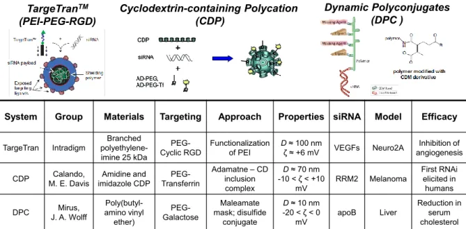
Notable cationic polymer-based delivery vehicles are illustrated in Figure 1.7 along with details regarding the nanoparticle characteristics, in vivo models, and efficacy. PEI was functionalized an RGD ligand, targeting integrins on tumor endothelium, through a PEG spacer to provide a functional cationic polymer capable of condensing, protecting, and delivering siRNA to target cells while exhibiting colloidal stability and reduction of surface charge and aspecific cellular uptake.63 Cyclodextrin-containing polycations featuring amidine and imidazole ionizable groups were mixed with adamantane-terminated PEG, PEG-transferrin, and siRNA to produce nanoparticles capable of effective RNAi in vivo, demonstrating the first gene silencing in human subjects with melanoma.64 A series of
Dynamic Polyconjugates (DPC ) Cyclodextrin-containing Polycation (CDP) TargeTranTM (PEI-PEG-RGD)
System Group Materials Targeting Approach Properties siRNA Model Efficacy
TargeTran Intradigm
Branched polyethylene-imine 25 kDa
PEG-Cyclic RGD
Functionalization of PEI
D≈ 100 nm
ζ ≈ +6 mV VEGFs Neuro2A
Inhibition of angiogenesis CDP M. E. DavisCalando, imidazole CDPAmidine and Transferrin
PEG-Adamatne–CD inclusion complex
D ≈ 70 nm
-10 < ζ< +10
mV RRM2 Melanoma
First RNAi elicited in humans DPC J. A. WolffMirus,
Poly(butyl-amino vinyl ether) PEG-Galactose Maleamate mask; disulfide conjugate
D≈ 10 nm
-20 < ζ< 0 mV
apoB Liver
22
amphipathic amino poly(alkyl vinyl ether)s were pursued for establishing endosomolytic materials capable of transfection.65 After determining the ideal alkyl chain length in amphipathic polycations, an approach to mask the membrane-lytic behavior was established by reversibly conjugating maleic anhydride derivatives.66 Subsequently, conjugation of siRNA through a labile bond to amphipathic polycations functionalized with PEG and ligand-functionalized PEG enabled in vivo RNAi in mice liver toward the reduction of serum cholesterol.66
Figure 1.10 Structures of DOPE and DOTAP lipids as well as an illustration of a multilamellar lipoplex with nucleic acid sandwiched between bilayers.67
23
the nucleic acid and cationic liposome, leading to self-assembly of lipid bilayers between which the nucleic acid is present for multilamellar lipoplexes(Figure 1.10).69