CHARACTERIZATION OF THE RELATIONSHIP BETWEEN KSHV AND THE HOST IMMUNE RESPONSE
Sean Michael Gregory
A dissertation submitted to the faculty of the University of North Carolina at Chapel Hill in partial fulfillment of the requirements for the degree of Doctor of Philosophy in the department of Microbiology and Immunology.
Chapel Hill 2011
Approved By:
ii ABSTRACT
SEAN MICHAEL GREGORY: Characterization of the relationship between KSHV and host immune response.
(Under the direction of Dr. Blossom Damania)
All viruses must evade detection by the host immune response to establish productive infection. Kaposi’s sarcoma-associated herpesvirus (KSHV) employs numerous sophisticated mechanisms that enable it to remain undetected. The coordinated innate and adaptive immune responses suppress KSHV infection and keep the virus in a latent, symbiotic relationship with the host. However, loss of immune suppression leads to uncontrolled viral replication, and increased potential for KSHV-associated malignancy. We have uncovered novel mechanisms KSHV utilizes to tip the balance of immune control and viral persistence in its favor.
Once infection has ensued, KSHV persists in a latent state within a cell. Using latently infected cells, KSHV reactivation from latency was shown to occur in response to activation of toll-like receptors (TLR) 7 and 8. TLRs sense pathogens and induce an inflammatory state to eliminate the threat. These results suggest that KSHV senses its environment in response to activation of innate immune receptors such as TLRs to initiate viral replication as means of survival.
iii
during primary infection in human monocytes and reactivation in latently infected cells resulted in increased cytokine secretion and decreased viral fitness. Hence, viral pathogenesis may be strongly affected by NLRs in the absence of Orf63.
iv DEDICATION
v
ACKNOWLEDGEMENTS
To my advisor, Dr. Blossom Damania, thank you for believing in me when I continuously questioned my ability and for giving me the confidence to believe in myself. I am constantly amazed at your work ethic, intelligence and enthusiasm day in and day out. It truly is inspiring. I know that whenever I needed help your door or inbox were always open.
The Damania lab members, both past and present, made lab fun and exciting and a place where we can discuss anything from science to sports. Thanks to former lab member Tamara Moyo (my “sister”), who was a model for me in how to be a successful graduate student. Thank you to John West, my rotation mentor, with whom I enjoyed a sense of normalcy in a world full of pressure, expectation and varied personalities. And a special thank you to Patrick Dillon and Aadra Bhatt, two friends that I can always rely on for help inside and out of lab. To Prasanna Bhende for always giving objective and sincere advice, I will miss our conversations.
I am very grateful to my committee members, Drs. Dirk Dittmer, Mark Heise, Nancy Raab-Traub and Jenny Ting. You have all been instrumental in my graduate work, which was not limited to just committee meetings. Each member influenced me with their support and intelligence, whether it was research suggestions, seminars and collaborations all contributed to my success. Also, I want to especially thank Mark Heise for his help during the preliminary exam. I know I would not be graduating without your support throughout the exam.
vi
TABLE OF CONTENTS
LIST OF FIGURES ix
LIST OF ABBREVIATIONS xi
Chapter One: INTRODUCTION
Kaposi’s sarcoma 2
Discovery of Kaposi’s sarcoma-associated herpesvirus 5
Other KSHV-associated malignancies 6
Herpesviridae family of viruses 8
Kaposi’s sarcoma-associated herpesvirus 10
Transmission 10
Epidemiology 10
Virion biology 10
KSHV lifecycle 11
Virus Entry 11
Latency 12
Lytic lifecycle 13
Reactivation from latency 14
Innate immunity 14
Toll-like receptors 17
vii
RIG-I-like receptors 24
Aim-like-receptors 24
Strategies employed by pathogens to inhibit the host innate immune response 26
Viral pathogens that modulate NLRs 26
Dissertation objectives 28
References 30
Chapter Two: TOLL-LIKE RECEPTOR SIGNALING CONTROLS REACTIVATION OF KSHV FROM LATENCY
Abstract 53
Introduction 54
Results 56
Discussion 78
Materials and methods 80
Acknowledgments 84
References 85
Chapter Three: DISCOVERY OF A VIRAL NLR HOMOLOG THAT INHIBITS THE INFLAMMASOME
Abstract 92
Introduction 93
Results 94
Discussion 108
Materials and methods 129
References 135
viii
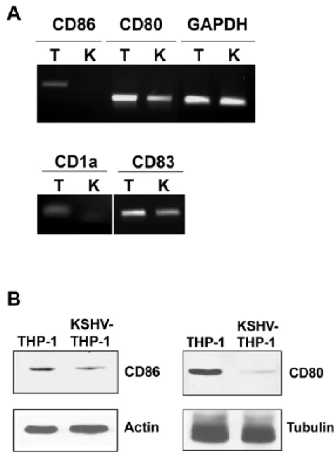
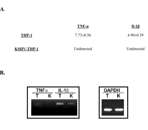
Chapter Four: LATENT KSHV INFECTION OF MONOCYTES DOWNREGULATES EXPRESSION OF COSTIMULATORY RECEPTORS OF ADAPTIVE IMMUNITY
Abstract 140
Introduction 141
Materials and methods 144
Results 148
Discussion 160
Acknowledgments 162
References 163
Chapter Five: GENERAL CONCLUSIONS
Conclusions 169
ix
LIST OF FIGURES Chapter One
1. Overview of innate immune signaling 16
Chapter Two 1. TLR stimulation and KSHV reactivation 58
2. TLR7/8, and not TLR4, reactivates latent KSHV 61
3. Single-stranded RNA activates an innate immune response 65
4. Single-stranded poly-U RNA activates KSHV replication 68
5. Whole genome profiling of KSHV after ssPoly-U treatment 72
6. TLR7/8 stimulation mediates KSHV reactivation to single-stranded RNA and VSV infection 76
Chapter Three 1. Orf63 is a viral homolog and inhibitor of NLRP1 96
2. Orf63 interacts with NLRP1 99
3. Orf63 inhibits NLRP1 inflammasome formation and is necessary for IL-1β inhibition during viral infection 103 4. Orf63 inhibits the NLRP3 inflammasome 106
Supplemental 1A. Alignment of KSHV Orf63 with NLRP1 109
Supplemental 1B. Alignment of KSHV Orf63 with NLRP1 110 Supplemental 2. KSHV Orf63 but not KSHV RTA inhibits NLRP1-
x
Supplemental 3: Orf63 does not interact with NLRP1 inflammasome
components ASC and caspase-1 113
Supplemental 4: The NBD of NLRP1 is required, but not sufficient, for
interactions with Orf63 114
Supplemental 5: Orf63-N and Orf63∆N mutants are capable of inhibiting
NLRP1 activity 115
Supplemental 6: Orf63 inhibits the interaction of procaspase-1 with NLRP1and NLRP1
oligomerization 117
Supplemental 7: NOD2 NBD is required for interactions with Orf63,
and Orf63 does not interact with NOD1 119
Supplemental 8: Purity of primary human monocytes purified from
healthy donors 120
Supplemental 9: Orf63 inhibits IL-1β expression during KSHV
reactivation 121
Supplemental 10: NLRP1 inhibits KSHV reactivation from latency and
production of infectious progeny virus 123 Supplemental 11: Orf63 inhibits NLRP3 in a dose-dependent manner 125 Supplemental 12: Orf63 inhibits the NLRP3 inflammasome 126 Supplemental 13: Rhesus monkey rhadinovirus (RRV) Orf63 demonstrates
homology to NLRP1 128
Chapter Four
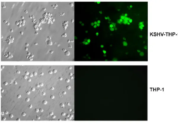
1: KSHV infection in THP1 cells 149
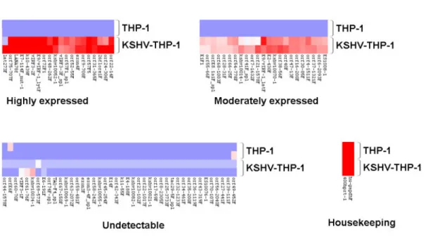
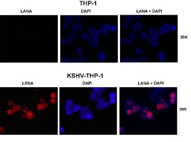
2: KSHV gene expression profile of KSHV-THP-1 cells 151 3: Expression of KSHV LANA by immunofluorescence staining in
KSHV-THP-1 compared to uninfected THP-1 cells 154
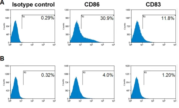
4: KSHV-THP-1 cells exhibit reduced CD86 and CD83 costimulatory
molecule expression 156
xi
LIST OF ABBREVIATIONS AIDS Acquired autoimmune deficiency syndrome AIM2 Absent in melanoma 2
ALR AIM2-like receptor APC Antigen presenting cell
ASC Apoptotic speck-like adaptor protein ATP Adenosine triphosphate
BCBL Body cavity based lymphoma BHK Baby hamster kidney
BIR Baculovirus inhibition of apoptosis protein repeat domain BMDM Bone-marrow derived macrophage
BSA Bovine serum albumin
CARD Caspase-activation and recruitment domain CD Castleman’s disase
CDC Centers for Disease Control
CHOP Cyclophosmide, doxorubicin, vincristine and prednisone CIITA MHC class two transactivator
cPOP cellular pyrin-only protein CT Cycle threshold
CTL Cytotoxic T cell
xii
DC-SIGN Dendritic Cell-Specific Intercellular adhesion molecule-3-Grabbing Non- integrin
DMSO Dimethyl sulfoxide DNA Deoxyribonucleic acid DTT Dithiothreitol
E Early
EBV Epstein-Barr virus
EMCV Encephalomyocarditis virus
FADD Fas-Associated protein with death domain FBS Fetal bovine serum
FITC Fluorescein isothiocyanate
FLICE FADD-like interleukin-1 beta-converting enzyme FLIP FLICE inhibitory protein
GAPDH Glyceraldehyde-3-phosphate-dehydrogenase GFP Green fluorescent protein
GPCR G-protein coupled receptor
HAART Highly active antiretroviral therapy HCMV Human cytomegalovirus
HDAC Histone deacetylase HHV8 Human herpesvirus 8
HIV Human immunodeficiency virus HSV Herpes simplex virus
HV Hyaline variant IAV Influenza A virus
xiii IFA Immunofluorescence assay
IFN Interferon
IL Interleukin
IRAK Interleukin-1 receptor-associated kinase IRF Interferon regulatory factor
KS Kaposi’s sarcoma
KSHV Kaposi’s sarcoma-associated herpesvirus
L Late
LANA Latency associated nuclear antigenLANA LPS Lipopolysaccharide
LRR Leucine-rich receptor
MAPKK Mitogen-activated protein kinase kinase MAVS Mitochondrial antiviral signaling MCD Multicentric CD
MHC Major histocompatibility complex MMLV Moloney murine leukemia virus MOI Multiplicity of infection
MSM Men who have sex with men MVA Modified Vaccinia ankara
MyD88 Myeloid differentiation associated factor 88 NaB Sodium Butyrate
NBD Nucleotide binding domain NFDM Non-fat dry milk
NLR Nucleotide binding and leucine-rich repeat receptor NTC Non-template control
xiv ORF Open reading frame
PAMP Pathogen-associate molecular pattern PAN Poly-adenylated nuclear RNA
PC Plasma cell
PCR Polymerase chain reaction PEL Primary effusion lymphoma
PKR Double-stranded RNA protein kinase PMA Phorbol myristate acetate
PRRs Pattern recognition receptor
PTLD Post-transplant lymphproliferative disorder
PYD Pyrin domain
RDA Representation differential analysis RFP Red fluorescent protein
RGD Arginine glycine aspartic acid RIP Receptor interacting protein RNA Ribonucleic acid
ROS Reactive oxygen species
RTA Replication and transcription activator RT-PCR Real-time PCR
SDS Sodium dodecyl sulfate STING Stimulator of interferon genes TAB TAK1-binding proteins
TAK1 Transforming growth factor β-activating kinase 1
TBK1 TRAF-family-member-associated NF-kB activator-binding kinase TIR Toll-interluekin-1(IL-1) receptor
xv TNF Tumor necrosis factor
TPA 12-O-tetradecanoylphorbol-13-acetate TRAF tumor necrosis factor associated factors
TRIF TIR-domain-containing adaptor protein inducing interferon (IFN-β) TRITC etramethyl Rhodamine Isothiocyanate
CHAPTER ONE
2 Kaposi’s Sarcoma
In 1872, the Austro-Hungarian dermatologist Moritz Kaposi published the first report on several clinical cases of middle to late-aged Jewish male patients presenting with skin lesions
characterized as “idiopathic multiple pigmented sarcoma of the skin” (105). These lesions were found mainly on the feet and described as “flat, with a firm elastic consistency, and they are sometimes swollen, like a cavernous hemangioma. They stand isolated, and become raised when they enlarge, or they cluster and remain more flat”. Kaposi goes on to note that, histologically, cells in the lesion cluster and exhibit a yellow-brown to black pigment. The previously uncharacterized disorder would later be named Kaposi’s sarcoma (KS), after its discoverer. It would also take greater than a hundred years to discover the etiological agent of the disease, Kaposi’s sarcoma associated herpesvirus (KSHV) (40).
3
AIDS epidemic brought about increased awareness of KS and further characterization of the various types of KS.
There are four epidemiological types of KS: classic, endemic, iatrogenic, and AIDS- or epidemic-KS. Classic KS is historically a disease that affects elderly men of Mediterranean and eastern European descent (105). This form of the disease is typically a painless disorder, with slow developing skin lesions commonly found on the legs (178, 179). Due to the slow nature of classic KS, the majority of patients do not need treatment, while spontaneous remission has been documented (24). In the rare case when progression does occur, it remains confined to the original lesions. Classic KS is not considered a life threatening illness (178, 179).
The endemic-African form of KS affects HIV-negative hosts including children and is typically more aggressive than classic KS (219). There are two subtypes of endemic KS, which are divided by geographical, age and pathobiology factors (100). The first subtype resembles classic KS in that it is moderately aggressive with localized lesions, however, substantial edema is sometimes observed. In addition, in sub-Saharan African countries Uganda, Sudan, Democratic Republic of Congo, Rwanda, Burundi, Malawi, Eastern Zaire and the Cameroon coast, adults aged 25-50 years old are mostly affected (47, 199). In contrast, the second subtype of KS affects children younger than 10 years of age of central, eastern and southern Africa. This pediatric form of endemic KS presents as a lymphadenopathy and, in general, is considerably more serious than the first type with death
occurring within two years, and endemic KS is increasing in incidence (76, 205).
Iatrogenic, or hospital-acquired KS, follows immunosuppression often as a result of treatment for organ transplantation, very often after renal transplants (44, 74, 93). Recipients of solid-organ transplants are up to 1000-times more likely to develop KS compared to healthy controls (70, 135). Iatrogenic KS develops as a result of immunosuppression and opportunistic infection associated with organ transplant. If there is internal organ involvement, the prognosis is considerably poor (157). The clinical characteristics of iatrogenic KS are similar to classic KS, and reduction in
4
The most severe form is epidemic- or AIDS-associated KS, which, in addition to skin and lymph node involvement, may spread to the lungs, gastrointestinal tract, liver or spleen (2). Epidemic KS is the leading AIDS-defining malignancy (182). Epidemic KS is most often associated with homosexual men in the early 1980s, and as mentioned, more recently with spread of HIV in Africa (75). With the introduction of highly active antiretroviral therapy (HAART), KS incidence dropped precipitously in the late 1990s, which is attributable to reconstitution of the host immune system that is otherwise suppressed by active HIV infection (3, 34, 61, 62, 198). With increased life-span attributed to HAART, there is accumulating evidence demonstrating higher incidence of non-AIDS-defining cancers such as Hodgkin’s lymphoma, lung cancer, hepatocellular carcinoma, and breast cancer found among HIV infected people (182). However, KS still remains the most frequent malignancy in the HIV positive in the USA and in many parts of Africa, KS is the most common cancer in the entire population (31).
KS lesions are composed of multiple cell types including inflammatory mononuclear cell infiltrate, however, KS is defined by a classic spindle-shaped lymphatic endothelial cell, or so called spindle cell (45). KS lesions are multifocal and exhibit marked neovasculature with irregularly shaped endothelium-lined vascular spaces. The tumor is generally polyclonal at early stages, but evolution to a monoclonal neoplasia has been documented (163).
Discovery of Kaposi’s sarcoma-associated herpesvirus (KSHV)
5
for CMV, only that it was often, but not always, found in these lesions. A little bit later, and after many unsuccessful attempts to identify the causative agent, a clue would come from an
epidemiological study conducted by Beral and colleagues at the Centers for Disease Control (CDC) (18). Beral’s epidemiological study of homosexual and bisexual AIDS patients demonstrated a 20-fold higher KS incidence compared to immunosuppressed hemophilic men, and although it did not identify the infectious agent, the study provided support for an infectious cofactor in the development of KS other than human immunodeficiency virus and enabled continued research into the true cause of KS. Because attempts to culture the infectious agent had failed, researchers were becoming
convinced an HIV factor, specifically the tat protein, may be a reason for development or progression of KS in HIV-1 infected people since Tat-depleted conditioned media blocked development and growth of AIDS-KS endothelial cells (68). In addition, various other non-culturable agents such as bacteria and papillomaviruses were suspected to be cofactors (138). Then, several years later, two researchers, Patrick Moore and Yuan Chang, using a newly developed method to identify differences between two complex genomes called representational differential analysis (RDA), showed that 25 of 27 KS lesions contained viral DNA sequences similar to but unique enough from other human
6 Other KSHV-associated malignancies
KSHV is commonly associated with two B cell disorders: multicentric Castleman’s disease (MCD) and primary effusion lymphoma (PEL) or body cavity-based lymphoma (BCBL). First noted in 1956 by Benjamin Castleman, Castleman’s disease (CD) was described as a unicentric proliferative tumor of the lymphatic system, which is a mild disorder requiring minimal treatment (33). CD is characterized by three variants: hyaline vascular (HV), plasma cell (PC), or a mixed HV/PC type (77, 110). HV CD is generally benign in nature, however, MCD and specifically PC MCD is highly aggressive with patients demonstrating enlarged lymph nodes, endothelial cell proliferation, and associated with high fatality generally from autoimmune hemolytic anemia or secondary cancer. Although MCD is considered to be a polyclonal plasmablast of an immature phenotype originating from naïve B-cells, monoclonal populations of PC MCD have been observed (92, 164). HIV positive patients with MCD are nearly always coinfected with KSHV versus 50% KSHV positive for HIV negative MCD patients (60, 192). High levels of interleukin-6 (IL-6), a cytokine inducing B-cell proliferation and differentiation, were associated with MCD, and indeed treatment with anti-IL-6 antibody showed a positive effect (15, 146, 149). It is worth noting that KSHV itself expresses a viral homolog of human IL-6, vIL-6, which suggests a dependence on this cytokine and supported by therapeutic treatment with anti-IL-6 antibodies (137). Treatment of KSHV-associated MCD with ganciclovir, a viral replication inhibitor, is very effective suggesting lytic replication is necessary for disease progression (32). Conventional chemotherapy as well as treatment with anti-CD20 antibody are also moderately effective in the setting of MCD (145, 150).
7
vincristine and prednisone (CHOP) is the current first-line therapy for PEL, however only 43% of patients demonstrate brief remission (188). Recent therapeutic efforts have yielded positive results based on molecular analyses of the pathways upon which PEL depends, for example
phosphatidylinositol 3-kinase-Akt-mTOR signaling pathway, or based on providing antiviral drugs at the same time as viral replication inhibitors (20, 113). However, since PEL is relatively rare, analysis of clinical responses to these regimens is limited.
Herpesviridae Family of Viruses
By the end of the twentieth century, it was becoming increasingly clear that KS in both HIV-negative and HIV-positive individuals was likely because of an infectious agent (18, 148). The discovery of KSHV in 90% of AIDS-associated KS by Drs. Yuan Chang and Patrick Moore in the mid-1990s was the penultimate event in a long line of studies to find the etiological agent of KS. Their seminal finding as well as follow-up studies demonstrated that DNA isolated from an AIDS-related lymphoma that was continuously infected showed that the DNA did not hybridize to other herpesvirus’ DNA including EBV or CMV (139). However, sequence analysis confirmed that the new agent belonged to the herpesvirus family of human DNA tumor viruses, and would now be classified as the 8th human herpesvirus or HHV8.
8
lead to development of keratitis, a disease of the cornea that can result in corneal scarring and blindness, and in rare cases HSV-1 infection can lead to meningitis (133). VZV causes varicella or chickenpox upon infection and replication in the skin (156). Later in life, latent VZV can reactivate resulting in shingles, which can cause severe pain and long-term debilitation (152). The
betaherpesvirus members include cytomegalovirus (CMV), Roseolovirus (HHV-6) and HHV-7. CMV infection is an asymptomatic infection, however, CMV mononucleosis occurs in rare cases (156). Congenital CMV infection is a serious disorder of newborns that damages the central nervous system and is thought to be the number one cause of sensorineural deafness and brain damage in the US (154). Roseolovirus and HHV-7 are sometimes associated with Exanthem subitum, which is an infantile disease normally resulting in fever and systemic rash (30). Finally, the human
gammaherpesviruses include EBV (HHV-4) and KSHV (HHV-8). EBV is primarily associated with infectious mononucleosis as well as several malignancies such as nasopharyngeal carcinoma, post-transplant lymphoproliferative disease (PTLD), Burkitt’s lymphoma and Hodgkin’s lymphoma (51, 186, 200). It is important to note that these malignancies are often found in the context of immune suppression, and other herpesvirus diseases often develop in this context.
9 Kaposi’s Sarcoma-Associated Herpesvirus
Transmission
Transmission of KSHV was originally thought be sexual in nature since KSHV was detected much more frequently in men who have sex with men (MSM) compared to other HIV-associated diseases and KSHV was detected in semen and the female genital tract (18, 28, 56, 211). Moreover, KSHV prevalence was higher in persons infected with STDs as well as correlated with number of sexual partners in homosexual men (109, 129). Nevertheless, detection of high levels of KSHV in saliva of MSM compared to semen, lack of heterosexual sexual transmission, as well as evidence that KSHV infection in children in endemic areas suggested that KSHV was not definitively a STD (63, 80, 130, 155, 190, 210, 215). Thus, KSHV infection is most likely transmitted via infected saliva consistent with horizontal transmission and can also be transmitted by infected blood used in transfusions (11). Several cofactors are likely to influence transmission including geographical and environmental differences, exposure to water-born parasites as well as host genetic factors (64, 131, 176, 185, 204).
Epidemiology
KSHV is globally distributed with a low seroprevalence in the general population,
particularly in developed nations; however, in Africa and Mediterranean regions KSHV is endemic. In the U.S., KSHV is detected anywhere from 0 to 23% with a more likely estimate in the range of 0 to 10% (78, 96, 109, 123). However, in the MSM population in the U.S, KSHV positivity rises to 50-60%. Detection is heavily influenced by the methodology since different immunological assays are subject to false positives or have high stringencies (67). In sub-Saharan Africa, incidence of KSHV ranges from 12.5% to as high as 82% with considerable geographical variation, with the high
10
Virion Biology
KSHV is a linear double-stranded DNA virus, like all herpesviruses, of about 165Kb, which encodes for greater than 80 open reading frames as well as several virally encoded micro-RNAs (27, 121, 161). The KSHV genome is flanked by multiple tandem repeats of GC-rich DNA (121). There are regions of homology between herpesviruses with large blocks of conserved genes, but many regions of genes unique to KSHV are found, which are designated with a K in their nomenclature e.g., K1 and K15. KSHV DNA is encapsidated by a icosahedral capsid containing 162 capsomeres surrounded by a proteinacious layer called the tegument, which incorporates viral proteins into the virion for structural purposes, but also for release during infection for modulating host protein synthesis, immune responses and initiating viral gene expression (140, 141). The outer-most layer of the virion consists of viral glycoproteins and host derived double-membrane lipid bilayer (10, 69, 193, 212).
KSHV Lifecycle
Virus Entry
KSHV establishes productive infection in a variety of cell types, including B cells, monocytes, endothelial cells, plasmacytoid and conventional dendritic cells, keratinocytes and epithelial cells, which indicate that KSHV likely has broad tropism in vivo (37, 208). However, KSHV-infected PEL cells from patient biopsies are the only naturally derived cell lines for use in vitro. Initial cellular adsorption has been shown to be facilitated by heparan sulfate (HS), which likely serves as an attachment factor in this process and may explain the wide tropism of the virus due the ubiquitous expression of HS (6, 21). Once initial attachment is aided by HS, several putative
11
integrins likely mediate entry (37). Two additional surface receptors were shown to play a role in KSHV entry: the 12-transmembrane glutamate/cysteine exchange transporter protein xCT in adherent cells, and dendritic cell-specific intercellular adhesion molecule 3 (ICAM-3)-grabbing nonintegrin (DC-SIGN) in dendritic cells, macrophages and B cells (103, 168, 169). However, none of these receptors are complete in mediating entry and it is likely additional receptors facilitate entry, and these are likely to be cell specific, thus giving KSHV the ability to infect multiple cell types.
Once KSHV has initiated the infectious process, it most likely continues into the cell through endocytosis. Several lines of evidence suggests this is the case in B cells, monocytes, fibroblasts, epithelial cells and endothelial cells (4, 6, 90, 99, 165, 166, 169). Once inside the endosome, the viral envelop fuses with the lipid membrane of endocytic vesicles, releasing the viral capsid and tegument proteins into the cytoplasm (37). From there, assisted by dynein motors, KSHV is trafficked down microtubules to nuclear membrane pores, through which the viral DNA is released into the nucleus and initiation of viral gene expression occurs. Infection is associated with concomitant induction of cellular gene expression, which is thought to be manipulation on behalf of the virus to facilitate infection (187).
Latency
After primary infection of naïve cells, viral DNA is released into the nucleus at which time a period of viral replication may ensue, though, in most instances the viral genome is circularized to form an episome and initiation of the dormant phase. Within several days, the episomal viral DNA has become attached to host chromosomes through the viral protein latency-associated nuclear antigen (LANA) (48). LANA is one of several transcripts that can be detected during latency, which include viral cyclin (vCyclin), viral fas-associated death domain (FADD)-like
12
facilitates replication of viral DNA during cellular DNA replication and transmission to daughter cells after mitosis.
Lytic Lifecycle
Lytic replication proceeds through temporal regulation of gene transcription in three distinct stages: immediate early (IE), early (E) and late (L) transcription. Interestingly, a single protein or lytic switch protein, replication and transcription activator (RTA/Orf50), is responsible for initiation of lytic replication (89). Thus, RTA is the earliest detected transcript in lytically replicating cells and as its name indicates RTA activates immediate early gene transcription through an N-terminal DNA-binding domain and C-terminal transactivation domain that binds RTA-responsive elements in viral promoters (191). Signals that activate RTA are key to determining what governs reactivation and viral replication.
IE proteins are characterized by transcription in the presence of cycloheximide, which inhibits protein synthesis. Very few IE proteins are known and, like RTA, mainly serve as transactivators of E genes, including Orf45, Orf50, and K8 (120, 183). Orf45, however, has immunomodulatory capabilities in that it inhibits IRF-7-dependent induction of type I interferon through blocking IRF-7 phosphorylation and nuclear translocation (218).
The next stage in lytic replication is the transcription of E genes, which are defined by their susceptibility to cycloheximide treatment and not dependent on viral DNA synthesis. Most E proteins serve to facilitate viral DNA replication. These include a thymidine kinase, DNA polymerase,
ribonucleotide reductase, and various DNA binding proteins. A key E protein is KSHV Orf57 which plays a critical role in blocking RNA splicing, nuclear-cytoplasmic transport of unspliced RNA, and mRNA polyadenylation (128). Once E gene products have been synthesized to sufficient levels, DNA replication begins at two sites of origin of replication (ori-lyt) in a rolling circle mechanism at
13
Transcription of L genes begins once DNA replication is underway, as demonstrated by inhibitors of viral lytic DNA synthesis such as ganciclovir. L proteins are generally virus structural proteins of the capsid and envelope as well as proteins localized to the tegument. Accumulation of L proteins begins the process of assembly of progeny capsids within the nucleus. Newly formed capsids move out of the nucleus and acquire viral proteins of the tegument and finally the glycoprotein studded lipid envelope is wrapped around the tegument and capsid. At this point, mature progeny virions lyse the cells during release from the infected cells, subsequently capable of repeating the infectious process in a naive cell.
Reactivation from Latency
In vivo, reactivation from latency is required for life-long viral persistence and transmission. In cultured PEL cells, a small percentage (1-5%) of cells will undergo spontaneous reactivation from latency. Reactivation can be initiated by several chemical stimuli including
12-O-tetradecanoylphorbol-13-acetate (TPA), histone deacetylase (HDAC) inhibitors and biologically relevant stimuli hypoxia, interferon-γ, and hormones epinephrine/norepinephrine (38, 39, 53, 214). In addition, superinfection with KSHV or coinfection with different viruses such as CMV or HIV initiates reactivation from latency. It is possible that a common pathway is required or reactivation may occur in response to different triggers through unique pathways. The molecular mechanisms of KSHV reactivation in several of these settings remain incompletely characterized.
Innate Immunity
14
pathogenic event. The adaptive or acquired immune response is a delayed mechanism, occurring between 7-10 days post-infection, consisting of cell proliferation and somatic gene editing to tailor the immune response to a particular pathogen. Effector molecules upregulated by the innate response play a necessary role in shaping adaptive immunity in strength, duration, cell type involvement, specificity and trafficking to sites of infection. B and T cell receptors undergo a process of receptor editing, or honing, to ensure that B and T cells effectively recognize pathogen-derived peptides on the surface of antigen presenting cells (APCs), which are activated by the innate immune response, to eliminate the pathogenic threat. Hence, the innate immune response is critical in being able to sense the pathogen, containing the infection through immediate release of antipathogen effector molecules, as well as initiate the adaptive immune response.
The innate immune system is composed of several families of germline-encoded pattern recognition receptors (PRRs), which recognize pathogen-associate molecular patterns (PAMPs) such as nucleic acids, lipids, and proteins unique to bacteria, viruses and fungi (107). PRRs are found on nearly every cell type in both peripheral and lymphoid tissues, but are highly expressed in APCs monocytes, macrophage, dendritic cells and B cells. There are three main families of PRRs: Toll-like receptors (TLRs), Retinoic acid-like receptors (RIG-I), and the nucleotide binding and
15
16
Toll-like Receptors
There are 10 functional TLRs in humans, and they can be found on the surface or located within the endosome of APCs. The toll family was discovered in Drosophila and found to be capable of mediating immune responses to fungus (122). Later, toll homologs would be found in mammalian cells and shown to be vital in innate immunity (134). TLRs are single transmembrane proteins consisting of a leucine-rich repeat (LRR) domain, which is found on the outside of a cell, or within the endosome. The LRR domain is responsible for binding PAMPs and facilitates heterotypic interactions between different TLRs. TLRs 1, 2, 4, 5, and 6 are found on the cell surface and detect PAMPs such as bacterial and fungal cell wall constituents lipopolysaccharide (LPS), peptidoglycan, and bacterial flagellum, while endosomal TLRs 3, 7, 8, and 9 generally detect virally derived single-stranded or double-single-stranded RNA and DNA (106).
Recognition of each TLR by its cognate ligand initiates a signal transduction cascade
beginning with a toll-interleukin-1(IL-1) receptor (TIR) domain located on the cytoplasmic side of the cellular or endosomal membranes. Next, downstream TLR signaling is facilitated by interactions between the TIR domain and the adaptor protein myeloid differentiation associated factor eighty-eight (MyD88) (23). TLR3 exclusively and TLR4 occasionally utilize another adaptor protein called TIR-domain-containing adaptor protein inducing interferon (IFN-β) (TRIF). MyD88 signaling converges on one of IL-1 receptor associated kinases (IRAKs) including IRAK1, 2, or 6, to which a variety of tumor necrosis factor associated factors (TRAFs) are associated, facilitating signal
17
NF-kB. Phosphorylated IKKα in turn phosphorylates interferon regulatory factor 7 (IRF 7), causing IRF-7 to homodimerize and translocate to the nucleus to upregulate type I IFN expression. In addition, downstream of TLR4, TRAF3 associates with the kinase IKKε and TRAF-family-member-associated NF-kB activator-binding kinase (TBK1) to phosphorylate IRF7 and IRF3, which
heterodimerize and regulate IFN gene expression. TLR 3 and 4 signaling through TRIF leads to the association of receptor interacting protein (RIP) and eventual activation of canonical p50/p65 NF-kB pathways and upregulation of NF-kB inducible cytokines. Thus, each TLR is capable of signaling to IRF or NF-kB transcription factors through related pathways and ultimately modulate expression of host antipathogen genes.
Nucleotide-binding, oligomerization and leucine-rich repeat proteins
Recent research has uncovered an emerging role for the NLR family of innate immune sensors in various human diseases from metabolic disorders, cancer, autoinflammatory conditions and autoimmune disease (52). The association with human disease suggests that dysregulation of innate immunity is a key factor in disease progression.
There are over 22 genes encoding for NLRs in humans, most of which remain
18
apoptosis protein repeat domain (BIR). Adenosine triphosphate (ATP) is necessary for the molecular oligomerization exhibited by NLRs, and as a result, they are characterized by a centrally located nucleotide-binding domain (NBD). The last feature of the NLR family is a variable number of LRRs, which is considered to function in ligand sensing and negative regulation in the inactivated state.
With the exception of CIITA, NLR function is hypothesized to be sensing PAMPs and, more recently, danger associated molecular patterns (DAMPs) as well as environmental irritants. Similar to TLRs, NLRs such as NOD1 and NOD2 interact with RIP2 to activate the transcription factors IRFs 3 and 7, NF-kB and MAPK and the eventual upregulation of IL-6, TNF, pro-IL-1β and pro-IL-18 as well as antiviral type I IFN. Moreover, several NLRs such as NLRX1 and NLRC5 can inhibit NF-kB and type I IFN signaling, suggesting this pathway has evolved mechanisms to regulate its pathways for control of the innate immune response. Still other NLR family members regulate innate immunity through the formation of large molecular complexes called inflammasomes.
Inflammasomes, by definition, are responsible for generation of biologically active IL-1β and IL-18. IL-1β and IL-18 are capable of mediating infection and activation of NF-kB through secretion and binding to their cognate receptors, IL-1 (IL-1R) and IL-18 (IL-18R). Inflammasomes generally consist of oligomerization of a particular NLR, recruitment of procaspase-1 and apoptotic-associated speck-like protein containing a CARD domain (ASC). To date, several NLR family members have been shown to form inflammasomes including NLRP1, 3, NLRC4 and, more recently, several non-NLR PRRs have been shown to form inflammasomes and therefore active IL-1β and IL-18. These include absent in melanoma 2 (AIM2) and retinoic acid inducible gene-I (RIG-I).
19
pathogens activate NLRs, only a description of the viral activators of inflammasomes is highlighted below.
The most well characterized inflammasome is the NLRP3 inflammasome, which consists of six to seven molecules of NLRP3, ASC and procaspase-1. In response to PAMPS, NLRP3 self oligomerizes and associates with ASC through PYD-PYD interactions, and ASC with procaspase-1 through CARD-CARD interactions. The first viral activator of the NLRP3 inflammasome was Sendai virus (SeV) and influenza A virus (IAV) (104). Using macrophages deficient in NLRP3, infection with SeV or IAV resulted in loss of detection of the p20 subunit of caspase-1 indicating that NLRP3 played a role in sensing SeV and IAV leading to caspase-1 activation and IL-1β. Later, a role for NLRP3 in sensing IAV infection in vivo would be confirmed by showing that mice lacking NLRP3 had higher mortality and reduced IL-1β and that NLRP3 inflammasome dependent responses were critical for development and proliferation of CD4, CD8 T cells and mucosal and systemic IgG immune responses (7, 97). It is important to note that activation of NLRP3 was attributed to
generation of reactive oxygen species (ROS) in response to viral infection, suggesting that, since IAV also activates TLRs 7 and 8, NLRP3 may respond secondarily to infection and not necessarily to a viral PAMP itself. In this regard, it is hypothesized that NLRs may sense intracellular changes in response to viral infection such as generation of ROS through free thioredoxin-interacting protein (TXNIP) interacting with NLRP3, or cathepsin B in response to lysosomal disruption after
20
likely regulate host immunity (162). The NLRP3 inflammasome mediates innate immunity to two DNA viruses in addition to the RNA viruses described, including adenovirus and modified Vaccinia Ankara (MVA). THP-1 acute monocytic leukemia cells treated with shRNAs to knockdown NLRP3 showed a significant reduction in IL-1β in response to MVA infection, and cross-talk between TLRs 2 and 6 again suggest multiple sensors of the innate immune system contribute to the host response to viral infection (54). This was confirmed using NLRP3-/- bone-marrow derived macrophages
(BMDM). Adenovirus is a non-enveloped DNA virus, as opposed to MVA, which is an enveloped DNA virus. Adenovirus was shown to activate IL-1β processing and secretion in THP1 cells, and that this was dependent on the viral DNA itself (142). Using NLRP3 deficient peritoneal macrophages, adenoviral infection-induced IL-1β maturation was significantly inhibited, and later NLRP3 was shown to induce immunity to adenovirus in vivo albeit a complete reduction was not observed indicating additional, redundant sensor for adenovirus infection likely exist. This is not surprising given that multiple sensors are known to recognize a single virus. However, more recently, an NLRP3-independent role was described for adenoviral infection, and instead, β3-integrins were suggested to be the key pathway in activation of proinflammatory responses irrespective of DNA sensing (55) while two additional reports indicates NLRP3 is necessary (13, 14). Herpesviruses, like MVA, are a family of large, enveloped DNA viruses, which establish lifelong latent infection in the human host. Varicella-zoster virus (VZV) is the etiological agent of chickenpox (varicella) and shingles (herpes zoster). In THP1 cells, melanoma cells, and skin xenografts, the NLRP3
inflammasome was induced after VZV infection resulting in recruitment of ASC and caspase-1 (147). NLRC5 is ubiquitously expressed and its highest levels are detected in brain, lung and
21
expression suggesting it may play a role in antiviral responses to herpesviruses. At the same time, another report emerged suggesting NLRC5 sensed SeV in THP1 cells and primary human dermal fibroblasts, suggesting NLRC5 was capable of innate immunity to RNA and DNA viruses, similar to NLRP3 (144). It remains unclear whether both NLRs sense PAMPs specific to viral infection or a secondary event such as ROS generation or ion imbalance. Treatment of NLRC5 deficient bone marrow derived dendritic cells, macrophages and peritoneal exudate cells with activators of various inflammasomes did not alter IL-1β processing and secretion, suggesting NLRC5 may have a unique activator and does not play a role in the activation of other inflammasomes (117). Still others have described a negative regulatory role for NLRC5 in response to viral infection. NLRC5, in response to LPS treatment interacts with IKKα and IKKβ to inhibit phosphorylation and NF-kB activation (50). Alternatively, knockdown of NLRC5 resulted in increased innate immune responses, specifically, RIG-I and MDA-5 were activated in NLRC5’s absence. Benko and colleagues also identified NLRC5 as a negative regulator of innate immune responses in over-expression studies in HEK293T cells, with NF-kB, AP-1, and type I IFN signaling suppressed (17). Moreover, they found that suppression of NLRC5 leads to upregulation of TNF, IL-6, and IL-1β. It remains to be seen what effect activation of NLRC5 has in vivo in response to HCMV and SeV and whether these conflicting reports are due to cell type specificity or inflammasome versus non-inflammasome function.
22
interaction between MAVS and NOD2 was dependent on the LRR and NBD domain of NOD2, which is somewhat counter intuitive considering both molecules contain CARD domains, and that MAVS interacts with RIG-I through CARD-CARD interactions. Nevertheless, this study firmly established two distinct roles for NOD2, first in response to bacterial ligands NOD2 activates NF-kB and proinflammatory pathways, whereas activation by viral infection leads to signaling through MAVS and an antiviral response consisting of type I IFN induction. This work established discrete roles for NLRs in antiviral immunity and suggests that in different contexts, NLRs will act differently to tailor the host response to the invading pathogen. In support of a role for NOD2 in antiviral
immunity, Dugan et al, demonstrated that NOD2 interacted with 2’-5’-oligoadenylate synthetase type 2 (OAS2), which is important in recognition and elimination of many viral infections through RNase L-mediate degradation of viral RNA (59).
23
Rig-I-Like Receptors
While RLRs have been shown to mediate antiviral immune responses to many viruses, here we will describe recent reports suggesting a novel role for RLRs, specifically RIG-I, in formation of an inflammasome-like structure and, by definition, production of IL-1β and IL-18. Formation of inflammasomes as defined by recruitment of procaspase-1 and production IL-1β and IL-18 has been expanded to include non-NLR innate immune sensors. In addition, RIG-I associates with NLR family member NOD2, indicating that relationships between innate immune molecule families is important for host immunity. WT BMDMs infected with VSV demonstrate significant IL-1β secretion, however, RIG-I deficient BMDM show no IL-1β secretion (162). Furthermore, VSV infection induced the proteolytic processing of procaspase-1 to caspase-1 as determined by immunoblotting for the p10 subunit of caspase-1. The production of pro-IL-1β was found to be dependent on MAVS, CARD9 and BCL-10. Interestingly, RIG-I associated with ASC to activate caspase-1 and production of active IL-1β, independently of NLRP3. MDA-5, a RLR family member, did not form an inflammasome after infection with VSV or ECMV, a known activator of MDA-5. Another group found that RIG-I signaling lead to secreted active IL-1β and IL-18 after infection with IAV, which was dependent on caspase-1 (173). Thus, RIG-I has the potential to activate NF-kB, type I IFN and inflammasome innate immune responses. It is important to note that activation of
24
Aim-like Receptors
Several groups identified a novel cytoplasmic sensor capable of detecting dsDNA (197). An interferon inducible 200 (IFI200/HIN200) family member, called absent in melanoma two (AIM2) was found to recruit ASC and procaspase-1 to form an inflammasome in response to Vaccinia virus, murine cytomegalovirus (MCMV), and various bacteria (26, 72, 73, 95, 102, 170, 175). The IFI200 family consists of four members in humans (IFI16, MNDA, IFX and AIM2). Importantly, IFI200 family members have N-terminal PYD domains, which facilitate interactions with the adaptor protein ASC. AIM2 binds DNA through the HIN-200 domain, thus confirming its ability to sense
cytoplasmic DNA. In mice lacking AIM2, MCMV titers were higher than WT mice, and this was correlated with increased pyroptosis, a caspase-1-dependent cell death process. The function of AIM2 in inflammasome activation is entirely independent of traditional antiviral responses including type I IFN as well as independent of NLR inflammasomes. However, AIM2, in coordination with stimulator of interferon genes (STING), is capable of inducing type I IFN responses through TBK1 and IRF3 activation.
25
DNA, one could speculate it may also be important for detection of nuclear DNA, either viral or cellular. HCMV ppUL83 protein utilizes IFI16 for initiation of viral transcription (49). Thus, the nuclear and cytoplasmic localization of IFI16 may determine its role in either host immunity or transcriptional regulation.
Strategies Employed by Pathogens to Inhibit Host Innate Immune Response
As cellular sensors are discovered and their mechanisms of host immunity elucidated, so too are pathogenic inhibitors capable of blocking these sensors. As host immunity has evolved to counter the ever-present threat of infection, pathogens have co-evolved to counter this response. In some cases, multiple inhibitors of a pathway are necessary for infection. With the emerging importance of NLRs in pathogen sensing, recent work has focused on uncovering pathogen-encoded inhibitors of the NLR response. Indeed, several viral encoded inhibitors are known to inhibit various
inflammasomes at multiple levels, and bacterial inhibitors have also been characterized. The discovery of pathogenic inhibitors of NLRs underscores the importance of this arm of the host immune response in combating infection.
Viral Pathogens that Modulate NLRs
DNA viruses like poxviruses and herpesviruses have large genomic coding capacities, and are known to express viral homologs of cellular proteins, which serve diverse functions including
inhibiting host immunity, apoptosis, promoting cell proliferation, blocking differentiation and cell migration.
26
M013 results in a severely attenuated virus with reduced replication in monoctyes and lymphocytes with increased active caspase-1 and secreted IL-1β. Similarly, another poxvirus, Shope fibroma virus, expresses a viral POP, gp013L, that colocalized and associated with ASC (57). In HEK293 cells overexpressing NLRP3 inflammasome components, gp013L inhibited secreted IL-1β.
The first inhibitor described was a Cowpox protein crmA. CrmA blocks caspase-1 by directly interacting with and preventing proteolytic processing of procaspase-1 to active caspase-1, resulting in reduced IL-1β secretion (171). In the absence of crmA, poxvirus lesions in chicken allantoic membranes were decreased, however, in vivo there was no difference in IL-1β levels, suggesting additional mechanisms exist to suppress caspase-1 or IL-1β. Cellular serpin proteinase inhibitor 9 (PI-9) blocks pro-caspase-1 processing to attenuate inflammation, therefore, poxviruses seem to have taken advantage of this cellular mechanism of attenuation for their advantage. Indeed, several other poxvirus encoded serpins would be characterized. Vaccinia virus gene B13R encodes a protein homologous to crmA, SPI-2, that blocks IL-1β processing in THP1 cells, but is dispensable for viral infection in vivo (111, 112). Myxoma virus also encodes a caspase-1 inhibitor, Serp2, which when deleted, results in attenuation of the virus in vivo. Serp2 cannot compliment a crmA deleted Cowpox virus, suggesting crmA is more important for viral infection (159, 201). Since poxviruses express a wide variety of cellular homologs, it therefore is not surprising that they also express IL-1β and IL-18 scavenger receptors (vIL-1βR/18R) capable of blocking signaling of their respective cellular
homologs. This also suggests that redundancies in blocking specific antiviral pathways are important and that a deficiency in one viral inhibitor that shows little to no effect on viral fitness or
inflammation should not be surprising (57, 112, 159, 167, 171, 201). It is worth noting that cellular BCL-2 and BCL-xl, two antiapoptotic proteins, have been shown to inhibit the NLRP1
27
Additional viruses have been shown to encode inhibitors of inflammasome sensors. H1N1 IAV protein NS1 inhibits the export of host mRNA as well as block innate immunity. NS1 contains a RNA-binding and dimerization domain, which when mutated, induced significantly more active IL-1β and IL-18 compared to control virus (195). Increased inflammation resulted in apoptosis in macrophages, most likely due to caspase-1 dependent cell death since caspase-1 levels were elevated in the absence of NS1. However, since NS1 has many other roles in innate immune response
pathways, it is likely that type I IFN as well as other inflammatory cytokines induced by the NS1 target double-stranded protein kinase receptor (PKR) contributed to apoptosis (91). This result points to the significance of NS1 in inflammasome signaling, and based on extensive work showing IAV activates inflammasomes, the relationship between IAV and NLRs is likely to be important in better understanding pathogenesis and disease.
Dissertation Objectives:
1) Determine the effect of TLR stimulation on the KSHV lifecycle
KSHV establishes life-long latency in the host. Hence, this virus is able to evade the innate immune response during primary infection. We hypothesized that TLR activation of the host innate immune response upon secondary infection of a latently KSHV-infected cell could result in either dampening of the innate immune response to the incoming secondary pathogen or reactivation of the virus from latency. In this objective, we proposed to investigate whether TLR activation reactivated KSHV from latency.
2) Characterize the role of Orf63 in inhibiting NLR innate immune responses
NLRs are a family of receptors similar to TLRs in the ability to detect and eliminate invading pathogens. NLRs activate caspase-1 and the inflammasome-associated proinflammatory cytokines, IL-1β and IL-18, and several NLRs mediate upregulation of type I interferon and additional
28
discovered a novel viral homolog of cellular NLRP1encoded by KSHV called Orf63. We hypothesize that Orf63 is a virulence protein that promotes KSHV survival through inhibition of NLRP1 innate immune signaling.
3) Evaluate the effect of KSHV latency on immune response costimulatory molecules in monocytes
29 REFERENCES
1. Abdul-Sater, A. A., N. Said-Sadier, V. M. Lam, B. Singh, M. A. Pettengill, F. Soares, I. Tattoli, S. Lipinski, S. E. Girardin, P. Rosenstiel, and D. M. Ojcius. 2010. Enhancement of reactive oxygen species production and chlamydial infection by the mitochondrial Nod-like family member NLRX1. J 285:41637-45. Epub 2010 Oct 19.
2. Ablashi, D. V., L. G. Chatlynne, J. E. Whitman, Jr., and E. Cesarman. 2002. Spectrum of Kaposi's sarcoma-associated herpesvirus, or human herpesvirus 8, diseases. Clin Microbiol Rev. 15:439-64.
3. Aboulafia, D. M. 1998. Regression of acquired immunodeficiency syndrome-related pulmonary Kaposi's sarcoma after highly active antiretroviral therapy. Mayo Clin Proc. 73:439-43.
4. Akula, S. M., P. P. Naranatt, N. S. Walia, F. Z. Wang, B. Fegley, and B. Chandran. 2003. Kaposi's sarcoma-associated herpesvirus (human herpesvirus 8) infection of human fibroblast cells occurs through endocytosis. J Virol. 77:7978-90.
5. Akula, S. M., N. P. Pramod, F. Z. Wang, and B. Chandran. 2002. Integrin alpha3beta1 (CD 49c/29) is a cellular receptor for Kaposi's sarcoma-associated herpesvirus (KSHV/HHV-8) entry into the target cells. Cell. 108:407-19.
6. Akula, S. M., F. Z. Wang, J. Vieira, and B. Chandran. 2001. Human herpesvirus 8 interaction with target cells involves heparan sulfate. Virology. 282:245-55.
7. Allen, I. C., M. A. Scull, C. B. Moore, E. K. Holl, E. McElvania-TeKippe, D. J. Taxman, E. H. Guthrie, R. J. Pickles, and J. P. Ting. 2009. The NLRP3 inflammasome mediates in vivo innate immunity to influenza A virus through recognition of viral RNA. Immunity. 30:556-65. Epub 2009 Apr 9.
30
9. Arnoult, D., F. Soares, I. Tattoli, C. Castanier, D. J. Philpott, and S. E. Girardin. 2009. An N-terminal addressing sequence targets NLRX1 to the mitochondrial matrix. J Cell Sci.
122:3161-8.
10. Asher, Y., M. Heller, and Y. Becker. 1969. Incorporation of lipids into herpes simplex virus particles. J Gen Virol. 4:65-76.
11. Bagni, R., and D. Whitby. 2009. Kaposi's sarcoma-associated herpesvirus transmission and primary infection. Curr Opin HIV AIDS. 4:22-6.
12. Barbera, A. J., J. V. Chodaparambil, B. Kelley-Clarke, V. Joukov, J. C. Walter, K. Luger, and K. M. Kaye. 2006. The nucleosomal surface as a docking station for Kaposi's sarcoma herpesvirus LANA. Science. 311:856-61.
13. Barlan, A. U., P. Danthi, and C. M. Wiethoff. 2010. Lysosomal localization and mechanism of membrane penetration influence nonenveloped virus activation of the NLRP3
inflammasome. Virology. 412:306-14. Epub 2011 Feb 18.
14. Barlan, A. U., T. M. Griffin, K. A. McGuire, C. M. Wiethoff, A. U. Barlan, P. Danthi, and C. M. Wiethoff. 2011. Adenovirus membrane penetration activates the NLRP3 inflammasome Lysosomal localization and mechanism of membrane penetration influence nonenveloped virus activation of the NLRP3 inflammasome. J 85:146-55. Epub 2010 Oct 27.
15. Beck, J. T., S. M. Hsu, J. Wijdenes, R. Bataille, B. Klein, D. Vesole, K. Hayden, S. Jagannath, and B. Barlogie. 1994. Brief report: alleviation of systemic manifestations of Castleman's disease by monoclonal anti-interleukin-6 antibody. N Engl J Med. 330:602-5.
16. Belanger, C., A. Gravel, A. Tomoiu, M. E. Janelle, J. Gosselin, M. J. Tremblay, and L. Flamand. 2001. Human herpesvirus 8 viral FLICE-inhibitory protein inhibits Fas-mediated apoptosis through binding and prevention of procaspase-8 maturation. J Hum Virol. 4:62-73.
17. Benko, S., J. G. Magalhaes, D. J. Philpott, and S. E. Girardin. 2010. NLRC5 limits the activation of inflammatory pathways. J Immunol 185:1681-91.
18. Beral, V., T. A. Peterman, R. L. Berkelman, and H. W. Jaffe. 1990. Kaposi's sarcoma among persons with AIDS: a sexually transmitted infection? Lancet. 335:123-8.
31
20. Bhatt, A. P., P. M. Bhende, S. H. Sin, D. Roy, D. P. Dittmer, and B. Damania. 2010. Dual inhibition of PI3K and mTOR inhibits autocrine and paracrine proliferative loops in PI3K/Akt/mTOR-addicted lymphomas. Blood. 115:4455-63. Epub 2010 Mar 18.
21. Birkmann, A., K. Mahr, A. Ensser, S. Yaguboglu, F. Titgemeyer, B. Fleckenstein, and F. Neipel. 2001. Cell surface heparan sulfate is a receptor for human herpesvirus 8 and interacts with envelope glycoprotein K8.1. J Virol. 75:11583-93.
22. Boshoff, C., T. F. Schulz, M. M. Kennedy, A. K. Graham, C. Fisher, A. Thomas, J. O. McGee, R. A. Weiss, and J. J. O'Leary. 1995. Kaposi's sarcoma-associated herpesvirus infects endothelial and spindle cells. Nat Med. 1:1274-8.
23. Bowie, A. G., and L. Unterholzner. 2008. Viral evasion and subversion of pattern-recognition receptor signalling. Nat Rev Immunol. 8:911-22.
24. Brooks, J. J. 1986. Kaposi's sarcoma: a reversible hyperplasia. Lancet. 2:1309-11.
25. Bruey, J. M., N. Bruey-Sedano, F. Luciano, D. Zhai, R. Balpai, C. Xu, C. L. Kress, B. Bailly-Maitre, X. Li, A. Osterman, S. Matsuzawa, A. V. Terskikh, B. Faustin, and J. C. Reed. 2007. Bcl-2 and Bcl-XL regulate proinflammatory caspase-1 activation by interaction with NALP1. Cell. 129:45-56.
26. Burckstummer, T., C. Baumann, S. Bluml, E. Dixit, G. Durnberger, H. Jahn, M. Planyavsky, M. Bilban, J. Colinge, K. L. Bennett, and G. Superti-Furga. 2009. An orthogonal proteomic-genomic screen identifies AIM2 as a cytoplasmic DNA sensor for the inflammasome. Nat Immunol. 10:266-72. Epub 2009 Jan 21.
27. Cai, X., S. Lu, Z. Zhang, C. M. Gonzalez, B. Damania, and B. R. Cullen. 2005. Kaposi's sarcoma-associated herpesvirus expresses an array of viral microRNAs in latently infected cells. Proc Natl Acad Sci U S A. 102:5570-5. Epub 2005 Mar 30.
28. Calabro, M. L., J. R. Fiore, A. Favero, A. Lepera, A. Saracino, G. Angarano, T. F. Schulz, and L. Chieco-Bianchi. 1999. Detection of human herpesvirus 8 in cervicovaginal secretions and seroprevalence in human immunodeficiency virus type 1-seropositive and -seronegative women. J Infect Dis. 179:1534-7.
29. Carbone, A., E. Cesarman, M. Spina, A. Gloghini, and T. F. Schulz. 2009. HIV-associated lymphomas and gamma-herpesviruses. Blood. 113:1213-24. Epub 2008 Oct 27.
30. Caselli, E., and D. Di Luca. 2007. Molecular biology and clinical associations of
32
31. Casper, C. 2008. New approaches to the treatment of human herpesvirus 8-associated disease. Rev Med Virol. 18:321-9.
32. Casper, C., W. G. Nichols, M. L. Huang, L. Corey, and A. Wald. 2004. Remission of HHV-8 and HIV-associated multicentric Castleman disease with ganciclovir treatment. Blood. 103:1632-4. Epub 2003 Nov 13.
33. Castleman, B., L. Iverson, and V. P. Menendez. 1956. Localized mediastinal lymphnode hyperplasia resembling thymoma. Cancer. 9:822-30.
34. Cattelan, A. M., M. L. Calabro, P. Gasperini, S. M. Aversa, M. Zanchetta, F. Meneghetti, A. De Rossi, and L. Chieco-Bianchi. 2001. Acquired immunodeficiency syndrome-related Kaposi's sarcoma regression after highly active antiretroviral therapy: biologic correlates of clinical outcome. J Natl Cancer Inst Monogr.:44-9.
35. CDC. 1981. Kaposi's sarcoma and Pneumocystis pneumonia among homosexual men--New York City and California. MMWR Morb Mortal Wkly Rep. 30:305-8.
36. Cesarman, E., Y. Chang, P. S. Moore, J. W. Said, and D. M. Knowles. 1995. Kaposi's sarcoma-associated herpesvirus-like DNA sequences in AIDS-related body-cavity-based lymphomas. N Engl J Med. 332:1186-91.
37. Chandran, B. 2009. Early events in Kaposi's sarcoma-associated herpesvirus infection of target cells. J Virol 84:2188-99.
38. Chang, J., R. Renne, D. Dittmer, and D. Ganem. 2000. Inflammatory cytokines and the reactivation of Kaposi's sarcoma-associated herpesvirus lytic replication. Virology. 266:17-25.
39. Chang, M., H. J. Brown, A. Collado-Hidalgo, J. M. Arevalo, Z. Galic, T. L. Symensma, L. Tanaka, H. Deng, J. A. Zack, R. Sun, and S. W. Cole. 2005. beta-Adrenoreceptors reactivate Kaposi's sarcoma-associated herpesvirus lytic replication via PKA-dependent control of viral RTA. J Virol. 79:13538-47.
33
41. Chang, Y., P. S. Moore, S. J. Talbot, C. H. Boshoff, T. Zarkowska, K. Godden, H. Paterson, R. A. Weiss, and S. Mittnacht. 1996. Cyclin encoded by KS herpesvirus. Nature. 382:410.
42. Chen, N., K. E. Nelson, F. J. Jenkins, V. Suriyanon, A. Duerr, C. Costello, V. Robison, and L. P. Jacobson. 2004. Seroprevalence of human herpesvirus 8 infection in Northern Thailand. Clin Infect Dis. 39:1052-8. Epub 2004 Sep 13.
43. Chisholm, S. T., G. Coaker, B. Day, and B. J. Staskawicz. 2006. Host-microbe interactions: shaping the evolution of the plant immune response. Cell. 124:803-14.
44. Civati, G., G. Busnach, B. Brando, M. L. Broggi, C. Brunati, G. P. Casadei, and L. Minetti. 1988. Occurrence of Kaposi's sarcoma in renal transplant recipients treated with low doses of cyclosporine. Transplant Proc. 20:924-8.
45. Cockerell, C. J. 1991. Histopathological features of Kaposi's sarcoma in HIV infected individuals. Cancer Surv. 10:73-89.
46. Collenberg, E., T. Ouedraogo, J. Ganame, H. Fickenscher, G. Kynast-Wolf, H. Becher, B. Kouyate, H. G. Krausslich, L. Sangare, and D. M. Tebit. 2006. Seroprevalence of six different viruses among pregnant women and blood donors in rural and urban Burkina Faso: A comparative analysis. J Med Virol. 78:683-92.
47. Cook-Mozaffari, P., R. Newton, V. Beral, and D. P. Burkitt. 1998. The geographical
distribution of Kaposi's sarcoma and of lymphomas in Africa before the AIDS epidemic. Br J Cancer. 78:1521-8.
48. Cotter, M. A., 2nd, and E. S. Robertson. 1999. The latency-associated nuclear antigen tethers the Kaposi's sarcoma-associated herpesvirus genome to host chromosomes in body cavity-based lymphoma cells. Virology. 264:254-64.
49. Cristea, I. M., N. J. Moorman, S. S. Terhune, C. D. Cuevas, E. S. O'Keefe, M. P. Rout, B. T. Chait, and T. Shenk. 2010. Human cytomegalovirus pUL83 stimulates activity of the viral immediate-early promoter through its interaction with the cellular IFI16 protein. Journal of Virology 84:7803-14. Epub 2010 May 26.
50. Cui, J., L. Zhu, X. Xia, H. Y. Wang, X. Legras, J. Hong, J. Ji, P. Shen, S. Zheng, Z. J. Chen, and R. F. Wang. NLRC5 negatively regulates the NF-kappaB and type I interferon signaling pathways. Cell. 141:483-96.
34
52. Davis, B. K., H. Wen, and J. P. Ting. 2010. The Inflammasome NLRs in Immunity, Inflammation, and Associated Diseases. Annu Rev Immunol:5.
53. Davis, D. A., A. S. Rinderknecht, J. P. Zoeteweij, Y. Aoki, E. L. Read-Connole, G. Tosato, A. Blauvelt, and R. Yarchoan. 2001. Hypoxia induces lytic replication of Kaposi sarcoma-associated herpesvirus. Blood. 97:3244-50.
54. Delaloye, J., T. Roger, Q. G. Steiner-Tardivel, D. Le Roy, M. Knaup Reymond, S. Akira, V. Petrilli, C. E. Gomez, B. Perdiguero, J. Tschopp, G. Pantaleo, M. Esteban, and T. Calandra. 2009. Innate immune sensing of modified vaccinia virus Ankara (MVA) is mediated by TLR2-TLR6, MDA-5 and the NALP3 inflammasome. PLoS Pathog. 5:e1000480. Epub 2009 Jun 19.
55. Di Paolo, N. C., E. A. Miao, Y. Iwakura, K. Murali-Krishna, A. Aderem, R. A. Flavell, T. Papayannopoulou, and D. M. Shayakhmetov. 2009. Virus binding to a plasma membrane receptor triggers interleukin-1 alpha-mediated proinflammatory macrophage response in vivo. Immunity. 31:110-21. Epub 2009 Jul 2.
56. Diamond, C., M. L. Huang, D. H. Kedes, C. Speck, G. W. Rankin, Jr., D. Ganem, R. W. Coombs, T. M. Rose, J. N. Krieger, and L. Corey. 1997. Absence of detectable human herpesvirus 8 in the semen of human immunodeficiency virus-infected men without Kaposi's sarcoma. J Infect Dis. 176:775-7.
57. Dorfleutner, A., S. J. Talbott, N. B. Bryan, K. N. Funya, S. L. Rellick, J. C. Reed, X. Shi, Y. Rojanasakul, D. C. Flynn, and C. Stehlik. 2007. A Shope Fibroma virus PYRIN-only protein modulates the host immune response. Virus Genes. 35:685-94. Epub 2007 Aug 4.
58. Duan, X., L. Ponomareva, S. Veeranki, R. Panchanathan, E. Dickerson, and D. Choubey. 2011. Differential Roles for the Interferon-inducible IFI16 and AIM2 Innate Immune Sensors for Cytosolic DNA in Cellular Senescence of Human Fibroblasts. Mol Cancer Res 2011:6.
59. Dugan, J. W., A. Albor, L. David, J. Fowlkes, M. T. Blackledge, T. M. Martin, S. R. Planck, H. L. Rosenzweig, J. T. Rosenbaum, and M. P. Davey. 2009. Nucleotide oligomerization domain-2 interacts with 2'-5'-oligoadenylate synthetase type 2 and enhances RNase-L function in THP-1 cells. Mol Immunol. 47:560-6. Epub 2009 Oct 23.
35
61. Dupin, N., V. Rubin De Cervens, I. Gorin, V. Calvez, E. Pessis, M. Grandadam, C. Rabian, J. P. Viard, J. M. Huraux, and J. P. Escande. 1999. The influence of highly active antiretroviral therapy on AIDS-associated Kaposi's sarcoma. Br J Dermatol. 140:875-81.
62. Dupont, C., E. Vasseur, A. Beauchet, P. Aegerter, H. Berthe, P. de Truchis, D. Zucman, E. Rouveix, and P. Saiag. 2000. Long-term efficacy on Kaposi's sarcoma of highly active antiretroviral therapy in a cohort of HIV-positive patients. CISIH 92. Centre d'information et de soins de l'immunodeficience humaine. Aids. 14:987-93.
63. Engels, E. A., J. O. Atkinson, B. I. Graubard, G. M. McQuillan, C. Gamache, G. Mbisa, S. Cohn, D. Whitby, and J. J. Goedert. 2007. Risk factors for human herpesvirus 8 infection among adults in the United States and evidence for sexual transmission. J Infect Dis. 196:199-207. Epub 2007 Jun 4.
64. Engels, E. A., E. Clark, L. M. Aledort, J. J. Goedert, and D. Whitby. 2002. Kaposi's sarcoma-associated herpesvirus infection in elderly Jews and non-Jews from New York City. Int J Epidemiol. 31:946-50.
65. Engels, E. A., R. M. Pfeiffer, J. J. Goedert, P. Virgo, T. S. McNeel, S. M. Scoppa, and R. J. Biggar. 2006. Trends in cancer risk among people with AIDS in the United States 1980-2002. Aids. 20:1645-54.
66. Engels, E. A., M. D. Sinclair, R. J. Biggar, D. Whitby, P. Ebbesen, J. J. Goedert, and J. L. Gastwirth. 2000. Latent class analysis of human herpesvirus 8 assay performance and infection prevalence in sub-saharan Africa and Malta. Int J Cancer. 88:1003-8.
67. Engels, E. A., D. Whitby, P. B. Goebel, A. Stossel, D. Waters, A. Pintus, L. Contu, R. J. Biggar, and J. J. Goedert. 2000. Identifying human herpesvirus 8 infection: performance characteristics of serologic assays. J Acquir Immune Defic Syndr. 23:346-54.
68. Ensoli, B., G. Barillari, S. Z. Salahuddin, R. C. Gallo, and F. Wong-Staal. 1990. Tat protein of HIV-1 stimulates growth of cells derived from Kaposi's sarcoma lesions of AIDS patients. Nature. 345:84-6.
69. Epstein, M. A. 1962. Observations on the mode of release of herpes virus from infected HeLa cells. J Cell Biol. 12:589-97.
36
71. Faustin, B., Y. Chen, D. Zhai, G. Le Negrate, L. Lartigue, A. Satterthwait, and J. C. Reed. 2009. Mechanism of Bcl-2 and Bcl-X(L) inhibition of NLRP1 inflammasome: loop domain-dependent suppression of ATP binding and oligomerization. Proc Natl Acad Sci U S A. 106:3935-40. Epub 2009 Feb 17.
72. Fernandes-Alnemri, T., J. W. Yu, P. Datta, J. Wu, and E. S. Alnemri. 2009. AIM2 activates the inflammasome and cell death in response to cytoplasmic DNA. Nature. 458:509-13. Epub 2009 Jan 21.
73. Fernandes-Alnemri, T., J. W. Yu, C. Juliana, L. Solorzano, S. Kang, J. Wu, P. Datta, M. McCormick, L. Huang, E. McDermott, L. Eisenlohr, C. P. Landel, and E. S. Alnemri. 2010. The AIM2 inflammasome is critical for innate immunity to Francisella tularensis. Nat 11:385-93. Epub 2010 Mar 28.
74. Franceschi, S., and M. Geddes. 1995. Epidemiology of classic Kaposi's sarcoma, with special reference to mediterranean population. Tumori. 81:308-14.
75. Friedman-Kien, A. E. 1981. Disseminated Kaposi's sarcoma syndrome in young homosexual men. J Am Acad Dermatol. 5:468-71.
76. Friedman-Kien, A. E., and B. R. Saltzman. 1990. Clinical manifestations of classical, endemic African, and epidemic AIDS-associated Kaposi's sarcoma. J Am Acad Dermatol. 22:1237-50.
77. Frizzera, G. 1988. Castleman's disease and related disorders. Semin Diagn Pathol. 5:346-64.
78. Gao, S. J., L. Kingsley, M. Li, W. Zheng, C. Parravicini, J. Ziegler, R. Newton, C. R. Rinaldo, A. Saah, J. Phair, R. Detels, Y. Chang, and P. S. Moore. 1996. KSHV antibodies among Americans, Italians and Ugandans with and without Kaposi's sarcoma. Nat Med. 2:925-8.
79. Garber, A. C., M. A. Shu, J. Hu, and R. Renne. 2001. DNA binding and modulation of gene expression by the latency-associated nuclear antigen of Kaposi's sarcoma-associated
herpesvirus. J Virol. 75:7882-92.
37
81. Giraldo, G., and E. Beth. 1972. [Kaposi's sarcoma: presence of herpes type virus in tissue culture in 5 different cases. A possible new model in the study of viruses associated with human tumors]. C R Acad Sci Hebd Seances Acad Sci D. 275:289-92.
82. Giraldo, G., E. Beth, and F. M. Buonaguro. 1983. Kaposi's sarcoma: a natural model of interrelationships between viruses, immunologic responses, genetics, and oncogenesis. Antibiot Chemother. 32:1-11.
83. Giraldo, G., E. Beth, P. Coeur, C. L. Vogel, and D. S. Dhru. 1972. Kaposi's sarcoma: a new model in the search for viruses associated with human malignancies. J Natl Cancer Inst. 49:1495-507.
84. Giraldo, G., E. Beth, and F. Haguenau. 1972. Herpes-type virus particles in tissue culture of Kaposi's sarcoma from different geographic regions. J Natl Cancer Inst. 49:1509-26.
85. Giraldo, G., E. Beth, W. Henle, G. Henle, V. Mike, B. Safai, J. M. Huraux, J. McHardy, and G. deThe. 1978. Antibody patterns to herpesviruses in Kaposi's sarcoma. II. Serological association of American Kaposi's sarcoma with cytomegalovirus. Int J Cancer. 22:126-31.
86. Giraldo, G., E. Beth, and E. S. Huang. 1980. Kaposi's sarcoma and its relationship to
cytomegalovirus (CMNV). III. CMV DNA and CMV early antigens in Kaposi's sarcoma. Int J Cancer. 26:23-9.
87. Giraldo, G., E. Beth, F. M. Kourilsky, W. Henle, G. Henle, V. Mike, J. M. Huraux, H. K. Andersen, M. R. Gharbi, S. K. Kyalwazi, and A. Puissant. 1975. Antibody patterns to herpesviruses in kaposi's sarcoma: serological association of european kaposi's sarcoma with cytomegalovirus. Int J Cancer. 15:839-48.
88. Gottlieb, G. J., A. Ragaz, J. V. Vogel, A. Friedman-Kien, A. M. Rywlin, E. A. Weiner, and A. B. Ackerman. 1981. A preliminary communication on extensively disseminated Kaposi's sarcoma in young homosexual men. Am J Dermatopathol. 3:111-4.
89. Gradoville, L., J. Gerlach, E. Grogan, D. Shedd, S. Nikiforow, C. Metroka, and G. Miller. 2000. Kaposi's sarcoma-associated herpesvirus open reading frame 50/Rta protein activates the entire viral lytic cycle in the HH-B2 primary effusion lymphoma cell line. J Virol. 74:6207-12.