Original Article
Glutaminase inhibitor compound 968 inhibits cell
proliferation and sensitizes paclitaxel in ovarian cancer
Lingqin Yuan1,2*, Xiugui Sheng1*, Leslie H Clark2, Lu Zhang1,2, Hui Guo1,2, Hannah M Jones2, Adam K Willson2,
Paola A Gehrig2,3, Chunxiao Zhou2,3, Victoria L Bae-Jump2,3
1Department of Gynecologic Oncology, Shandong Cancer Hospital Affiliated to Shandong University, Shandong Academy of Medical Sciences, Jinan, Shandong, China; 2Division of Gynecological Oncology, 3Lineberger Comprehensive Cancer Center, University of North Carolina, Chapel Hill, NC, USA. *Equal contributors and co-first authors.
Received May 31, 2016; Accepted September 14, 2016; Epub October 15, 2016; Published October 30, 2016
Abstract: Objective: Our overall goal was to investigate the anti-tumor activity of the glutaminase 1 (GLS1) Inhibitor compound 968 in ovarian cancer cells. The human ovarian cancer cell lines, HEY, SKOV3 and IGROV-1 were used. Cell proliferation was assessed by MTT assay after treatment with compound 968. Cell cycle progression and Annexin V expression were evaluated using Cellometer. Western blotting was performed to determine changes in GLS1, cellular stress and cell cycle checkpoints. Reactive oxygen species (ROS) and glutamate dehydrogenase
(GDH) activity were assessed by ELISA assay. Compound 968 significantly inhibited cell proliferation and the expres -sion of GLS1 in a dose-dependent manner in all three ovarian cancer cell lines. Compound 968 induced G1 phase cell cycle arrest and apoptosis. Treatment with compound 968 increased ROS levels and induced the protein expres-sion of calnexin, binding immunoglobulin protein (BiP) and protein kinase RNA-like endoplasmic reticulum kinase (PERK). Deprivation of glutamine increased the sensitivity of cells to paclitaxel, and compound 968 sensitized cells to the anti-proliferative effects of paclitaxel. Compound 968 inhibited cell growth in ovarian cancer cells through induction of G1 phase cell cycle arrest, apoptosis and cellular stress, suggesting that targeting GLS1 provide a novel therapeutic strategy for ovarian cancer.
Keywords: Glutaminase, compound 968, apoptosis, cellular stress, ovarian cancer
Introduction
Ovarian cancer is the deadliest gynecologic malignancy, with an estimated 22,280 cases and 14,240 deaths predicted for 2016 [1]. The majority of ovarian cancers are diagnosed at a
late stage, largely due to non-specific symp -toms and the lack of a reliable screening test. The mainstay of treatment for ovarian cancer is cytoreductive surgery (to a goal of no gross residual tumor) followed by paclitaxel and plati-num based doublet chemotherapy. This treat-ment strategy provides effective tumor control in more than 80% of patients; however, most patients will develop disease recurrence and drug resistance. Thus, new treatment options for ovarian cancer are urgently needed.
Glutamine is the most abundant nonessential amino acid. It exists in every protein in the
human body whose primary function involves energy metabolism, reduction-oxidation hom- eostasis, macromolecule synthesis and signal-ing [2, 3]. Although glucose is the major energy source to support cancer cell growth, glutamine contributes to oxidative phosphorylation and glycolysis energy-forming pathways in cancer cells [4]. Glutaminase is an amidohydrolase and
rate-limiting enzyme that catalyzes the first
step in the glutaminolysis of glutamine to gluta-mate in reactions that either donate theamide nitrogen to biosynthetic pathways or release it as ammonia. Glutaminase exists as two iso-forms, glutaminase 1 (GLS1) and glutaminase
2 (GLS2), and were originally identified in the
with tumor growth and invasive activities in a number of cancer types [6]. Indeed, most can-cer cells cannot survive in vitro in the absence of an exogenous glutamine supply, indicating that cancer cells are glutamine dependent [5, 7]. Inhibition of glutaminolysis or GLS1 activity can induce cell apoptosis and decrease tumor growth in cancer cells and animal models of cancer [5, 6, 8, 9]. Our previous study demon-strated that glutamine restriction results in inhibition of cell growth, induction of apoptosis, G1 phase cell cycle arrest and increased reac-tive oxygen species (ROS) production through alterations in the mTOR pathway in ovarian can-cer cells [10].
Compound 968 is a small molecule that acts as an allosteric regulator of GLS1 and inhibits the activity of KGA and GAC. Several groups have reported anti-tumor activity for compo- und 968 in lymphoma, breast cancer, glio- blastoma and lung cancer in vitro [11-15]. Furthermore, compound 968 has been shown to block oncogenic transformation induced by
various Rho GTPases in fibroblasts, without
toxic effects on normal cells [11, 16]. Thus, inhibition of GLS1 by compound 968 or other GLS1 inhibitors may provide a new thera- peutic strategy for the treatment of different types of cancer, including ovarian cancer. In the current study, we aimed to address the anti-tumorigenic effects and underlying mec- hanisms of compound 968 in ovarian cancer cells.
Materials and methods
Cell Culture and reagents
The human ovarian cancer cell lines HEY, SKOV3 and IGROV-1 were used. The HEY and IGROV-1 cell lines were maintained in RPMI-1640 medium supplemented with 5% and 10% fetal bovine serum (FBS), respectively. The SKOV3 cell line was maintained in DMEM/ F12 medium supplemented with 10% FBS. To study the effects of compound 968, we used Gibco DMEM/F-12(1:1) medium with L-Gluta- mine and 15 mM Hepes (Catalog Number: 11039), containing 5% HyClone Characteri- zed FBS and supplied with varying concentra-tions of compound 968. The media was sup- plemented with 100 U/ml penicillin and 100 ug/ml streptomycin. The cells were cultured in
a humidified 5% CO2 at 37°C.
Compound 968 was purchased from Calbio- chem (Billerica, MA). 3-(4,5-Dimethyl-2-thiazo- lyl)-2, 5-diphenyl-2H-tetrazolium bromide (M-
TT), RNase A, 2’, 7’-Dichlorofluorescin diace -tate (DCFH-DA) and Paclitaxel were purchased from Sigma-Aldrich (St. Louis, MO). The GDH assay kit was bought from BioVision (Milpitas, CA). The Annexin V FITC kit was purchased from Biolegend (San Diego, CA). The anti-gluta-minase (GLS1) antibody was purchased from Abcam (Cambridge, MA), and all the other an- tibodies were obtained from Cell Signaling (Danvers, MA). Enhanced chemiluminescence (ECL) detection reagents were purchased from GE Health care (Piscataway, NJ). All other che- micals were purchased from Sigma-Aldrich (St. Louis, MO).
Cell proliferation assay
The ovarian cancer cell lines, HEY, SKOV3 and IGROV-1, were treated with media containing different concentrations of compound 968 (0, 2, 5, 10, 25, 50 uM) for 5 days after seeding cells at 3000 cells/well in 96-well plates in their culture media for 5 h. The media was refreshed at day 3. Cell proliferation was mea-sured by adding 5 ul MTT solution (5 mg/ml) per well for an additional incubation time of 1 h. The MTT reaction was terminated through the replacement of the media by 100 ul DMSO. Viable cell densities were determined by mea-suring absorbance of metabolic conversion of the colorimetric dye at 570 nm. Each experi-ment was performed in triplicate and repeated three times to assess for consistency of results. Cell cycle analysis
The effect of compound 968 on cell cycle progression was assessed using Cellometer (Nexcelom, Lawrence, MA). Cells were plated at a density of 1.5 × 10⁵ cells/well in 6-well plates
for 5 h, and then treated with varying concen-trations of compound 968 (0, 5, 10, 25 uM) for 48 h. Cells were collected by 0.05% trypsin (Gibco Grand Island, NY), washed with
phos-phate-buffered saline (PBS) solution, fixed in a
(PI) staining solution (2 mg/ml PI, 0.1 mg/ml Azide and 0.05% Triton X-100) was added to each tube and incubated for 10 min in the dark. The cells were assessed by Cellometer. The results were analyzed using FCS4 express software (Molecular Devices, Sunnyvale, CA). Each experiment was repeated at least twice for consistency of response.
Annexin V assay
The effect of compound 968 on cell apoptosis was detected by using the Annexin-V FITC kit.
Briefly, 1.75 × 10⁵ cells/well were seeded into
6-well plates for 5 h, and then the cells were treated with different concentrations of com-pound 968 (0, 5, 10, 25 uM) for 24 h. The cells were collected by 0.25% trypsin without EDTA. After washing with PBS, cells were resuspend-ed in 100 ul Annexin-V and PI dual-stain solu-tion (0.1 ug Annexin-V FITC and 1 ug PI) for 15 min in the dark and detected by Cellometer. The results were analyzed by FCS4 express software. Each experiment was repeated at least twice for consistency of response.
Reactive oxygen species (ROS) assay
Intracellular reactive oxygen species (ROS) pro-duction was detected using DCFH-DA. After treatment of the cells with different concentra-tions of compound 968 for 24 h, 10 ul of 200 uM DCFH-DA was added into the media and
mixed gently. The fluorescence intensity was
measured at an excitation wavelength of 485 nm and an emission wavelength of 530 nm using a plate reader (Tecan, Morrisville, NC). Data were normalized based on the viable cell counts measured by the MTT assays. Each experiment was performed in triplicate and repeated three times for consistency of results. Glutamate dehydrogenase (GDH) activity assay
Intracellular GDH activity was measured by using the GDH assay kit. HEY cells were trea- ted with compound 968 for 24 h. Cell lysates were prepared in cold GDH lysis buffer. Ten to 20 ul of cell lysates were transferred into a new 96-well plate, then GDH assay buffer was added until the total volume was 50 ul. Next, 100 ul of reaction mix (82 ul GDH assay buffer, 8 ul GDH developer and 10 ul glutameate) was added to each well. The concentration of GDH was measured at wavelength of 450 nm in a
plate reader after incubating for 3 min at 37°C. The results were normalized on the basis of the total protein concentration of each sample. The experiment was performed in triplicate and repeated twice to assess for consistency of results.
Western blot analysis
Total protein was extracted from ovarian can-cer cells using RIPA buffer (Boston Bioproducts,
Ashland, MA), and the protein was quantified with the BCA assay kit (Thermo Scientific,
Rockford, IL). Protein samples with equal load-ing (30 ug) were separated by 10-12% SDS-PAGE and transferred onto polyvinylidene
fluoride (PVDF) membranes. The membranes
were blocked with 5% nonfat milk and then incubated with a 1:1000 dilution of primary antibodies for overnight at 4°C. The membra- nes were washed and incubated with a sec- ondary peroxidase-conjugated antibody for 1 h at room temperature. The membranes were developed using enhanced ECL at Alpha Innotech Imaging System (Protein Simple, Santa Clara, CA). After developing, the mem-branes were re-probed using an antibody
against α-tubulin to confirm equal loading. The
bands’ intensity were measured and
normal-ized to α-tubulin. Each experiment was
repe-ated at least twice for consistency of results. Statistical analysis
Data are expressed as mean ± standard error. Data were compared using the two-tailed Student’s t test with P<0.05 considered sta-
tistically significant. Analysis of synergy was
calculated using CalcuSyn software (Biosoft, Cambridge, UK). Combination index (CI)<1 indi-cates synergism, CI=1 indiindi-cates additive eff- ects and CI>1 indicates antagonism.
Result
Compound 968 inhibited ovarian cancer cells proliferation
(Figure 1A). Furthermore, all cell lines were inhibited with a concentration of compound
968 as low as 2 uM. The half maxi-mal inhibitory concentrations (IC 50) were 8.9 ± 1.1 uM, 29.1 ± 4.1 uM and 3.5 ± 1.15 uM for HEY, SKOV3 and IGROV-1, respectively. We tested the effect of compound 968 on the expression of GLS1 after 24 h of treatment. The GLS1 antibody recognizes two isoen-zymes of glutaminase: glutaminase 1 (GLS1) and glutaminase (GAC). We found decreased expression of GLS1 after treatment with com-pound 968 in all three cell lines (Figure 1B). Given that GDH
con-verts glutamate to α-ketoglutarate
in glutamine catabolism, we next examined if GDH activity was aff- ected after treatment with com-pound 968 in HEY cells. Treatment
with compound 968 significantly
decreased the activity of GDH in the HEY cell line after 24 h treatment. These data suggest that compound 968 has the ability to inhibit cell proliferation and reduce glutamine metabolism in ovarian cancer cells (Figure 1C).
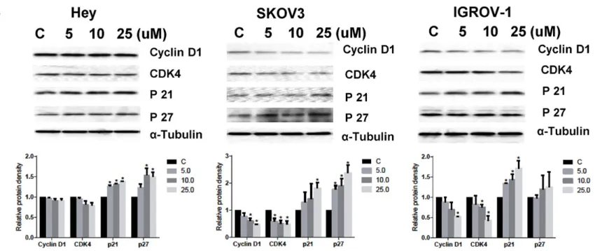
Compound 968 induced cell cycle G1 arrest
Studies indicate that glutamine is necessary for cell cycle progression [17]. We examined the effect of compound 968 on cell cycle pro-gression in all three ovarian cancer cell lines and found that treatment with compound 968 for 48 h result-ed in G1 phase cell cycle arrest. In addition to an increased proportion of cells in G1 phase, we noted a concomitant decreased proportion of cells in S phase (Figure 2A). Western blotting showed compound 968 treatment caused reduced pro-tein expression of CDK4 and cyclin D1 with increased expression of p21 and p27 (Figure 2B). These results are consistent with the effects of glutamine metabolism on cell cycle changes.
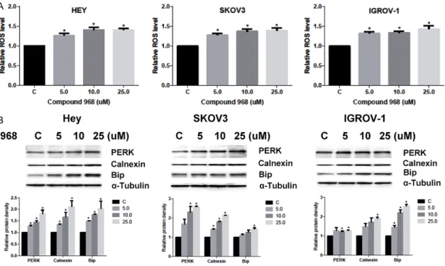
Compound 968 induced cell stress Figure 1. Compound 968 inhibited ovarian cancer cells proliferation.
The ovarian cancer cell lines, HEY, SKOV3 and IGROV-1, were treated in DMEM/F12 medium containing 2.5 mM L-Glutamine and supplied with varying concentrations of compound 968 (0, 2, 5, 10, 25, 50 uM) for 5 days. Cell proliferation was assessed by MTT assay. Compound 968 inhibited cell proliferation in a dose dependent manner (A). Com-pound 968 reduced GLS1 (KGA and GAC) expression after 24 h treat-ment (B). HEY cells were treated with compound 968 for 24 h, GDH activity was measured by ELISA assay (C) (*P<0.05). Data are shown as mean + SEM of three experiments.
and oxidative stress [4, 10], we tested the effects of compound 968 on the production of intracellular reactive oxygen species (ROS) and protein expression of endoplasmic reticulum stress. We treated all three ovarian cancer cell lines with compound 968 for 24 h. We found
that treatment with compound 968
signifi-cantly increased ROS production in a dose dependent manner (Figure 3A). Western blot-ting showed the protein expression of prot- ein kinase-like endoplasmic reticulum kinase (PERK), calnexin and binding immunoglobulin protein (BiP) increased after treatment with compound 968 (Figure 3B), suggesting that
compound 968 induces significant cellular
stress in ovarian cancer cells.
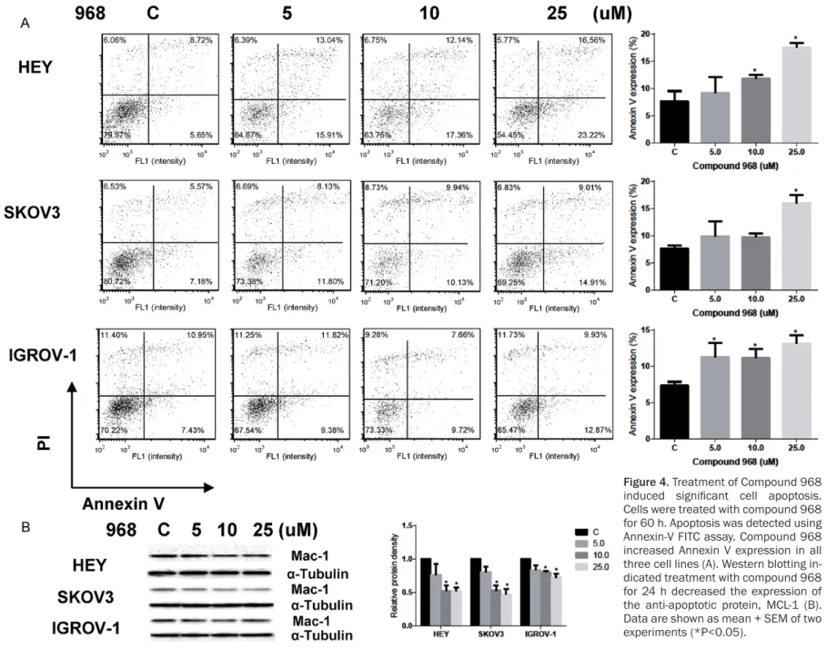
Compound 968 induced cell apoptosis
The effect of compound 968 on apoptosis was evaluated in all three ovarian cancer cells. We treated the cell lines with varying concentra-tions of compound 968 for 60 h, and found
sig-nificantly increased expression of Annexin V
with increasing concentrations of compound 968 (Figure 4A). We performed western blot-ting and found decreased expression of MCL-1,
a member of the Bcl-2 family, after 24 h treat-ment with compound 968 (Figure 4B). These results demonstrate that treatment with com-pound 968 results in increased apoptosis in ovarian cancer cell lines.
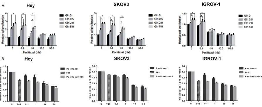
Compound 968 sensitized paclitaxel in ovarian cancer cells
Inhibition of glutaminolysis has been previously shown to sensitize paclitaxel in paclitaxel-resis-tant breast cancer cells [18]. We assessed the effect of glutamine and compound 968 on sen-sitivity to paclitaxel in ovarian cancer cell lines. As expected, increasing glutamine concentra-tions resulted in increased cell proliferation and decreased cell sensitivity to low dose pacli-taxel in all three ovarian cancer cell lines (Figure 5A). MTT assays demonstrated that the combi-nation of 5 uM of compound 968 with paclitax-el at varying concentrations resulted in syner-gistic inhibitory effects in all three ovarian can-cer cell lines after 72 h treatment (Figure 5B, CI<1). These results suggest that compound 968 treatment increased the sensitivity of the ovarian cancer cells to paclitaxel.
Figure 4. Treatment of Compound 968
induced significant cell apoptosis.
Discussion
We found that compound 968 inhibits cell pro-liferation, induces G1 phase cell cycle arrest, and initiates apoptotic cell death in the HEY, SKOV3 and IGROV-1 ovarian cancer cell lines in a dose dependent manner. Increased ROS pro-duction was also seen with increasing doses of compound 968. Moreover, low concentrations of glutamine resulted in increased sensitivity of ovarian cancer cells to paclitaxel, while
com-pound 968 treatment resulted in significant
synergy when combined with paclitaxel to inhib-it ovarian cancer cell growth. These results sup-port a role for targeting GLS1 or inhibiting gluta-mine metabolism as a valuable strategy for the treatment of ovarian cancer [6, 10, 13].
Glutamine is a major source of carbon and energy to promote cell growth and viability in cancer. The activity of glutaminase (GLS) and glutamine levels correlate with cancer cell liferation in vitro [19]. High level of GLS1 pro-tein expression is associated with worse overall survival in patients with ovarian cancer and silencing of GLS1 via siRNA in glutamine depen-dent SKOV3 cell-bearing mice results in a
sig-nificant reduction in tumor weight and tumor
volume when compared to controls [20]. The depletion of glutamine results in inhibition of cell proliferation, induction of G1 phase cell cycle arrest, and apoptosis through mTOR/S6 pathway in ovarian cancer cells. GLS knock-down results in reduced mitochondrial mem-brane potential in glioma cells, suggesting that mitochondrial dysfunction plays a role in induc-ing apoptosis with inactivity of GLS [21]. Recent studies indicate that the response to glutamine deprivation in cancer cells is dependent on their different genetic and epigenetic back-ground [9, 13].
The mechanisms of action of compound 968’s inhibition on cancer cell proliferation are less well characterized. Treatment of MDA-MB-231 cells with compound 968 resulted in downr- egulation of 11 anti-apoptotic genes, upregu- lation of several critical tumor suppressor genes (including VHL, MGMT and FHIT), and alternations of the expression for genes rela- ted to cell cycle progression, suggesting that inhibition of cell growth by compound 968 was related to induction of apoptosis and cell cycle arrest in breast cancer cells. These chan- ges in gene expression were accompanied by
an increase in histone H4K16ac and a decrease in H3K4me3 suggesting that altered regulation of cell cycle progression through histone
modi-fications may also play a role [13, 22, 23]. In
this study, we evaluated the impact of com-pound 968 on cell cycle arrest and apoptosis in three ovarian cancer cell lines. Our results show that compound 968 inhibited cell growth through induction of G1 phase cell cycle arrest and augmented Annexin-V expression, along with decreased expression of CDK4, cyclin D1 and MCL-1, and increased p21 and p27 expres-sion in all three cell lines. These results are consistent with experiments showing depriva-tion of glutamine causes apoptosis and cell cycle arrest in some types of cancer [10, 24-26].
Deprivation of glutamine in cell culture can increase ROS production and induce endop- lasmic reticulum stress [10]. Glutamine met- abolism protects against oxidative stress by increasing reduced glutathione (GSH) levels, an antioxidant, and providing a source of nico-tinamide adenine dinucleotide phosphate (NADPH) [27]. Depletion of GLS1 by shRNA exhibited lower levels of both GSH and oxidi- zed glutathione (GSSG) [21], indicating that GLS1 activity was involved in the process of oxidative stress. Treatment with BPTES, an allosteric inhibitor of glutaminase, in lung can-cer cells resulted in increased ROS levels
through inhibition of glucose flow into the
induced by compound 968. The complexity of cellular oxidative stress and endoplasmic reti- culum stress and the exact mechanisms by which compound 968 induces both stresses provide opportunities for further investigation. Hence, in combination with the above results, we believe compound 968 inhibits cell prolifer-ation through multiple pathways including ROS formation, cell cycle arrest and apoptosis. Paclitaxel is a standard chemotherapeutic agent for the treatment of ovarian cancer. It has been reported that paclitaxel can increase glutamine uptake and GLS1 expression in breast cancer cells. High expression of GLS1 and upregulation of glutamine metabolism are associated with paclitaxel resistant breast cancer cells and knockdown of GLS1 by siRNA results in sensitization of paclitaxel-resistant breast cancer cells to paclitaxel [18]. Gluta- mine deprivation creates synthetic lethality to paclitaxel in breast and prostate cell lines bearing K-Ras-mutations [26]. Targeting GLS1 by CB-839, a novel selective glutaminase in-
hibitor, significantly inhibited tumor cell growth
in vitro and in vivo [25, 31, 32]. CB-839 in com-bination with paclitaxel largely increased the sensitivity to paclitaxel in xenograft models of triple-negative breast cancer [31]. After treat-ing the ovarian cancer cell lines with different concentrations of glutamine in combination with paclitaxel, we observed that low concen-trations or depletion of glutamine considerably increased the sensitivity of the cells to low doses of paclitaxel in all three cell lines. We also found that the combination of compound
968 and paclitaxel significantly and synergisti -cally increased the sensitivity of the ovarian cancer cell lines to paclitaxel. Together, our results indicate that glutaminase appears to be a promising therapeutic target for ovarian can-cer and worthy of further investigation.
Acknowledgements
This work was generously supported by the Steelman Fund (Bae-Jump VL). This work was also supported by National Natural Science Foundation of China (81372778, Sheng X and Zhou C).
Disclosure of conflict of interest None.
Address correspondence to: Drs. Chunxiao Zhou and Victoria L Bae-Jump, Division of Gynecologic
Oncology; Lineberger Comprehensive Cancer Cen- ter, University of North Carolina, Chapel Hill, NC 27599, USA. Tel: 919-966-3270; E-mail: czhou@ med.unc.edu (CXZ); Tel: 843-4899; Fax: 919-843-5387; E-mail: [email protected] (VLB)
References
[1] Siegel RL, Miller KD and Jemal A. Cancer sta-tistics, 2016. CA Cancer J Clin 2016; 66: 7-30. [2] Le A, Lane AN, Hamaker M, Bose S, Gouw A,
Barbi J, Tsukamoto T, Rojas CJ, Slusher BS, Zhang H, Zimmerman LJ, Liebler DC, Slebos RJ, Lorkiewicz PK, Higashi RM, Fan TW and Dang CV. Glucose-independent glutamine me-tabolism via TCA cycling for proliferation and survival in B cells. Cell Metab 2012; 15: 110-121.
[3] Mates JM, Segura JA, Martin-Rufian M, Cam -pos-Sandoval JA, Alonso FJ and Marquez J. Glu-taminase isoenzymes as key regulators in metabolic and oxidative stress against cancer. Curr Mol Med 2013; 13: 514-534.
[4] Daye D and Wellen KE. Metabolic reprogram-ming in cancer: unraveling the role of gluta-mine in tumorigenesis. Semin Cell Dev Biol 2012; 23: 362-369.
[5] Katt WP and Cerione RA. Glutaminase regula-tion in cancer cells: a druggable chain of events. Drug Discov Today 2014; 19: 450-457. [6] Szeliga M, Bogacinska-Karas M, Rozycka A,
Hilgier W, Marquez J and Albrecht J. Silencing of GLS and overexpression of GLS2 genes co-operate in decreasing the proliferation and vi-ability of glioblastoma cells. Tumour Biol 2014; 35: 1855-1862.
[7] Lukey MJ, Wilson KF and Cerione RA. Thera-peutic strategies impacting cancer cell gluta-mine metabolism. Future Med Chem 2013; 5: 1685-1700.
[8] Mohamed A, Deng X, Khuri FR and Owonikoko TK. Altered glutamine metabolism and thera-peutic opportunities for lung cancer. Clin Lung Cancer 2014; 15: 7-15.
[9] Hensley CT, Wasti AT and DeBerardinis RJ. Glu-tamine and cancer: cell biology, physiology, and clinical opportunities. J Clin Invest 2013; 123: 3678-3684.
[10] Yuan L, Sheng X, Willson AK, Roque DR, Stine JE, Guo H, Jones HM, Zhou C and Bae-Jump VL. Glutamine promotes ovarian cancer cell prolif-eration through the mTOR/S6 pathway. Endocr Relat Cancer 2015; 22: 577-591.
[12] Kahlert UD, Cheng M, Koch K, Marchionni L, Fan X, Raabe EH, Maciaczyk J, Glunde K and Eberhart CG. Alterations in cellular metabo-lome after pharmacological inhibition of Notch in glioblastoma cells. Int J Cancer 2016; 138: 1246-1255.
[13] Simpson NE, Tryndyak VP, Pogribna M, Beland FA and Pogribny IP. Modifying metabolically sensitive histone marks by inhibiting gluta-mine metabolism affects gene expression and alters cancer cell phenotype. Epigenetics 2012; 7: 1413-1420.
[14] Simpson NE, Tryndyak VP, Beland FA and Pogribny IP. An in vitro investigation of meta-bolically sensitive biomarkers in breast cancer progression. Breast Cancer Res Treat 2012; 133: 959-968.
[15] Katt WP, Ramachandran S, Erickson JW and Cerione RA. Dibenzophenanthridines as inhibi-tors of glutaminase C and cancer cell prolifera-tion. Mol Cancer Ther 2012; 11: 1269-1278. [16] Wang JB, Erickson JW, Fuji R, Ramachandran
S, Gao P, Dinavahi R, Wilson KF, Ambrosio AL, Dias SM, Dang CV and Cerione RA. Targeting mitochondrial glutaminase activity inhibits on-cogenic transformation. Cancer Cell 2010; 18: 207-219.
[17] Moncada S, Higgs EA and Colombo SL. Fulfill -ing the metabolic requirements for cell prolif-eration. Biochem J 2012; 446: 1-7.
[18] Fu A, Yu Z, Song Y and Zhang E. Silencing of glutaminase 1 resensitizes Taxol-resistant breast cancer cells to Taxol. Mol Med Rep 2015; 11: 4727-4733.
[19] Sappington DR, Siegel ER, Hiatt G, Desai A,
Penney RB, Jamshidi-Parsian A, Griffin RJ and
Boysen G. Glutamine drives glutathione syn-thesis and contributes to radiation sensitivity of A549 and H460 lung cancer cell lines. Bio-chim Biophys Acta 2016; 1860: 836-843. [20] Yang L, Moss T, Mangala LS, Marini J, Zhao H,
Wahlig S, Armaiz-Pena G, Jiang D, Achreja A, Win J, Roopaimoole R, Rodriguez-Aguayo C, Mercado-Uribe I, Lopez-Berestein G, Liu J, Tsu-kamoto T, Sood AK, Ram PT and Nagrath D. Metabolic shifts toward glutamine regulate tu-mor growth, invasion and bioenergetics in ovarian cancer. Mol Syst Biol 2014; 10: 728. [21] Martin-Rufian M, Nascimento-Gomes R, Higue
-ro A, Crisma AR, Campos-Sandoval JA, Gomez-Garcia MC, Cardona C, Cheng T, Lobo C, Segu-ra JA, Alonso FJ, Szeliga M, Albrecht J, Curi R, Marquez J, Colquhoun A, Deberardinis RJ and Mates JM. Both GLS silencing and GLS2 over-expression synergize with oxidative stress against proliferation of glioma cells. J Mol Med (Berl) 2014; 92: 277-290.
[22] Serrano L, Martinez-Redondo P, Marazuela-Duque A, Vazquez BN, Dooley SJ, Voigt P, Beck DB, Kane-Goldsmith N, Tong Q, Rabanal RM,
Fondevila D, Munoz P, Kruger M, Tischfield JA
and Vaquero A. The tumor suppressor SirT2 regulates cell cycle progression and genome stability by modulating the mitotic deposition of H4K20 methylation. Genes Dev 2013; 27: 639-653.
[23] Grandy RA, Whitfield TW, Wu H, Fitzgerald MP,
VanOudenhove JJ, Zaidi SK, Montecino MA, Lian JB, van Wijnen AJ, Stein JL and Stein GS. Genome-Wide Studies Reveal that H3K4me3
Modification in Bivalent Genes Is Dynamically
Regulated during the Pluripotent Cell Cycle and Stabilized upon Differentiation. Mol Cell Biol 2015; 36: 615-627.
[24] Yuneva M, Zamboni N, Oefner P,
Sachidanan-dam R and Lazebnik Y. Deficiency in glutamine
but not glucose induces MYC-dependent apop-tosis in human cells. J Cell Biol 2007; 178: 93-105.
[25] Jacque N, Ronchetti AM, Larrue C, Meunier G, Birsen R, Willems L, Saland E, Decroocq J, Ma-ciel TT, Lambert M, Poulain L, Hospital MA, Su-jobert P, Joseph L, Chapuis N, Lacombe C, Moura IC, Demo S, Sarry JE, Recher C, Mayeux P, Tamburini J and Bouscary D. Targeting gluta-minolysis has antileukemic activity in acute myeloid leukemia and synergizes with BCL-2 inhibition. Blood 2015; 126: 1346-1356. [26] Saqcena M, Mukhopadhyay S, Hosny C,
Al-hamed A, Chatterjee A and Foster DA. Blocking anaplerotic entry of glutamine into the TCA cycle sensitizes K-Ras mutant cancer cells to cytotoxic drugs. Oncogene 2015; 34: 2672-2680.
[27] Shanware NP, Mullen AR, DeBerardinis RJ and Abraham RT. Glutamine: pleiotropic roles in tu-mor growth and stress resistance. J Mol Med (Berl) 2011; 89: 229-236.
[28] Ulanet DB, Couto K, Jha A, Choe S, Wang A, Woo HK, Steadman M, DeLaBarre B, Gross S, Driggers E, Dorsch M and Hurov JB. Mesenchy-mal phenotype predisposes lung cancer cells to impaired proliferation and redox stress in response to glutaminase inhibition. PLoS One 2014; 9: e115144.
[29] Sanders YY, Liu H, Zhang X, Hecker L, Bernard K, Desai L, Liu G and Thannickal VJ. Histone
modifications in senescence-associated resis -tance to apoptosis by oxidative stress. Redox Biol 2013; 1: 8-16.
[31] Gross MI, Demo SD, Dennison JB, Chen L, Chernov-Rogan T, Goyal B, Janes JR, Laidig GJ, Lewis ER, Li J, Mackinnon AL, Parlati F, Rodri-guez ML, Shwonek PJ, Sjogren EB, Stanton TF, Wang T, Yang J, Zhao F and Bennett MK. Anti-tumor activity of the glutaminase inhibitor CB-839 in triple-negative breast cancer. Mol Can-cer Ther 2014; 13: 890-901.