THE ROLE OF CELLULAR AND CHEMICAL SIGNALING WITHIN THE NUCLEUS ACCUMBENS IN VALUE-BASED DECISION MAKING BEHAVIORS
Jonathan Adam Sugam
A dissertation submitted to the faculty of the University of North Carolina at Chapel Hill in partial fulfillment of the requirements for the degree of Doctor of Philosophy in the
Department of Psychology.
Chapel Hill 2013
Approved By: Regina M. Carelli Todd E. Thiele
Rita A. Fuchs Lokensgard Mark Hollins
ii ©2013
iii ABSTRACT
JONATHAN ADAM SUGAM: The role of cellular and chemical signaling within the nucleus accumbens in value-based decision making behaviors
(Under the direction of Regina M. Carelli)
iv
vi
ACKNOWLEDGEMENTS
vii PREFACE
viii
TABLE OF CONTENTS
LIST OF TABLES ...x
LIST OF FIGURES ...xi
LIST OF ABBREVIATIONS ...xiii
Chapter I. GENERAL INTRODUCTION ...1
Associative learning and decision making ...2
Nucleus accumbens circuitry ...6
The nucleus accumbens and goal-directed action ...11
The mesolimbic dopamine system ...14
Role of mesolimbic dopamine activity in associative learning and decision making ...19
Goals of this dissertation ...23
Specific aims ...24
II. NUCLEUS ACCUMBENS NEURONS ENCODING OF REWARD RELATED INFORMATION TRACKS RISK TAKING BEHAVIOR ...27
Introduction ...29
Methods ...32
Results ...39
Discussion ...54
ix
Introduction ...63
Methods ...66
Results ...73
Discussion ...83
IV. OPTOGENETIC STIMULATION OF PHASIC DOPAMINE RELEASE IN THE NUCLEUS ACCUMBENS AND VALUE BASED DECISION MAKING ...90
Introduction ...92
Methods ...95
Results ...106
Discussion ...123
V. GENERAL DISCUSSION ...132
Summary of experiments ...,,132
General discussion and relevance of findings ...135
Future directions ...142
Concluding remarks ...145
x
LIST OF TABLES Table
2.1 Distribution of accumbens cellular activity
by trial type ...43 2.2 Distribution of accumbens cellular activity
xi
LIST OF FIGURES Figure
1.1 Simplified schematic of afferent and efferent
connections of the nucleus accumbens ...9 2.1 Schematic representation of risky decision making task ...35 2.2 Individual differences in risky decisions making ...40 2.3 Nucleus accumbens neurons are activated during
different components of the risky decision making task ...42 2.4 Nucleus accumbens neurons display cue-selective
encoding for safe versus risk cue presentations ...45 2.5 Nucleus accumbens neurons display prepress-selective
encoding for safe versus risk lever presses ...47 2.6 Reward omission processing in the nucleus accumbens...49 2.7 Individual risk preferences related to reward
omission processing...51 2.8 Histological verification of recording array wires
in the nucleus accumbens core and shell ...53 3.1 Schematic representation of delay discounting task ...67 3.2 Choice behavior during delay discounting task ...74 3.3 Dopamine release encodes the relative value of
cue presentations during delay discounting ...76 3.4 Differential dopamine release is correlated with
response allocation in delay discounting ...78 3.5 Dopamine signaling during choice trials on the
delay discounting task ...80 3.6 Dopamine release to delayed reward delivery ...81 3.7 Anatomical distribution of electrode recording sites
xii
4.2 Schematic representation of decision making task ...103 4.3 Channelrhodopsin expression in dopaminergic cells ...107 4.4 Optical stimulation of dopamine neurons promotes
phasic dopamine release ...110 4.5 Optical stimulation of dopamine terminals in the nucleus
accumbens is sufficient to promote goal-directed behavior...114 4.6 Lever press training for TH::Cre(+/-) versus control rats ...115 4.7 Delay and magnitude discounting for TH::Cre(+/-)
and control groups ...117 4.8 Optical stimulation of dopamine terminals modulates delay
xiii
LIST OF ABBREVIATIONS
ANOVA Analysis of variance BLA Basolateral amygdala ChR2 Channelrhodopsin CR Conditioned response CS Conditioned stimulus FR Fixed ratio
FSCV Fast-scan cyclic voltammetry LH Lateral hypothalamus
MSN Medium spiny neuron NAc Nucleus accumbens OFC Orbitofrontal cortex
PCA Principal component analysis PEH Peri-event histogram
PFC Prefrontal cortex
SEM Standard error of the mean SN Substantia nigra
CHAPTER 1 INTRODUCTION
2
integrates this information and impacts behavior through its connections with motor-related regions (Zahm and Brog, 1992a; Zahm, 1999), supporting the theory that the NAc is a critical site of convergence for reward-related decision making. While a large amount of work has contributed to the characterization of NAc signaling and its critical role in decision making (Cardinal et al., 2001; Cardinal and Howes, 2005; Ghods-Sharifi and Floresco, 2010; Stopper and Floresco, 2011; Stopper et al., 2013), the functional contributions of these signals, and how they may be causally linked to controlling appropriate behavioral responding, has not been extensively studied. Therefore, the experiments outlined in this dissertation seek to further characterize the role of NAc cellular signaling and the associated dopaminergic contributions to decision making based on several factors, including subjective value, reward devaluation/impulsive choice, and the causal link between this signaling and appropriate responding. Here, a brief introduction is provided of the exhaustive literature on the role of the NAc, and mesolimbic dopamine system, in reward processing and goal-directed behavior. First, this chapter will provide a review of the overall relevance of the processes that govern learning and choice behavior. Next, the cellular and systems-level mechanisms underlying neural communication within the NAc is discussed, with emphasis on its dopaminergic input from the VTA. Finally, these ideas will be integrated in order to examine theoretical and empirical links between dopamine release in the NAc, NAc neural activity, and reward-related decision making.
Associative learning and decision making
3
and consume rewards. Within this framework there are two general types of associative processes that have developed. The first type of learning is stimulus-outcome learning (known as Pavlovian or classical conditioning) in which an organism learns to associate a previously neutral stimulus (conditioned stimulus CS) with a biologically significant outcome such as food (the unconditioned stimulus US). Following several pairings of the CS with the US, the CS gains biological significance and can then influence ongoing behavior towards collecting resources (Pavlov and Anrep, 1927; Dickinson, 1980; Rescorla, 1988b). Pavlovian stimuli are presented non-contingently to the organism such that behavioral actions are not required to produce the outcome. Importantly, this type of learning is dependent on several factors that influence the ability to associate predictive cues with appropriate outcomes including the identity and value of the US, the identity of the predictive CS, the contingency between the CS and US, and the temporal relationship between the CS and US, among other factors (Rescorla, 1968, 1969; Rescorla, 1988b; Rescorla, 1988a). As such, Pavlovian conditioning is not simply a reflex, but instead reflects a complex understanding of the relationship of the motivational state to distinct and important stimuli in the environment.
4
vigor of responding are dependent on several factors related both to the action and the outcome including, the amount of responding necessary to produce the outcome, the frequency with which the outcome is presented, the identity and value of the outcome, among other factors (Ferster and Skinner, 1957). Importantly, action-outcome responding can be differentiated from habitual responding for outcomes. The performance of habitual actions depends on the association between a predictive stimulus and associated action, regardless of the outcome, while action-outcome responding is mediated by the associations between the action and the consequences of action, and thus rely on separate brain circuits. In particular action-outcome behaviors are dependent on the NAc while habitual responding is dependent on the dorsal striatum (Everitt and Robbins, 2005; Takahashi et al., 2007; Everitt et al., 2008; Dezfouli and Balleine, 2012). Further, unlike habitual responding, action-outcome responses are sensitive to changes in the value of outcomes (Adams, 1982) and are sensitive to changes in the causal relationship between the action and outcome delivery (Dickinson, 1998). Maladaptive behaviors, such as drug addiction, are often characterized as a shift from action-outcome associations to habitual actions (Robbins and Everitt, 1999; Everitt et al., 2001; Everitt and Robbins, 2005; Everitt et al., 2008).
5
behavioral responding on their own (termed conditioned reinforcers), such that animals will perform operant actions for this cue delivery, even in the absence of the primary reward itself (Zimmerman, 1957). Numerous studies have also shown that Pavlovian cues can potentiate operant responding, even when there is no association between the cue and the response, a behavioral effect known as Pavlovian-to-instrumental transfer (PIT) (Estes, 1948; Rescorla and Solomon, 1967; Saddoris et al., 2011). In a PIT task, animals are first trained that a cue predicts a positive outcome. Next, animals are separately trained that a behavioral response also leads to reinforcement. When animals are engaged in behavioral responses, presentation of the reward paired cue functions to invigorate responding, increasing behavioral response rates, suggesting that the cue also holds some motivational value.
6
performance results in learning (i.e. representations of future actions are updated to optimize future choices) (Rangel et al., 2008). This type of value-based decision making can be modeled in humans and animals by exposing organisms to situations in which there is a choice between rewards of different value. For example, humans (Coffey et al., 2003; Green and Myerson, 2004; Hariri et al., 2006; Prévost et al., 2010) and animals (Cardinal et al., 2001; Roesch et al., 2007; Roesch et al., 2009; Day et al., 2010) show similar patterns of choice behavior based on the time the organism spends waiting for the reward, choosing the larger option less often as the delay to reward increases. Similar patterns of discounting are also seen when organisms must choose between rewards based on reward cost and risk (St Onge and Floresco, 2008; Floresco and Whelan, 2009; Simon et al., 2009; Day, 2010; Day et al., 2010; Gan et al., 2010; Prévost et al., 2010; Sugam et al., 2012). These results demonstrate that organisms use cost-benefit analysis to guide selection between actions to maximize resources, even when both actions are rewarded.
Nucleus accumbens circuitry
Cellular and chemical composition of the nucleus accumbens: In order to learn
7
Kawaguchi, 1997) with a large radially projecting dendritic tree (about 250μm in diameter) (Preston et al., 1980; Groves, 1983; Gerfen, 1988). These cells have axons that project from the NAc to areas such as the substantia nigra, ventral pallidum, and lateral hypothalamus to influence behavior (Gerfen, 1988; Kawaguchi, 1997; Zahm, 1999). Importantly, the NAc is not a homogeneous structure as MSNs have specific characteristics that define a complex circuitry. For example, immunohistomchemical markers reveal that MSNs contain enkephalin, dynorphin, substance P, and neurotensin, and the specific type of marker predict the separate pathway and output structure projections of each MSN (Meredith, 1999). Further control of this specific circuitry comes from the dense dopaminergic projection from the VTA. The majority of MSNs express either D1-like or D2-like receptors, with very few expressing both (17% coexpress both in the NAc shell and 6% coexpress both in the NAc core) (Bertran-Gonzalez et al., 2008). D1-like labeled MSNs expressing dynorphin and D2-like labeled MSNs expressing enkephalin, and thus may represent separate projection systems. As such dopamine may be playing a specific function in modulating certain projection pathways from the NAc (Le Moine and Bloch, 1995).
8
the likelihood of burst firing of MSNs (Cacciapaglia et al., 2011), classifying dopamine as a key neuromodulator of neuronal function in the NAc (Goto and Grace, 2005).
The other 5% of neurons in the NAc are considered local circuit or interneurons and are of two main types: cholinergic interneurons or GABAergic neurons (Groves, 1983; Meredith, 1999). The cholinergic interneurons are much larger in size (35 µm diameter soma) with radially emanating dendrites, and dendrites that are mostly devoid of spines. Importantly, these cholinergic interneurons can be differentiated from classic MSNs based both on morphology and firing rate. The baseline firing rate for MSNs is typically 1-3Hz exhibiting phasic bursts of activity while cholinergic interneurons display tonic firing rates and are the source of acetylcholine within the NAc (Kawaguchi et al., 1995; Meredith, 1999). GABAergic interneurons make up the rest of the neurons within the NAc. These neurons comprise at least three different populations; parvalbumin, calretinin, or somatostatin and neuropeptide Y positive populations and are differentiated from MSNs based on their tonic activity with brief high frequency bursts. Further, the oscillatory behavior between these GABAergic interneurons and MSNs is critical for mediating normal MSN activity (Berke et al., 2004).
Afferent and efferent connections of the nucleus accumbens: The NAc has been proposed to
9
hippocampus (Brog et al., 1993), as well as thalamic regions (Finch, 1996; MacAskill et al., 2012) Further, the NAc receives a dense dopaminergic input from the VTA (approximately 85% of VTA dopamine neurons project to the NAc) (Fields et al., 2007). In turn, the NAc sends efferent projections to nuclei that organize motor behavior including the ventral pallidum and subthalamic nucleus (Nauta et al., 1978).
10
modulated by VTA activity (Brady and O'Donnell, 2004). Thus, NAc neurons are in a prime position to coordinate associative learning from afferents with action selection in downstream motor targets.
Structural and functional subregions of the nucleus accumbens: the core and shell:
11
pallidum (ventromedial district) and VTA (Zahm, 1999). These dissociable afferent and efferent projection patterns suggest that the core and shell differentially participate in reward related behaviors (Saddoris et al., 2012).
The nucleus accumbens and goal-directed action
Given the functional connectivity of the NAc as a corticolimbic motor integration center, early studies sought to inactivate or lesion the NAc to evaluate how disruptions of this portion of the circuit affected motivated responding. This early work found an important role for the NAc in contributing to appetitive and consummatory phases of reward responding (Stratford and Kelley, 1997; Berridge and Robinson, 1998; Kelley, 2004). In particular, studies found that infusion of GABAA and GABAB agonists into the NAc shell induced feeding behaviors (likely through connections with the lateral hypothalamus) (Stratford and Kelley, 1997; Stratford and Kelley, 1999; Kelley, 2004) while administration of glutamate antagonists into the NAc (specifically the AMPA antagonist DNQX) also functioned to stimulate feeding behaviors (Stratford et al., 1998; Kelley, 2004). This data suggests that inhibition of NAc activity, and thus a disinhibition of downstream motor areas, was critical for the initiation of food related foraging behaviors. Interestingly, these early studies on the role of NAc activity in food intake found that the NAc shell was predominantly involved in these consummatory phases of reward delivery, again suggesting both a structural and functional division of the NAc core and shell. Recent evidence using optogenetic techniques has provided further evidence that activity of mesolimbic dopamine neurons are also a driving force of reward consumption behaviors (van Zessen et al., 2012).
12
technique, researchers have been able to record NAc neural activity in an intact animal as the animal performs reward related behaviors, therefore giving functional insight into how the NAc is critically involved in encoding reward related behaviors. When examining NAc neural activity during reward consumption, researchers first found that NAc neurons show decreases in firing during this period (Roitman et al., 2005; Taha and Fields, 2005a, 2006; Wheeler et al., 2008; Ambroggi et al., 2011), suggesting the functional link between NAc activity and the earlier pharmacological studies that showed that decreasing neural activity in the NAc induced feeding. This suggests that NAc neurons function to “gate” foraging or consummatory behaviors. Specifically reducing neural activity within the NAc releases motor areas from strong GABAergic inhibitions, releasing the “gate” on reward consumption behaviors. In support of this theory, another study found that individual ventral pallidum neurons (the downstream target of NAc projections) show increases in firing rate during reward consumption (Tindell et al., 2006).
13
learning and decision making processes, NAc neural encoding not only needs to track reward deliveries, but also the predictive stimuli associated with these rewards. In support, electrophysiological recordings have shown that NAc neurons also display increased and/or decreased cell firing to cues that predict future rewards (Carelli, 2000, 2004; Nicola et al., 2004a; Roitman et al., 2005; Day et al., 2006; Jones et al., 2010a; Saddoris et al., 2011), suggesting that the NAc is able to encode the association between reward predictive cues and positive outcomes. Further, ventral striatal neurons display differential activity to cue presentation based on the value of future outcomes (Schultz et al., 1992; Cromwell and Schultz, 2003; Kim et al., 2009; Roesch et al., 2009; Day et al., 2011), suggesting an ability to track specific features associated with cue presentations. Finally, NAc neurons also show phasic activity during the execution of behavioral responses (Carelli, 2002; Hollander and Carelli, 2005; Day et al., 2011; Saddoris et al., 2011), supporting a direct role for the NAc as a limbic-motor integrator.
14
actions, an intact NAc circuitry is necessary for learning the associations between cues and outcomes, actions and outcomes, and changes in reward value (Sokolowski and Salamone, 1998; Corbit et al., 2001; Cardinal et al., 2002; Cardinal and Cheung, 2005). Therefore, the NAc appears to be uniquely involved in associative processing of more complex situations rather than driving simple goal-directed actions. As such, researchers have begun to investigate how the NAc is implicated in behaviors in which organisms must use these complex associations, such as in value-based decision making. As discussed above, models of decision making expose subjects to cues that predict outcomes of different value, and animals are allowed to make choices between these different options. Damage to the NAc has repeatedly been shown to disrupt the ability of animals to show normal decision making in these experiments. For example, animals with lesions or temporary inactivation of the NAc were impaired in decisions based on both reward delay, effort and probability, choosing smaller, certain, immediate rewards much more often, even when this was the less advantageous option (Cardinal et al., 2001; Cardinal and Cheung, 2005; Cardinal and Howes, 2005; Floresco et al., 2007; Ghods-Sharifi and Floresco, 2010; Stopper and Floresco, 2011). Further, inactivation of the NAc shell resulted in the inability to choose the appropriate response when evaluating rewards of different magnitude, choosing the larger reward less often then controls (Stopper and Floresco, 2011). This evidence suggests the NAc plays a critical role in the association of reward related cues and behaviors in appropriate reward seeking, especially when presented with several options of different value.
The mesolimbic dopamine system
Anatomy of the VTA: Decades of research on the mesolimbic dopamine system have shown
15
guide appropriate behaviors (Schultz et al., 1997; Schultz, 1998; Waelti et al., 2001), and has recently been shown to be causally linked to cue-outcome learning (Stuber et al., 2008; Tsai et al., 2009; Zellner et al., 2009; Zellner and Ranaldi, 2010). The mesolimbic dopamine projections originate from dopamine cell bodies in the VTA which is ventral to the red nucleus and medial to the dopamine rich substantia nigra (SN). As the VTA lacks clear borders, it can be distinguished from the SN based on both the projection patterns of the dopamine neurons and a unique afferent projection from the lateral hypothalamic area (Nauta, 1958; Ikemoto, 2007). The VTA and SN comprise two of the main projection sites of dopamine neurons, and importantly provide two distinct circuits of dopamine projections. The SN comprises the nigro-striatal pathway of dopamine release and sends the majority of its projections to the dorsal striatum (caudate and putamen). Conversely, the VTA projects to diverse brain targets including the PFC, amygdala, hippocampus, ventral pallidum and NAc. Importantly, the majority of dopamine neurons from the VTA project to the NAc core and shell (Anden et al., 1964; Swanson, 1982; Fields et al., 2007; Ikemoto, 2007). Dopamine neurons comprise the majority of the VTA, while GABAergic neurons comprise a smaller population of cells that function to regulate dopaminergic cellular activity within the VTA (Kalivas et al., 1990; Olson and Nestler, 2007). Further, the VTA receives inputs back to dopamine neurons from the hypothalamus, dorsal raphe, NAc, pallidum, and amygdala (Watabe-Uchida et al., 2012).
Release patterns of mesolimbic dopamine neurons: Mesolimbic dopamine neurons display
16
neurons will show increased burst like activity which results in significant increases in dopamine release, known as “phasic” activity (Grace and Bunney, 1984; Grace, 1991; Chergui et al., 1993; Grace, 2000). Importantly, this phasic activation of dopamine neurons results in a robust, although transient, increase in dopamine concentration (Garris et al., 1994; Garris et al., 1999; Brady, 2004). Phasic burst firing of dopamine neurons and subsequent terminal release is dependent on glutamatergic activity within the VTA, specifically resulting from stimulation of NMDA receptors (Chergui et al., 1993; Sombers et al., 2009). NMDA receptor antagonism within the VTA results in the elimination of phasic release events within the NAc while not disrupting tonic baseline levels (Sombers et al., 2009). Importantly, phasic dopamine release is not confined to the synaptic cleft and is able to spill over into outlying areas, supporting the role of dopamine as a volume transmitter such that phasic release of dopamine can modulate relatively large territories of neural tissue (Rice and Cragg, 2008). Importantly, phasic dopamine release within the terminal region is highly variable across the microenvironment, suggesting heterogeneity in dopamine release (Venton et al., 2003).
17
in the NAc can also increase dopamine release, presumably through presynaptic mechanisms (Imperato et al., 1990; Howland, 2002). The duration of dopamine release at the level of the NAc is tightly regulated by the presence of dopamine transporters, which function to terminate dopamine signaling through reuptake mechanisms (Cragg and Rice, 2004). Dopamine transporters are highly expressed in both the NAc core and shell, although with greater density in the NAc core (Jones et al., 1996), supporting differential patterns of dopamine transmission within these regions. Dopamine transporters are also the site of action for several drugs of abuse, including cocaine and amphetamine, and blockade of dopamine transporters by these drugs functions to increase phasic dopamine levels and increases the time to dopamine reuptake (Addy et al., 2010).
Dopamine receptors: Once released from the neuron, dopamine can function at one of two
18
NAc to output regions based on receptor expression. Further, the different subtypes of receptors have a different affinity for dopamine, with D2-like receptors having a much higher affinity than D1-like receptors (Richfield et al., 1989; Missale et al., 1998). Therefore, in the presence of low levels of dopamine, such as during baseline tonic release periods, D2-like receptors are much more likely to be activated. In contrast, high concentration phasic burst events of dopamine release are much more likely to activate D1-like receptors. Both the anatomical and functional organization of dopamine receptors allow for the mesolimbic dopamine system to function in a highly dynamic manner.
Synaptic actions of dopamine within the nucleus accumbens: MSNs in the NAc receive
19
and LTD (Thomas et al., 2000; Ishikawa et al., 2013), and is dependent on which dopamine receptors are activated. This evidence suggests that dopamine release within the NAc can function to modulate specific synaptic connections, interacting with specific glutamatergic inputs depending on where dopamine neurons synapse, thus providing a dynamic system for the mediation of synaptic plasticity within specific target regions of the NAc.
Role of mesolimbic dopamine activity in associative learning and decision making
Several decades of research have tried to determine the precise role of mesolimbic dopamine activity in reward related behavior and have demonstrated that the blockade of dopamine receptors produced a decrease in goal-directed behavior for food and other rewards (Wise et al., 1978b; Wise et al., 1978a). Specifically, rats still worked for rewards with dopamine antagonists on board, however responding decreased across time. This suggested that dopamine functions to encode the “pleasurable” aspects of reward seeking. One of the leading preliminary hypotheses for dopamine function was the “anhedonia hypothesis” of reward. Proposed by Roy Wise in 1982, this hypothesis suggested that dopamine was the “rewarding” neurotransmitter and as such, dopamine release signaled rewards and this pleasure signal is what promoted goal-directed behaviors. Organisms would work to obtain rewards because the reward receipt “felt good” as a result of increased dopamine release (Wise, 1982; Wise, 2008). These findings initially led to the suggestion that dopamine
release in the NAc mediates the hedonic or “pleasure” aspects of rewarding stimuli, and,
in turn, that both natural and drug rewards could be defined by this common path of
activation. Further, this hypothesis suggested that neurological diseases that decrease
20
lines of evidence. For example, research has shown that dopamine mediates the “wanting” or how much an animal will work for a reward, but not how much an animal “likes” the reward. Specifically, dopamine depletions do not disrupt orofacial responses for hedonic rewards, suggesting that dopamine does not mediate the appetitive valence, hedonic value, or simply the “pleasure” associated with rewards (Berridge et al., 1989). Further, aversive stimuli have also been shown produce increases in dopamine release in the NAc and dorsal striatum, suggesting that the mesolimbic dopamine circuit is also important for aversive responding (Badrinarayan et al., 2012; Budygin et al., 2012; Lammel et al., 2012). Further, dopamine antagonists as well as lesions of the NAc disrupted reward related responding only when effort requirements were high, but not in simple response-outcome situations (Salamone et al., 2001; Salamone et al., 2002; Salamone et al., 2005). Taken together, these findings suggest that dopamine does not simply signal the hedonic or “pleasurable” aspects of reward-related behaviors, but instead supports a more complex role for the mesolimbic dopamine system in goal-directed behaviors.
21
during the reward period. Further, when a reward is unexpectedly omitted, there is a reduction in neural activity (Schultz et al., 1997). Schultz and colleagues believed that this dopamine signaling functioned as a “teaching signal” (Schultz et al., 1997) and as such this signaling is consistent with contemporary learning theory (Mirenowicz and Schultz, 1994; Waelti et al., 2001; Pan et al., 2005). Work from our laboratory has shown that this dopamine signal is transmitted to the terminal regions in the NAc, as electrochemical recordings of dopamine release in the NAc show similar patterns of activity to dopamine neural recordings during Pavlovian learning paradigms (Day et al., 2007). According to this hypothesis, activation of dopamine neurons during unexpected reward presentations signals an error in ongoing reward predictions. As cues come to predict future outcomes, the dopamine signaling shifts to these cues and acts as a predictor of future outcomes and no longer signals the reward delivery because this does not constitute a violation of reward predictions. By computing the difference between expected and actual outcomes, dopamine neurons are hypothesized to play a key role in reward-related learning. In support of its role in learning cue-outcome associations, pharmacological blockade of dopamine activity blocks the acquisition of Pavlovian learning (Di Ciano et al., 2001; Dalley et al., 2002; Zellner et al., 2009; Zellner and Ranaldi, 2010), while stimulation of phasic dopamine release is sufficient to promote associative learning (Tsai et al., 2009).
22
through receptor antagonists or dopamine lesion biases animals towards emitting more “impulsive” responses that lead to less desirable rewards, but require less effort (Sokolowski and Salamone, 1998; Salamone et al., 2001; Salamone et al., 2002; Ishiwari et al., 2004; Mingote et al., 2005), shorter latency to reward (Floresco et al., 2007) or higher probability of reinforcement (St Onge and Floresco, 2008; St. Onge et al., 2010). Further, dopamine neurons display dynamic encoding of reward value displaying increased activation for higher value rewards based on reward probability, delay, and magnitude (Fiorillo et al., 2003; Tobler et al., 2005; Kobayashi and Schultz, 2008). Value signaling of dopamine activity has also been observed in situations when animals are actively making decisions between two options, showing increased activity and dopamine release for the more valuable option (Roesch et al., 2007; Day et al., 2010; Sugam et al., 2012). This type of processing by dopamine neurons is hypothesized to be critical for decision making as it functions to broadcast information about reward value to striatal circuits that enable animals to maximize behaviors (Roesch et al., 2007; Day et al., 2010).
23
prediction is below the cost threshold the behavior is deemed worthwhile and the animal will perform the task. Conversely, options that are predicted to have low value will evoke much less dopamine release, and thus this signal of future value will fall above the cost threshold and will be deemed not worthwhile (Phillips et al., 2007). This model can also be applied to a situation in which animals are given concurrent choices with different values. Rather than serving as a threshold for performing a behavior or not, the dopamine system can be functioning to compare how two separate behaviors relate to the cost threshold and thus inform animals of which option is more worthwhile to guide choice behaviors. In support, dopamine neural activity and terminal release encode information about the best available option when animals are given a concurrent choice of options with different explicit value, irrespective of what the animal actually chooses (Roesch et al., 2007; Day et al., 2010; Sugam et al., 2012), thereby functioning to bias animals towards more valuable options. Thus, dopamine release in the NAc may play a key role in the evaluation of different responses and thus functions to promote appropriate action selection.
Goals of this dissertation
24
operant responses for rewards (Carelli et al., 2000; Carelli, 2002), and signal specific information during value-based decision making (Roesch et al., 2009; Day et al., 2011). Further, dopamine release within the NAc tracks cues that predict rewarding outcomes (Beyene et al., 2010; Day et al., 2010; Jones et al., 2010b; Sugam et al., 2012), operant responses for drug and natural rewards (Phillips et al., 2003b; Roitman et al., 2004), and is causally linked with behavioral conditioning (Tsai et al., 2009). However, little is known about how this NAc activity and dopamine signaling is implicated in more complex decision making behaviors based on subjective value, or as the value associated with reward predictive stimuli changes. Further, little is known about how dopamine release in the NAc terminals is causally linked with goal-directed behaviors and value-based decision making. The proposed studies seek to elucidate the specific role of NAc signaling and dopamine release in complex value-based decision making paradigms requiring animals to evaluate multiple aspects of reward value to make appropriate decisions.
Specific Aims:
25
extrinsic reward factors (Roesch et al., 2007; Day et al., 2010); however, little is known on how the NAc encodes the subjective or intrinsic value of behavioral responding. This aim will advance the existing literature by using a risky decision making task in which individual preferences determine if the risky or safe reward is more valuable. Individual NAc neurons will be monitored using in vivo electrophysiology to assess if NAc neurons differentially encode task related information about risk taking behaviors, including cue responding, behavioral choices, and reward deliveries.
26
reward delivery. As such, this study will clarify the role of phasic NAc dopamine release in encoding reward value as options change.
3 and 4. To determine if optical stimulation of dopamine terminals in the NAc is sufficient to promote motivated behavior, and guide value-based decision making.
CHAPTER 2
NUCLEUS ACCUMBENS NEURONS ENCODING OF REWARD RELATED INFORMATION TRACKS RISK TAKING BEHAVIOR
ABSTRACT
28
29 INTRODUCTION
Organisms must learn appropriate behaviors to secure the necessary resources for survival such as food, shelter, and mates. This behavior requires complex cost benefit decisions in which organisms evaluate potential risks and benefits of different courses of actions (Green and Myerson, 2004; Cardinal, 2006; Phillips et al., 2007; Rangel et al., 2008; Saddoris et al., 2013). Impairments in appropriate cost-benefit decision making is associated with several psychiatric disorders including drug and gambling addiction (Avanzi et al., 2004; Redish, 2004; Dodd et al., 2005; Schultz, 2011; Chang et al., 2012), as well as more complex disorders such as schizophrenia (Chang et al., 2012). As such, there is a growing interest in understanding how the brain encodes normal decision making, and how changes in this signaling may result in disordered decision making.
30
controls. Interestingly, lesioned animals avoided the risky lever, even when choosing it was more advantageous (Cardinal and Howes, 2005). Further, inactivation of the NAc biased animals away from larger rewards particularly when they were more uncertain (Stopper and Floresco, 2011). These observations suggest that NAc activity is critical for the evaluations of risks and making appropriate choices, and aberrations in this circuitry result in dysfunctional behaviors.
31
making decisions based on several factors including effort, delay, and magnitude (Roesch et al., 2009; Day et al., 2011).
32 METHODS
Animals
Male Sprague Dawley rats (n=17, Harlan Sprague Dawley, Indianapolis, IN), aged 90-120d and weighing 275-350g were used as subjects and were individually housed with a 12/12-h light/dark cycle. All experiments were conducted between 8:00am and 5:00pm. Animals were maintained at no less than 85% of pre-experimental bodyweights by food restriction (~10-15g of Purina laboratory chow each day in addition to approximately 1g of sucrose consumed during behavioral sessions) except during the post-operative recovery period when food was given ad libitum. All procedures were approved by the UNC Institutional Animal Care and Use Committee.
Surgery
33
via stainless steel screws and dental acrylic. All animals were allowed at least five post-operative recovery days before beginning training on the behavioral task.
Behavioral Training
34
35 Electrophysiological recordings
36
commercially available neurophysiological system (MAP System, Plexon, Inc., Dallas TX). Criteria for identifying different neurons on a single wire have been described in detail elsewhere (Chang et al., 1994; Nicolelis et al., 1997; Carelli et al., 1999). Briefly discrimination of individual waveforms corresponding to a single cell was accomplished using template analysis procedures provided by the neurophysiological software system. The template analysis procedure involves taking a ‘sample’ of the waveform and building a template of that extracellular waveform. Subsequent neurons that match this waveform are included as the same cell. Principal component regression of waveform data was conducted using the Offline Sorter Program (Plexon, Inc) to further separate waveforms recorded from the same microwire.
Neural Analysis
37
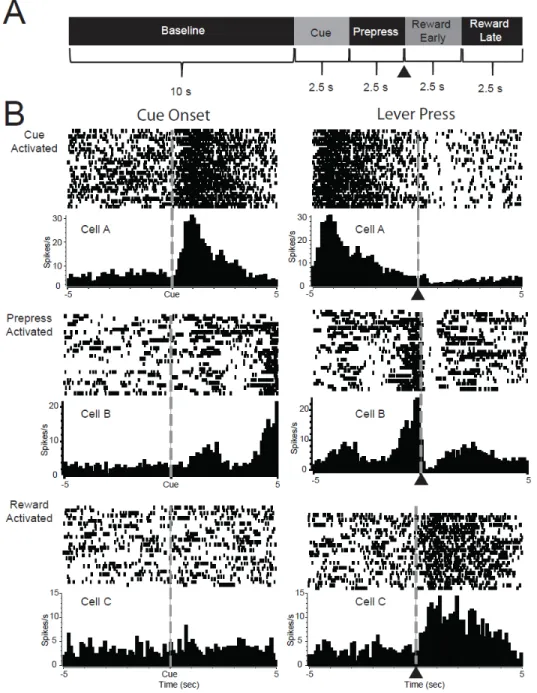
seconds prior to lever press (Prepress period), and two 2.5 second epochs during the reward period (Early Reward and Late Reward periods) (Figure 2.3A). In each analysis, a single cell was analyzed with bin as a repeated-measures factor (e.g., baseline bin vs. cue bin), and trial type (e.g., risk vs. safe; reward omission vs. large reward vs. small reward) as an independent-measures factor. To determine the type of phasic encoding, post-hoc Tukey tests were completed. We then determined the type of phasic encoding by comparing the firing during the bins: phasic cells showed significantly different firing during the effect period (cue, prepress, reward) compared to the baseline while nonphasic cells showed no differences between the effect periods or baseline bins. Selective cells were classified as cells that were phasic to only one trial type and/or cells that were phasic to both trial types that showed significantly different firing rates during each the trial types (e.g., risk vs. safe cue).
Encoding was evaluated as the percentage of the population of cells that encoded a particular event (e.g., risk cue) on each session and in each region (core or shell). Population analysis was conducted separately for cells in the nonpreferring, risk preferring, and safe preferring groups. Differences in the frequency or proportion of neuronal responses across different trial types, subregions, or reward preferences were examined using chi square analysis. All analyses were considered significant at α=0.05. Statistical and graphical analyses were conducted in Graphpad Prism 4 (Graphpad software, Inc.) and STATISTICA (StatSoft, Tulsa, OK).
Histology
38
39 RESULTS
Individual Differences in Risky Decision Making Behavior
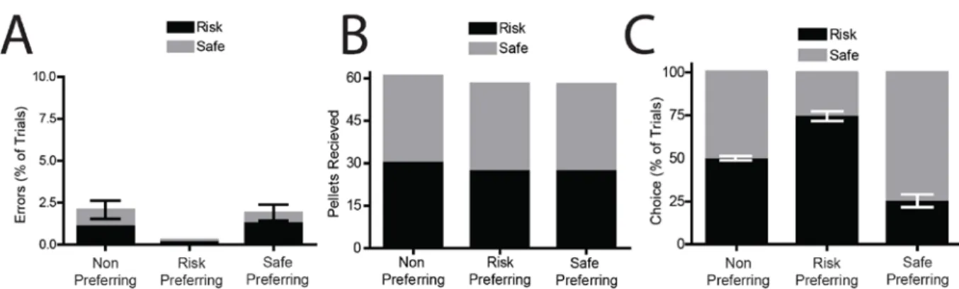
Several behavioral measures were used to verify that animals had acquired the task (Figure 2.1) and could discriminate between cues to guide behaviors. First, animals showed a significant decrease in the percentage of errors on risk and safe trials (F(24,384)=8.076,
P<0.0001) with a significant reduction in errors by session 5 compared to session 1 (Tukey’s HSD test, P<0.05 for sessions 5-25). During the recording session, the number of errors was not significantly different from 0 for any of the groups or trial types (Figure 2.2A; one sample t-test, comparison with theoretical mean of 0%, P>0.1 for all analyses).Further, across all three groups there was no significant differences in the number of reward pellets received for both cues (F(5,48)=0.6769, P=0.643), and the number of pellets received was not significantly different from the maximum possible (Figure 2.2B; one sample t-test, comparison with a theoretical mean of 30, P>0.15 for all comparisons). This data indicates that rats were able to use the cues to guide ongoing behaviors and select the appropriate response option that would be rewarded.
40
risk lever than chance (t(11)=9.299, P<0.001) while the remaining animals showing a safe preference(t(6)=6.367, P=0.0007, compared to chance).
Figure 2.2 Individual differences in risky decisions making. (A) Percentage of errors on Risk and Safe trials were not significantly different from 0 (P>0.1 for all comparisons), demonstrating behavioral discriminations between cues. (B) Reward pellets received during Risk and Safe trials. All groups of animals received maximum amounts of rewards (30 pellets for each trial type). (C) Response allocation on choice trials (as a percentage of choice) during recording sessions. 3 groups of rats were observed: non preferring rats chose both options equally (P=0.516), risk preferring rats chose the risk lever significantly more than chance (P<0.0001), safe preferring rats chose the safe lever significantly more than chance (P=0.0007).
Overview of nucleus accumbens neural activity during risky decision making
41
42
44
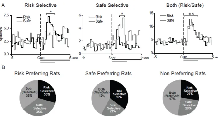
Cue activated neurons selectively encode risk versus safe options but not behavioral preferences
NAc neurons encode information about cues that predict rewarding outcomes (Nicola et al., 2004a; Day et al., 2006; Jones et al., 2010a; Saddoris et al., 2011), and selectively encode value related information during decision making (Roesch et al., 2009; Day et al., 2011). Therefore, here we examined how NAc cells encode information about risk versus safe options, and if encoding was related to each animal’s individual behavioral preference. Of the neurons that were activated (i.e., phasic) during risk trials, 61% showed decreases in activity during the cue period. Likewise, during safe trials 70% of cells were inhibited by the cue (see Table 2.1). This majority of inhibitory activity is consistent with the notion that NAc neurons primarily encode rewarding outcomes with decreases in firing rate (Roitman et al., 2005; Taha and Fields, 2006; Roitman et al., 2008; Wheeler et al., 2008). Further, there were no significant differences in the percentages of cells that displayed phasic activity across the three groups of animals, such that 36% of cells responded during cue presentations in the risk preferring group, 39% in the safe preferring group and 44% in the non preferring group (χ2
(2)=1.44, P=0.49).
45
percentages of neurons that were risk selective, safe selective, or both risk/safe in the three groups of rats (χ2
(4)=2.65, P=0.618), suggesting that NAc activity during cue presentations similarly encoded risk versus safe options in the three groups (Figure 2.4B). Further, there was no difference in the percentage of cells that were risk selective versus safe selective within each group of rats, indicating that neuronal responses were not modulated by the subjective value associated with each cue type. There were also no differences in the distribution of responses between the core and shell of the NAc during the cue period, nor were selective responses more concentrated in one region than another. As such, NAc core and shell data is presented together (Fisher’s exact test P>0.05 for all comparisons).
46
Response activated neurons selectively encode risk versus safe options but not behavioral preferences
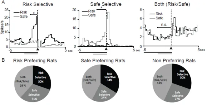
Previous research has shown that the NAc is critical for appropriate action selection (Cardinal et al., 2001; Cardinal and Howes, 2005; Cardinal, 2006; Phillips et al., 2007; Stopper and Floresco, 2011). We examined NAc neural activity during the 2.5s prior to lever press in our task, allowing for a functional measure of action selection processing within the NAc during risky decision making. A significantly greater proportion of NAc cells (229 cells 68%) were phasic during the prepress period compared to the cue period (χ2=52.25,
P<0.0001), supporting the role of the NAc in the processing of action selection. Similar to the cue period, the majority of prepress activated cells displayed phasic inhibitions (70% for both risk and safe levers; See Table 2.1). There were no significant differences between the three groups in the number of cells that were phasically activated during the prepress period (70% in the risk preferring group, 75% in the safe preferring group 67% in the nonpreferring group; χ2(2)=1.35, P=0.509), suggesting that the three groups are not differentially encoding response behaviors.
Similar to neural activity during the cue period, we found several populations of phasically active cells. A subset of cells showed selective activation prior to presses on the risk lever, another subset prior to presses on the safe lever, and a third group were similarly activated prior to both lever presses (Figure 2.5A). Of prepress activated cells, the majority (140 cells, 59%) displayed prepress selective encoding. While this encoding suggests that the NAc may be critical for action selection, it appears that NAc neural activity does not encode the subjective value associated with response selection. Specifically, there were no significant differences in the percentages of cells that were risk selective, safe selective or both risk/safe in the three groups (Figure 2.5B, χ2
47
period, similar percentages of cells were risk selective versus safe selective within each group of rats suggesting that the NAc does not differentially encode action selection based on individual preferences. Again there were no differences in the distribution of responses between the core and shell of the NAc during the prepress period, nor were selective responses more concentrated in one region than another so NAc core and shell data is presented together (Fisher’s exact test P>0.05 for all comparisons).
48
Accumbens neural activity encodes unexpected reward deliveries and omissions
For all neurons (regardless of preference), we first compared how the NAc encodes unexpected reward presentations and omissions. We found that similar percentages of neurons (roughly 30%) were phasic during large reward presentations, small reward presentations, and reward omissions (Table 2.2). Further, a larger percentage of cells displayed inhibitions compared to excitations during reward presentations. During omissions we found a larger percentage of excitations compared to inhibitions. Work in our lab and others has suggested that neurons in the NAc primarily encode rewarding outcomes with decreases in firing, while aversive outcomes are typically encoded as increases in firing (Roitman et al., 2005; Taha and Fields, 2006; Roitman et al., 2008). The current data support this finding, and extend the interpretation, such that reward omissions may be encoded as aversive events by the NAc. There was also a significant increase in the percentage of cells that encoded reward omissions from the early to late period (80 neurons (24%) to 107 neurons (33%), χ2
=4.992, P=0.026). Further, there was a significant shift in the ratio of cells that encoded excitations versus inhibitions. During the early period 49% of phasic cells encoded excitations while 51% encoded inhibitions. During the late reward period, 69% encoded excitations while 31% of phasic cells encoded inhibitions (χ2=7.143, P=0.0075). This suggests that omission of rewards may induce a negative affective state, particularly later during the reward period when animals are normally consuming the reward.
Neural responses to reward omissions and deliveries encode risk preferences
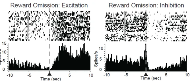
49
Figure 2.6 Reward omission processing in the nucleus accumbens. PEH and raster plot from representative reward omission activated NAc neurons. Data aligned to lever press (▲/dashed line). Grey circles in rasters denote cue onset. A subset of NAc neurons showed increased activity following lever press during the time period in which the animal expected the reward (left panel). Another subset of neurons exhibited decreases in activity following lever press during the time period in which the animal expected the reward (right panel). neural responding in the NAc core and shell to reward omissions and how this activity was correlated with behavioral preferences. First, there were no significant differences in the percentages of cells that were phasic during the omission period in either the NAc core (χ2
(2)=0.42, P=0.81) or NAc shell (χ2(2)=0.72, P=0.698) across the three groups. Cells responded with either an increase in activity during the reward period (excitation, Figure 2.6A left) or a decrease in activity during this period (inhibition, Figure 2.6A right).
50
individually to determine the relationship of excitation/inhibition ratio and risk preference. Importantly, we only included animals in this analysis with at least 5 cells recorded in the NAc core to ensure that animals with low numbers of cells did not bias our results. We found a negative correlation between the percentage of phasic cells that were excitatory and preference for the large risky lever (r2=.33, P=0.02; Figure 2.7B). That is, risk preferring rats exhibited less excitatory activity compared to safe preferring rats. As aversive outcomes are encoded with increases in excitations (Roitman et al., 2005; Wheeler et al., 2008), the current data suggest a differential processing of reward omission, a seemingly aversive event, between safe and risk preferring rats, such that increased encoding of aversion related to reward omission is associated with an increase in safe preferences.
51
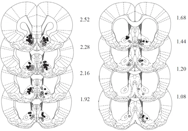
52 Electrode placement
53
54 DISCUSSION
NAc activity has been implicated in cue outcome learning (Day et al., 2006), reward related responding (Schultz et al., 1992; Nicola et al., 2004a; Nicola et al., 2004b; Taha and Fields, 2006; Wheeler et al., 2008; Jones et al., 2010a; Saddoris et al., 2011), and decision making (Roesch et al., 2009; Day et al., 2011). Here, we evaluated the role of NAc activity during risky decision making. As with previous reports using similar tasks (Roitman and Roitman, 2010; Sugam et al., 2012), we found that rats displayed individual preferences for safe versus risky options (risk preferring, safe preferring, and nonpreferring rats). In all groups of rats, NAc neurons exhibited phasic patterns of activity (excitations or inhibitions) relative to all aspects of the task (cue presentation, prepress period, and reward deliveries/omissions). Further, NAc neurons displayed selective activation, signifying differential encoding of risk versus safe options during each of these time periods. Somewhat unexpectedly, there were no differences in the percentages of cells that selectively encoded risk versus safe options during the cue or prepress period in the three groups. This suggests that NAc activity may not signal the subjective or intrinsic value associated with the different options, but instead may signal the expected value. The expected value is the prediction of future out comes based on the computation of several factors of reward value including the probability and magnitude. In support, we found that neural responding to reward outcomes was related to individual differences in risk taking behavior.
55
2004a; Day et al., 2006; Saddoris et al., 2011) Further, striatal neurons differentially encode cues that are predictive of rewards that require effort (Day et al., 2011), reward delay and magnitude (Schultz et al., 1992; Cromwell and Schultz, 2003; Roesch et al., 2009), the location of upcoming rewards (Taha and Fields, 2006), and reward valence (appetitive versus aversive) (Setlow et al., 2003; Roitman et al., 2005; Taha and Fields, 2005a; Wheeler et al., 2008). As such, we expected to see differences in cue related neuronal activity for the risk versus safe options based on risk preferences. Specifically, if the NAc encoded information about subjective value we expected that the three groups would differentially encode the risk versus safe cues, such that risk preferring rats have significantly greater percentage of selective cells encode the risk option, safe preferring rats have significantly greater percentage of selective cells encode the safe option, and nonpreferring rats have the greatest percentage of cells similarly encode both. Indeed we found that the majority of phasic neurons differentially encoded risk versus safe options, suggesting that the NAc does track these differences. Surprisingly, we found that there were similar proportions of cells that encoded the risk versus safe options in each of the three groups. Importantly, previous studies that showed that NAc neurons encode information about reward value during cue presentations (Schultz et al., 1992; Cromwell and Schultz, 2003; Roesch et al., 2009; Day et al., 2011) used rewards with different explicit value (i.e., situations in which choosing one reward is more advantageous than another) such as rewards of different size, delay, or effort. In these tasks, the expected value of one option is always more advantageous than the other option and therefore the expected and subjective value are conflated with one another.
56
shown that ventral striatal activity was correlated with the expected value associated with different cues based on the probability and magnitude of reward delivery. However, this value signaling was not correlated with individual risk attitudes, suggesting a primary role in encoding expected values irrespective of behavioral preferences (Tobler et al., 2007). Further, neurons in the dorsal striatum have been shown to encode information about subjective value during decision making behaviors (Samejima et al., 2005), while optical stimulation of dorsal striatal neurons prior to behavioral responses can modulate subjective value related decisions (Tai et al., 2012). As such, the dorsal versus ventral striatum may send competing value related information (subjective versus expected value) to movement related output structures to promote appropriate decision making (Nicola, 2007). The present findings are consistent with a role of the NAc in encoding expected value as the population encoding of risk versus safe options was similar for risk preferring, safe preferring, and nonpreferring animals.
57
differences across the three groups in the selectivity of responding, such that similar populations of neurons were risk selective and safe selective in risk, safe, and nonpreferring rats. Again, this suggests that NAc neural activity during the prepress period encoded information on the expected value of lever presses rather than subjective value.
58
value of action selection, and disruptions of this signaling result in maladaptive decision making.
59
Interestingly, we found the opposite pattern of responding in the NAc shell, an area that is normally linked with encoding reward valence (Roitman et al., 2008; Wheeler et al., 2011). Specifically, we found an increased percentage of neurons exhibiting excitatory activity during reward omissions in the risk preferring compared to safe preferring rats. A key difference between normal reward aversion tasks and the data reported here is that in our task the aversive event is reward omission, rather than the presentation of an aversive stimulus. The fact that there is no reward present may alter the way the NAc shell processes this information. Further, reward omission processing may function again as a gating signal within the NAc. For risk preferring rats, reward omissions may not be as aversive, and therefore increased excitations function to inhibit reward seeking behaviors. In contrast, in safe preferring rats, reward omissions appear highly aversive and thus the increased inhibitions during reward omissions may function to instruct the animal to continue seeking for rewards in a safe manner.
60
CHAPTER 3
ROLE OF PHASIC NUCLEUS ACCUMBENS DOPAMINE IN DELAY DISCOUNTING
ABSTRACT
62
63 INTRODUCTION
Learned associations between cues in the environment and positive outcomes are critical for making appropriate decisions. In order to maximize resources, organisms must evaluate the costs and benefits of different actions and choose the most valuable option available (Green and Myerson, 2004; Cardinal, 2006; Phillips et al., 2007; Rangel et al., 2008). This type of decision making engages a specific network of nuclei including the nucleus accumbens (NAc) and its dopaminergic input (Roesch et al., 2007; Roesch et al., 2009; Day et al., 2010; Day et al., 2011; Sugam et al., 2012). Subsecond dopamine release within the NAc is believed to modulate reward-seeking behaviors including those involving food and cocaine (Phillips et al., 2003b; Roitman et al., 2004), and track decisions based on effort, delay and risk (Day et al., 2010; Sugam et al., 2012). Importantly, the mesolimbic dopamine system functions as a prediction signal, displaying increased phasic activation to cues that reliably predict reward delivery (Mirenowicz and Schultz, 1994; Schultz et al., 1997; Waelti et al., 2001; Stuber et al., 2008; Tsai et al., 2009; Zellner and Ranaldi, 2010). This encoding provides a mechanism for the brain to track reward related information to mediate appropriate resource seeking behaviors. Phasic dopamine signaling of reward predictive cues is not only sufficient to promote behavioral conditioning (Tsai et al., 2009), but is also necessary for reward related learning (Sombers et al., 2009; Zellner et al., 2009).
64
Schultz, 2010). Further, dopamine release within the NAc encodes information related to reward costs, delays, and subjective value (Day et al., 2010; Gan et al., 2010; Sugam et al., 2012). However, dopamine signaling of reward value appears to be restricted to the NAc core (Day et al., 2010; Sugam et al., 2012; Saddoris et al., 2013). As such, this circuit provides a mechanism to encode the most valuable option available to the animal to bias decisions and promote adaptive behaviors. Finally, the mesolimbic dopamine projections to the NAc are necessary for appropriate value-based decision making as lesions or inactivation of it result in maladaptive choices (Cardinal et al., 2001; Salamone et al., 2001; Salamone et al., 2002; Cardinal and Howes, 2005; St Onge and Floresco, 2008; Ghods-Sharifi and Floresco, 2010; St. Onge et al., 2010; Stopper and Floresco, 2011; Stopper et al., 2013).
65
66 METHODS
Animals
Male Sprague Dawley rats (n=7 rats with 8 recording locations, Harlan Sprague Dawley, Indianapolis, IN), aged 90-120d and weighing 275-350g were used as subjects and were individually housed with a 12/12-h light/dark cycle. All experiments were conducted between 8:00am and 5:00pm. Animals were maintained at no less than 85% of pre-experimental bodyweights by food restriction (~10-15g of Purina laboratory chow each day in addition to approximately 1g of sucrose consumed during behavioral sessions) except during the post-operative recovery period when food was given ad libitum. All procedures were approved by the UNC Institutional Animal Care and Use Committee.
Behavioral Training
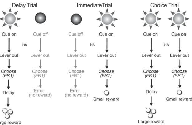
67
(10 trials of each). For Delay trials (Figure 3.1, left) a single cue light was illuminated over one lever for 5s, followed by extension of both levers. Responses (FR1) on
the lever below the illuminated cue light were rewarded with three sucrose pellets delivered after a delay. Lever presses on the other lever were not rewarded and counted as an error. The number of errors served as a behavioral measure of discrimination between the immediate and delayed cues. If the animal did not respond on either lever within 10s both
68
levers retracted and the trial was counted as an omission. Delay trials were critical since they informed the rat as to which delay was imposed during the upcoming free choice trials of that block. For Immediate trials (Figure 3.1, middle) the cue light above the second lever was illuminated for 5s, then both levers were extended into the chamber. Lever presses (FR1) on the lever under the illuminated cue light within 10 s were rewarded with a single sucrose pellet. Lever presses on the other lever were not rewarded and counted as an error. The next 10 trials within each block were Choice trials (Figure 3.1, right) in which both cue lights simultaneously illuminated for 5 s, and both levers were extended for 10 s. Once either lever was pressed, both levers retracted and the animal was rewarded based on the contingency of reinforcement for that block and chosen lever. Again, if the animal did not respond within 10 s the levers retracted and the trial was counted as an omission. Choice trials served as a measure of an animal’s overall sensitivity to the changes in reward delay across the session. During the first block/session, the delay to the large reward was 0s. In subsequent blocks, the delay to large reward was increased to 10s and 20s. Importantly, each trial was of fixed duration (60 s) so that reward choice did not influence how quickly a rat can complete the task, and choosing the smaller immediate reward did not lead to the next trial more quickly. Rats were trained for 25 sessions until stable behavior was observed. Following 25 training sessions, all rats were be prepared for electrochemical recording in the NAc core as described below. After recovery, animals underwent additional training sessions until behavior reached the presurgery baseline (at least 5 sessions).
Surgery
69
ketamine hydrochloride (100mg/kg) and xylazine hydrochloride (10mg/kg) mixture (intramuscular) and placed in a stereotaxic frame. A guide cannula (Bioanalytical Systems, West Lafayette, IN) was positioned dorsally to the NAc core (1.3 mm anterior, 1.3mm lateral from bregma). An Ag/AgCl reference electrode was placed contralateral to the stimulating electrode in the left forebrain. The bipolar stimulating electrode (Plastics 1 Inc., Roanoake, VA) was placed dorsally to the VTA (5.2 mm posterior, 1.0 mm lateral from bregma and 7 mm ventral from brain surface). Stainless steel skull screws and dental cement were used to secure all items. The bipolar stimulating electrode was lowered in 0.2 mm increments until physical responses to electrical stimulation diminished indicative of proper electrode placement. The stimulating electrode was then fixed with dental cement.
Fast-Scan Cyclic Voltammetry
Following surgery, animals were allowed one week to recover to presurgery body weight. Food restriction was then resumed to increase motivation during behavioral performance. On test day, a new carbon-fiber electrode, housed in the micromanipulator, was lowered into the NAc core and was used to measure dopamine changes during task performance. The potential of the carbon-fiber electrode was held at -0.4V versus the Ag/AgCl reference electrode. The carbon-fiber and Ag/AgCl electrodes were connected to a head-mounted voltammetric amplifier attached to a commutator (Crist Instrument Company, Hagerstown, MD). Prior to recording, the carbon fiber electrode was allowed to equilibrate for 20-30 minutes in the brain to minimize current drift.
70
electroactive within this potential range (including dopamine), producing a measurable change in current at the carbon-fiber. Specific analytes (including dopamine) were verified by plotting this change in current against the applied potential to produce a cyclic voltammogram (Heien et al., 2004; Heien et al., 2005). The stable contribution of current produced by oxidation and reduction of surface molecules on the carbon-fiber was removed by using a differential measurement (i.e., background subtraction) between a time when such signals were present but dopamine was not. For data collected during the behavioral session, this background period was obtained during the baseline window (10 s prior to cue onset). Following equilibration, dopamine release was electrically evoked by stimulating the VTA using a range of stimulation parameters (2-24 biphasic pulses, 20-60 Hz, 120µA, 2 ms per phase) to make sure that the carbon fiber electrode was placed close to dopamine release sites and to create a training set for principal component analysis. Animals then underwent task performance and electrochemical recordings were made continuously with 100 ms temporal resolution. A second computer and software system (Med Associates Inc) controlled behavioral events and sent digital outputs for each event to the voltammetry recording computer to be time stamped along with the electrochemical data. Following termination of the behavioral session, VTA stimulation was repeated to verify the stability of the electrode and ensure that the location of the electrode still supported dopamine release.
Signal Identification and Separation
71
constructed from representative, background-subtracted cyclic voltammograms for dopamine and pH, as previously described (Heien et al., 2004; Heien et al., 2005). The background period was obtained at the minima for the dopamine signal 5 s before event onset. This training set was used to perform principal component regression on data collected during the behavioral session. Principal components were selected such that at least 99% of the variance in the training set was accounted for by the model. All data presented here fit the resulting model at the 95% confidence level.
Data Analysis
All behavioral events occurring during training and electrochemical recordings were available for analysis. To determine if animals reliably acquired the delay discounting task we evaluated the number of errors and the number of omitted responses during the recording sessions. Further, we examined the ability of the animals to adjust behaviors as delays increased by using a repeated measures ANOVA to compare responses during choice trials for the large delayed reward across the three blocks of the session.
72
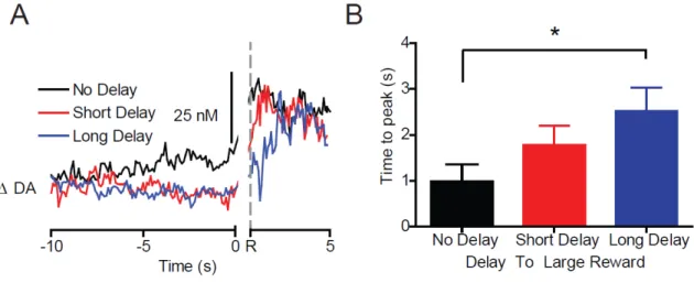
each event, with Tukey’s correction for multiple post-hoc comparisons. The effect of large reward delivery on dopamine signaling was evaluated using a 1-way repeated measures ANOVA comparing baseline to peak levels. A 1-way repeated measures ANOVA with Tukey’s post hoc test was used to evaluate the differences in the time to reach peak dopamine concentration following reward delivery for each delayed reward. All analyses were considered significant at α = 0.05. Statistical and graphical analysis was performed using
Graphpad Prism (Graphpad Software, Inc) and STATISTICA (Statsoft, Tulsa, OK). Histology