1363
Abstract:
Thrombosis is a leading cause of morbidity and mortality worldwide. Animal models are used to understand the pathological pathways involved in thrombosis and to test the efficacy and safety of new antithrombotic drugs. In this review, we will first describe the central role a variety of animal models of thrombosis and hemostasis has played in the development of new antiplatelet and anticoagulant drugs. These include the widely used P2Y12 antagonists and the recently developed orally available anticoagulants that directly target factor Xa or thrombin. Next, we will describe the new players, such as polyphosphate, neutrophil extracellular traps, and microparticles, which have been shown to contribute to thrombosis in mouse models, particularly venous thrombosis models. Other mouse studies have demonstrated roles for the factor XIIa and factor XIa in thrombosis. This has spurred the development of strategies to reduce their levels or activities as a new approach for preventing thrombosis. Finally, we will discuss the emergence of zebrafish as a model to study thrombosis and its potential use in the discovery of novel factors involved in thrombosis and hemostasis. Animal models of thrombosis from zebrafish to nonhuman primates are vital in identifying pathological pathways of thrombosis that can be safely targeted with a minimal effect on hemostasis. Future studies should focus on understanding the different triggers of thrombosis and the best drugs to prevent each type of thrombotic event. (Circ Res. 2016;118:1363-1379. DOI: 10.1161/ CIRCRESAHA.115.306823.)Key Words: animal models ■ anticoagulants ■ hemostasis ■ thrombosis ■ zebrafish
© 2016 American Heart Association, Inc.
Circulation Research is available at http://circres.ahajournals.org DOI: 10.1161/CIRCRESAHA.115.306823
Animal Models of Thrombosis From Zebrafish to
Nonhuman Primates
Use in the Elucidation of New Pathologic Pathways and the
Development of Antithrombotic Drugs
Pudur Jagadeeswaran, Brian C. Cooley, Peter L. Gross, Nigel Mackman
Circulation Research
Compendium on Thrombosis
Advances in Thrombosis and Hemostasis: An Introduction to the Compendium Global Burden of Thrombosis: Epidemiologic Aspects
Systems Analysis of Thrombus Formation
Animal Models of Thrombosis From Zebrafish to Nonhuman Primates
Platelet-Mediated Thrombosis: From Bench to Bedside Cross Talk Pathways Between Coagulation and Inflammation
Evolving Treatments for Arterial and Venous Thrombosis: Role of the Direct Oral Anticoagulants Evolving Treatments for Acute Ischemic Stroke
Gene Therapy for Coagulation Disorders
Jeffrey I. Weitz and John W. Eikelboom, Editors
Original received September 21, 2015; revision received November 10, 2015; accepted November 30, 2015.
From the Department of Biological Sciences, University of North Texas, Denton (P.J.); Department of Pathology and Laboratory Medicine (B.C.C.), and Department of Medicine (N.M.), University of North Carolina, Chapel Hill; and Department of Medicine, McMaster University, Hamilton, Ontario, Canada (P.L.G.).
Correspondence to Pudur Jagadeeswaran, PhD, Department of Biological Sciences, University of North Texas, Denton, TX 76203. E-mail [email protected]
H
emostasis is a physiological process that involves for-mation of a hemostatic clot at the site of vessel injury to prevent blood loss. In contrast, thrombosis is a pathologi-cal process, where thrombotic clots are formed within blood vessels and obstruct the flow of blood in the circulatory sys-tem.1–4 Thrombotic clots contain platelets as the major cellularcomponent and crosslinked fibrin as the main protein com-ponent. In addition, clots contain red blood cells and leuko-cytes. However, arterial and venous clots differ in the relative amounts of platelets and fibrin. Arterial thrombi most often form rapidly after rupture of atherosclerotic plaques and are platelet rich; these clots cause myocardial infarction and
stroke.5 Venous clots form in large veins, particularly in the
legs, and are fibrin-rich with high numbers of red blood cells and develop over hours to days.5–7
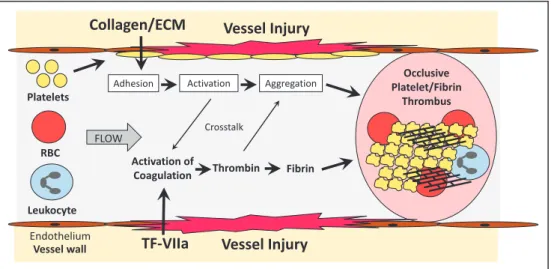
Vessel injury leads to rapid platelet activation by bind-ing of exposed collagen and deposited von Willebrand factor (vWF) to platelet receptors. In parallel, the clotting system is activated by exposure of tissue factor (TF) in the vessel wall and formation of the TF/factor VIIa complex (Figure 1). Factor XII of the intrinsic pathway has also been shown to contribute to thrombosis. Figure 2 shows simplified versions of the platelet and coagulation cascades. Platelets are activated by primary stimuli, such as collagen, vWF, and thrombin, and this leads to the release of secondary stimuli, such as throm-boxane A2 and ADP. Full activation of platelets ultimately leads to a change in the conformation of the integrin GPIIb/ IIIa (αIIbβ3) that allows binding of ligands, such as fibrinogen,
that mediate platelet aggregation. The coagulation cascade is composed of the extrinsic pathway (TF/factor VIIa), the in-trinsic pathway (factors XIIa, XIa, IXa, and VIIIa) and the common pathway (factors Va, Xa, and thrombin). Thrombin cleaves fibrinogen into fibrin monomers that self-polymerize and are subsequently crosslinked by the transglutaminase fac-tor XIIIa. Importantly, there is cross talk between the plate-let and coagulation cascades (Figures 1 and 2). For instance, activated platelets provide a thrombogenic surface for the assembly of various coagulation complexes, such as the te-nase complex (factor VIIIa/factor IXa) and the prothrombi-nase complex (factor Va/factor Xa). In addition, thrombin is a potent activator of human platelets via cleavage of protease-activated receptors 1 and 4 (PAR1 and PAR4). Figure 2 also shows the targets of currently approved antithrombotic drugs that are used to prevent and treat both arterial and venous thrombosis.
Animal models have played a central role in the identifica-tion of factors and pathways that drive thrombosis and in eval-uating the efficacy and safety of antithrombotic drugs. There are many reviews that have described in detail animal models of thrombosis and hemostasis, including zebrafish, rodents, and larger animals, such as nonhuman primates.8–20
In this review, we chose to focus on the use of animal models of thrombosis in the development of antithrombotic drugs, and in the identification of new pathological path-ways of thrombosis. The efficacy and safety of antithrom-botic drugs is evaluated in a variety of animal models of thrombosis and hemostasis before initiating phase I clini-cal trials. More basic studies of the thrombotic process are
performed in mouse models because of the availability of different transgenic mouse strains. These studies, particu-larly with venous thrombosis models, have identified new players in thrombosis, such as polyphosphates, neutrophil extracellular traps, and microparticles. Finally, we will de-scribe the emergence of zebrafish as a model of thrombosis and hemostasis and its potential to identify new factors in-volved in thrombosis.
Development of Animal Models
of Thrombosis
There are several general models of thrombosis originally developed in species larger than mice. In 1952, Wessler21
described a vein stasis model in which vein segments in dogs were clamped for >20 minutes to induce clot forma-tion. In 1976, Folts et al22 described an arterial injury model
that involved a 60% to 80% stenosis of the coronary artery of dogs. The coronary artery exhibited cyclic reductions in blood flow that were proposed to mimic changes that oc-cur in a stenosed coronary vessel. Administration of aspirin abolished the cyclic reductions in blood flow and reduced platelet aggregation. Variants of these models have been subsequently applied in a number species, including rab-bits, rats, and mice. Other models use a form of intraluminal injury, most often created by opening the vessel (eg, creat-ing an arteriotomy) and mechanically scratchcreat-ing or remov-ing the intima.23 In addition, thrombosis can be initiated by
placing a synthetic material inside the vessel, such as a su-ture.24 Another common model is to create an arteriovenous
shunt using a synthetic material that collects thrombus on its surface.25,26 This model offers several advantages because it
can be performed in a broad range or species—rats, rabbits, dogs, and nonhuman primates. In addition, it can be used for acute or more chronic treatment. There are variations in the vessels cannulated—carotid or femoral artery, jugular or femoral vein. A graft with an artificial surface can be added. A disadvantage of such a model is the limitations of clini-cal relevance in testing efficacy of the antithrombotic drugs targeting indications that this model does not simulate. For anticoagulants, these would include stroke prevention in atrial fibrillation and treatment and prevention of venous thrombosis. These various animal models of thrombosis have been used for many preclinical studies evaluating an-tithrombotic drugs.
There has been a lack of uniformity in the application of these various models, both within and across species, which has hindered standardization of any one model or group of models. The Folts model is arguably the one closest to simu-lating clinical arterial thrombosis but its application in various species and anatomic vessels leads to variability. Other mod-els have even greater variability and questionable relevance. For instance, the arteriovenous shunt model uses a synthetic surface to simulate thrombus formation (eg, polyethylene or Dacron) in a circuit going from arterial to venous pressure, that has little relevance to most forms of pathophysiologic thrombus formation. The main value in these models is in their ability to demonstrate thrombus inhibition in a vessel of comparable size to clinically thrombosing vessels.
Nonstandard Abbreviations and Acronyms
FDA Food and Drug Administration DVT deep vein thrombosis IVC inferior vena cava NDA New Drug Applications PAR protease-activated receptor TF tissue factor
TTO time to occlusion vWF von Willebrand factor
Use of Animal Models for the Approval
of New Antithrombotic Drugs
In the first part of this review, we will discuss the use of ani-mal models of thrombosis for the approval of antithrombotic drugs. Clearly, this is a large topic so we have focused on the use of models of thrombosis for approved Food and Drug Administration (FDA) applications of antithrombotic drugs (anticoagulants and antiplatelets) since 1997 at http://www. accessdata.fda.gov/scripts/cder/drugsatfda/index.cfm. The
FDA evaluates submissions on a compound for data from animal studies for 3 criteria: mechanism of action, efficacy on targeted biology, and safety pharmacology. For some drugs, the target is specific for a human protein, and thus nonhuman primates are the only suitable model for evaluating the effect.
We will summarize the thrombosis models used in the New Drug Applications (NDA) to demonstrate mechanisms of action and efficacy on targeted biology to obtain NDA ap-proval for the various drugs. Although nonhuman primates
Figure 1.Formation of an occlusive thrombus. After vessel injury platelets rapidly adhere to collagen and deposited von Willebrand Factor. The adhered platelets are activated by primary and secondary activators that lead to platelet aggregation mediated by various ligands, including fibrinogen. In parallel to platelet activation, the clotting system is activated by exposure of tissue factor (TF) in the vessel wall. In addition, factor XII may contribute to the activation of coagulation. Thrombin is the central protease of the coagulation cascade and cleaves fibrinogen to fibrin monomers that are crosslinked into a network by factor XIIIa. There is cross talk between the platelet and coagulation cascades. For instance, activated platelets provide a thrombogenic surface for the assembly of various coagulation protease complexes and thrombin is a potent activator of platelets by cleavage of protease activated receptors. Formation of an occlusive thrombosis will block blood flow.
Figure 2.Animal models of thrombosis are used in the development of antithrombotic drugs. The targets of both anticoagulant and antiplatelet drugs are shown. Platelet inhibitors include inhibitor of the primary activator and secondary activators. The protease-activated receptor 1 (PAR1) inhibitor vorapaxar blocks thrombin activation of platelets. Aspirin inhibits the generation of the secondary activator thromboxane A2. There are several inhibitors of the ADP receptor P2Y12 that are used clinically. Finally, inhibitors of integrin GPIIb/IIIa prevent platelet aggregation. Warfarin acts by inhibiting the generation of γ-carboxylation domains on several coagulation proteases. Unfractionated heparin (UF heparin) acts in an antithrombin-dependent manner to inhibit both thrombin (IIa) and factor Xa (FXa). Low-molecular-weight heparins are more selective for thrombin and the synthetic pentasaccharide fondaparinux only inhibits FXa. Direct thrombin inhibitor include parenteral drugs (bivalirudin and argatroban) and the oral inhibitor dabigatran etexilate (dabigatran). Three oral, direct FXa inhibitors have been developed. vWF indicates von Willebrand factor.
and pigs are often held up as the most human-like in their physiology, use of dogs, rabbits, and rats has been an accept-able practice for many NDA submissions. There are several caveats with using NDA to review animal models of throm-bosis. One caveat is that we will not describe the extensive pharmacokinetic and off-target safety studies that have been performed in many species, including nonhuman primates. In addition, published studies that were not included in the regu-latory filing will not be discussed. Finally, we have limited our presentation to NDAs for drugs that have been approved because this is what is accessible.
The drugs are divided into antiplatelet drugs and antico-agulants, and each group is presented in chronological order of submission because this gives some context to the choice of models. The primary reference is the FDA accessible NDA. The references cited are for the publications of the data in the NDA and are provided to add experimental detail. However, not all data in the NDAs are published but this can be found on the Website. The references also may contain additional thrombosis models that were not submitted in the NDA. There are also additional references of animal thrombosis models using the drugs, but if they are not part of the NDA then these are not reviewed. Antithrombotic drugs must be also assessed for safety using a variety of animal models of bleeding.27–29
The most common rodent bleeding model is the tail transec-tion model.30 Use of a template to standardize the diameter of
the tail that is cut improves the reproducibility of the model. Another mouse bleeding model is the saphenous vein model.31
The template skin bleeding test uses a standard skin cut and is used in nonhuman primates to assess bleeding.28 Other
bleed-ing tests used to assess antithrombotic drugs include the rabbit ear, cuticle bleeding, and the rat renal cortex template models.
Antiplatelets
Aspirin has been used for many years as an antiplatelet drug. More recently, several parenteral inhibitors of GPIIb/IIIa (αIIbβ3) have been developed (Figure 2).32 The first GPIIb/
IIIa inhibitor approved by the FDA was abciximab in 1994 followed by tirofiban and eptifibatide in 1998. The most re-cent developments in antiplatelet drugs include 2 new classes: P2Y12 inhibitors (clopidogrel, prasugrel, and ticagrelor) and a PAR-1 inhibitor (vorapaxar; Figure 2). These antiplatelet drugs are approved for acute treatment of myocardial infarc-tion, including with percutaneous coronary interventions, and, in patients with previous stroke, myocardial infarction, or pe-ripheral vascular disease, for prevention of recurrent thrombo-sis. For their registration trials, they were superior when added to aspirin, when compared with previously approved drugs in the same class or added to standard of care.
Abciximab and Tirofiban
The FDA NDA submissions for these 2 drugs are not available on the accessible Website.
Eptifibitide (1987; NDA 20–718)
The efficacy of eptifibitide was demonstrated in dogs where it reduced cyclic flow reductions in a major artery. In addition in dogs, coronary artery thrombi produced by an external elec-tric injury then lysed with tissue-type plasminogen activator
reoccluded less frequently after eptifibitide infusion, and even less frequently when lower doses of eptifibitide were admin-istered with hirudin. Finally, eptifibitide improved platelet count and platelet function after a hypothermic cardiopulmo-nary bypass in dogs. In baboons, eptifibitide reduced the ac-cumulation of 111In-oxine–labeled platelets on a Dacron graft
in a femoral arteriovenous shunt model. This effect was not increased by heparin or aspirin. The drug worked best when administered before graft placement. The effect of eptifibitide on template bleeding was also evaluated in dogs and baboons.
Clopidogrel (1997; NDA 20–839)
The efficacy of clopidogrel was demonstrated in rats using thrombosis models induced by inserting a metallic coil or an electric injury to arteries, and by venous stasis. These same models were used to demonstrate efficacy in rabbits. In ad-dition, clopidogrel reduced platelet accumulation in de-endo-thelialization carotid arteries in rabbits.33 Clopidogrel reduced
cyclic flow reductions in the left anterior descending artery induced by brief pinch injury to the artery with forceps and placing a constrictor around the injured portion of the vessel.34
The effect of clopidogrel on bleeding was evaluated in a rat tail model and a rabbit template model.
Prasugrel (2007; NDA 22–307)
Prasugrel reduced thrombosis in a rat model consisting of a ca-rotid to contralateral jugular arteriovenous shunt model using a polyethylene catheter with a silk thread inside.35 Prasugrel also
delayed time to occlusion (TTO) after electric injury to the rat carotid artery.36 Finally, prasugrel inhibited aggregation of
plate-lets that were isolated from the blood of cynomolgus monkeys dosed orally with prasugrel. The effect of prasugrel on bleeding was evaluated in a rat tail model; it is notable that the dose that caused 50% increase in bleeding was lower than clopidogrel.
Ticagrelor (2010; NDA 22–433)
Efficacy of ticagrelor was demonstrated in rats using a fer-ric chloride injury to the carotid artery.25 In dogs, ticagrelor
restored normal blood flow after injury of the femoral artery by squeezing it and partly obstructing it with an occluder (modified Folts model).37 In addition, ticagrelor was evaluated
in combination with aspirin or the direct thrombin inhibitor melagatran. The effect of ticagrelor on bleeding was evaluated in dogs using a template model. In addition, the submission also presented data on antithrombotic-bleeding efficacy by calculating the ratio of the IC50 of each effect and comparing this with other antiplatelet drugs.
Vorapaxar (2013; NDA 204886)
Vorapaxar is a molecule that binds to PAR-1 and blocks the binding of the tethered ligand released by thrombin cleavage. It has no activity in rodents. The FDA submission of vora-paxar did not include any efficacy data on the antithrombotic effects of vorapaxar. Instead, data on a similar compound, SCH602539, was presented and accepted as a bioequivalent. The efficacy of SCH602539 was demonstrated in cynomol-gus monkeys using a carotid artery modified Folts model.38
SCH602539 reduced cyclic flow reductions as well as a P2Y12 inhibitor, and the effect of combining the 2 drugs was more than synergistic.
Anticoagulants
Warfarin and heparin have been the mainstay of anticoagu-lant therapy for the past 50 years. Warfarin is administered orally and inhibits the formation of a Gla domain on several different coagulation proteases (factors VIIa, IXa, Xa, and thrombin; Figure 2). It is most often used for long-term an-ticoagulant therapy. Heparins are administered parenterally. Unfractionated heparin inhibits both factor Xa and throm-bin in an antithromthrom-bin-dependent manner (Figure 2). Low-molecular-weight heparins are more selective for factor Xa. The synthetic pentasaccharide fondaparinux is selective for factor Xa. Newer anticoagulants have been designed to direct-ly inhibit either factor Xa or thrombin. The first generation of these inhibitors was the thrombin inhibitor bivalirudin, which is administered parenterally to treat patients undergoing per-cutaneous coronary angioplasty. The next generation of these inhibitors included orally available inhibitors of either factor Xa (rivaroxaban, apixaban, and edoxaban) or thrombin (dabi-gatran etexilate; Figure 2). These drugs are approved for the prevention and treatment of venous thrombosis, and for the prevention of stroke in patients with atrial fibrillation.
Bivalirudin (1999; NDA 20–873)
Bivalirudin inhibited platelet and fibrin deposition as mea-sured in electron microscopy images of the thrombi in a rat carotid endarterectomy model.39 Bivalirudin also decreased
reperfusion time when given with tissue-type plasminogen ac-tivator as a thrombolytic to aortic thrombi in rats. Bivalirudin also decreased 111In-oxine–labeled platelet accumulation in
the brain after intracarotid injection of thrombin to rabbits. In this model, it was compared with aspirin and with heparin.40
Bivalirudin inhibited thrombosis and the frequency of subse-quent occlusions in a pig model where the carotid artery was repeatedly occluded with a clamp. In baboons (Papio anubis), bivalirudin reduced platelet and fibrin deposition in various versions of an exteriorized femoral arteriovenous access shunt model, including those with endarterectomized aortae, collagen-coated Gortex, Dacron, a 2-chambered device, and a chronic arteriovenous shunt.26 Bleeding was evaluated in the
same animals using a template bleeding model.
Dabigatran Etexilate (2010; NDA 22–512)
Dabigatran was given intravenously to rats where it reduced thrombus weight in a thromboplastin/stasis inferior vena cava (IVC) model.41,42 Rats were also dosed orally with dabigatran
etexilate. In rabbits, the weight of clots in the jugular vein induced by stenosis and polidocanol, a sclerosant, was also reduced by dabigatran.43 Bleeding was evaluated in rats using
a template tail bleeding model42; the efficacy of an activated
prothrombin complex concentrate and recombinant factor VIIa to reverse dabigatran bleeding was also demonstrated.44 Rivaroxaban (2011; NDA 202439 and 22406)
Rivaroxaban reduced thrombosis in a mouse model of ferric chloride injury to the carotid artery and prevented death af-ter injection of thromboplastin. In rats, rivaroxaban reduced thrombosis in a carotid to contralateral jugular45 arteriovenous
shunt model with a polyethylene catheter with a nylon thread inside. This evaluation also included combining rivaroxaban
with heparin, with low-molecular-weight heparin, with aspi-rin, with various nonsteroidal anti-inflammatory drugs, with clopidogrel, with clopidogrel and aspirin, and with warfarin. Studies in rats showed that rivaroxaban reduced the size of thrombi induced by an electrolytic or ferric chloride injury of the carotid artery.46 The drug was also shown to be effective
in an IVC stenosis and thromboplastin-induced hypercoagu-lability model.47 Rivaroxaban also reduced thrombosis in
rab-bits in a similar carotid to contralateral jugular arteriovenous shunt model, but with a polyurethane catheter with a larger nylon thread inside. Baboons (template), rats (tail bleeding), and rabbits (ear bleeding)47 were used to evaluate the effect
of rivaroxaban on bleeding. The efficacy of recombinant fac-tor VIIa and prothrombin complex concentrates and activated prothrombin concentrates on bleeding was evaluated in rats only.48
Apixaban (2011; NDA 202155)
Clot weights were reduced by apixaban in rats in an arterio-venous shunt model, and after ferric chloride injury to the carotid artery and to the IVC. Apixaban also reduced clot weight in an arteriovenous shunt model and maintained flow in an electrolytical injury carotid model49; the latter was also
combined with antiplatelet drugs. In dogs, apixaban reduced thrombus weight in an arteriovenous shunt model and delayed TTO after electric injury to the femoral artery.50 The effect of
apixaban on bleeding was evaluated in rats with a renal cortex template model and in rabbits with a cuticle bleeding model.
Edoxaban (2014; NDA 206316)
In the FDA NDA, the antithrombotic effectiveness of edoxa-ban was only demonstrated in rats. The models included an arteriovenous shunt model, and 2 IVC ligation models, one with double ligation and the other with partial ligation.51 In
the arteriovenous shunt model, edoxaban reduced thrombus protein content, whereas in the IVC models edoxaban reduced thrombus weight. Edoxaban also reduced thrombus weight in a venous thrombosis model induced by placing a platinum wire into the IVC. This was also compared with treatments with enoxaparin, fondaparinux, and warfarin. In a dissemi-nated intravascular coagulation model where thromboplastin is injected into the femoral vein, edoxaban normalized the amount of thrombin–antithrombin complexes and the platelet counts and the fibrinogen concentration. A subsequent publi-cation also included a study in rabbits using a model of throm-boplastin and jugular vein stenosis (modified Wessler model) to cause thrombosis.52 The effect of edoxaban on bleeding
was also only evaluated in rats using tail and plantar template bleeding models where it was compared with low-molecular-weight heparin.
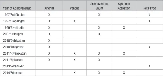
Summation of Findings From NDAs
Table 1 summarizes the different thrombosis models that have been used in the development of various antithrombotic drugs for NDA submission. Over the past 18 years several trends are apparent in the use of nonmurine animal models in FDA submissions for antithrombotic drugs. Over time, the use of nonhuman primates is becoming limited to cases where close similarity to humans is required. The use of dogs is also
becoming less prevalent (Table 2). The only mouse models used to demonstrate efficacy in the NDAs were applied for evaluating rivaroxaban in 2011. There also seems to be a drug class-effect in the animal models selected where later drugs in the same class have less testing in higher-order species. This is evident with edoxaban, the third factor Xa inhibitor approved, which only had efficacy data in rats in the NDA.
In these NDAs, the efficacies of antiplatelet drugs have been largely demonstrated in arterial models, such as variants on the Folts model. This model mimics many features impor-tant in preventing thrombosis in coronary interventions where the antiplatelet drugs that have been approved have shown efficacy. Interestingly, clopidogrel was also shown to reduce venous thrombosis in a mouse model.53 The most prevalent
model used to show efficacy of anticoagulants in the NDAs is the arteriovenous shunt model. Table 2 summarizes the dif-ferent animal models that have been used in the evaluation of antithrombotic drugs.
In these NDAs, the efficacy of the direct factor Xa inhibi-tors were demonstrated in both arterial and venous models (Table 1). The efficacy of dabigatran was not demonstrated in an artery-specific model in its NDA, whereas the efficacy
of bivalirudin was not demonstrated in a vein-specific model (Table 1). In conclusion, the nonmurine thrombosis models used in FDA NDAs for antithrombotic drugs that were eventu-ally approved used a variety of animal models of thrombosis to demonstrate efficacy. It should be noted that these models lack close mimicry to clinical scenarios, and are not necessar-ily closely correlated to the clinical indication for which the antithrombotic drug is later approved for.
Murine Models of Thrombosis
Mice are the most common animal species utilized as a re-search tool in thrombosis for several reasons. They are a mammalian system with many physiological similarities to humans with a wealth of biological information available for research, and they are economical to house and to manipu-late. Currently, the most far-reaching advantage of mice, over other mammalian species, is the ease with which their ge-nome can be manipulated and the availability of numerous transgenic, knockout, and knockin lines for a multitude of genes. Use of inbred mouse lines also reduces variability in experiments compared with outbred animals. In addition, se-quencing of the mouse genome has created a vast knowledge
Table 1. Types of Thrombosis Models Used to Support New Drug Application Submissions of Approved Antithrombotic Drugs
Year of Approval/Drug Arterial Venous
Arteriovenous Shunt
Systemic
Activation Folts Type
1997/Eptifibatide X X X
1997/Clopidogrel X X X
1999/Bivalirudin X X X
2007/Prasugrel X X
2010/Dabigatran X
2010/Ticagrelor X X
2011/Rivaroxaban X X X X
2011/Apixaban X X X
2013/Vorapaxar X
2014/Edoxaban X X X
Table 2. Animal Models Used to Support New Drug Application Submissions of Approved Antithrombotic Drugs
Year of Approval/Drug Rat Rabbit Dog Pig Nonhuman Primate
1997/Eptifibatide X X
1997/Clopidogrel X X X
1999/Bivalirudin X X X X
2007/Prasugrel X X
2010/Dabigatran X X
2010/Ticagrelor X X
2011/Rivaroxaban X X X
2011/Apixaban X X X
2013/Vorapaxar X
2014/Edoxaban X
base for this species. Therefore, mice have played a promi-nent role in research in hemostasis and thrombosis. Table 3 shows the variety of vessels that have been used in the differ-ent thrombosis models.
Arterial Thrombosis Models
The majority of thrombosis experiments in mice use the com-mon carotid artery because of the ease of access, dissection, and manipulation of a long, unbranched vessel segment. Transferring techniques learnt in rats,72 the models use either
ferric chloride54 or Rose bengal-plus-light55 to induce thrombus
formation. These models are relatively easy to learn and apply, have modest equipment needs, and yield similar end point out-comes that have shown strong discriminative use in thrombosis research. Both models rely on a free-radical–based mechanism of injury: ferric chloride presents an initially brief but persis-tent vessel wall injury via outer surface application at a defined time and concentration, with what seems to be a multimodal mechanism of injury to the blood vessel73–75; the Rose bengal
model requires continuous light activation of circulating Rose bengal at the site of laser illumination on the carotid artery, which confers free-radical–localized injury from the inside of the vessel, again by an incompletely understood mechanism. The time to flow cessation or flow below a chosen cutoff (TTO)
is determined with flow monitoring. Thus, acute thrombosis that leads to thrombotic occlusion is the operative clinical simulation. A more recent free-radical–based model uses elec-trolytic injury applied to the arterial surface,56,76 with intravital
fluorescence microscopy56 for image acquisition and off-line
quantitation of thrombus formation. Other models of throm-bus use mechanical injuries14,58,77 including a Folts-like model,
which includes a stenosis site,57 direct electric injury,62
intralu-minal collagen,61 microvascular anastomosis,63,78 or ultrasound
to cause disruption of atherosclerotic lesions.60 However,
out-come measures for these models have shown more variability, lowering their use and selection for research studies.
Uses of Arterial Thrombosis Models
These models have been used both for in vivo characterization of antithrombotic drugs (as described above) and for the con-tribution of individual gene–based influences on thrombotic responses. The acute nature and rapid onset of clinical arterial thrombosis leaves the murine arterial simulations as reason-able analogs for evaluating the acute clinical response. The murine models are of particular value for studying platelet responses under in vivo conditions, to evaluate agonists and inhibitors of platelet receptor responses, platelet activation, and the subsequent aggregatory response.
Table 3. Murine Models of Thrombosis by Vessel Type and Mechanism of Injury
Vessel Injury/Thrombosis Induction Specific Mechanism References
Arterial
Carotid Free-radical injury Ferric chloride Farrehi et al54
Carotid Free-radical injury Rose bengal+light Eitzman et al55
Carotid Free-radical injury Electrolysis with iron ions Cooley56
Carotid Mechanical Pinch Mangin et al57
Carotid Mechanical Hard, brief ligation Schulz et al58
Carotid Mechanical Endothelial wire/needle injury Cornelissen et al59
Carotid Mechanical Ultrasound (plaque disruption) Kuijpers et al60
Carotid Intraluminal collagen Adventitial collagen surface Cooley61
Carotid Heat Cautery Carmeliet et al62
Carotid Anastomosis Sutured repair Cooley and Daley63
Venous
IVC Stasis IVC ligation Myers et al64
IVC Low flow IVC stenosis Singh et al65
IVC Low flow+mild mechanical IVC stenosis+brief clamp injury Singh et al66
IVC, jugular, saphenous Free-radical injury Ferric chloride Wang et al67
Femoral, IVC Free-radical injury Electrolysis with iron ions Cooley56 and Cooley et al68
Femoral Mechanical Pinch Pierangeli et al69
Femoral Intraluminal collagen Adventitial collagen surface Cooley61
Femoral Anastomosis Sutured repair Cooley and Daley63
Microvessels
Cremasteric or mesenteric Free-radical injury Ferric chloride Denis et al70
Cremasteric or mesenteric Laser Heat/photochemical Falati et al71
IVC indicates inferior vena cava.
The initial characterization of the ferric chloride model was described by Farrehi et al54 and was used to show a
reduc-tion in thrombus formareduc-tion in plasminogen activator inhibi-tor-1 knockout mice compared with wild-type mice. Several versions of the model have been used, with descriptions for optimizing the methodology.17,79 Importantly, the degree of
in-jury can be modulated by the concentration of the ferric chlo-ride and the time of exposure. The model has been applied to demonstrate the roles of platelet aggregation inhibitors, such as aspirin, clopidogrel, ticagrelor, and other clinically approved or experimental compounds,56,58,80–82 on preventing
arterial thrombosis. In parallel to these studies, transgenic/ knockout mouse lines have been used to demonstrate the criti-cal role of different platelet receptors using this model. For example, PAR-3, PAR-4, or platelet P2Y12 receptor knockout mice have shown reduced thrombotic responses to ferric chlo-ride–induced thrombosis.59 Interestingly, combining PAR-3
or PAR-4 deficiency with a deficiency in P2Y12 mimic phar-maceutical inhibition of PAR-1 and P2Y12 in human platelets. Platelet adhesion receptor function in thrombosis has also been revealed,83,84 as has the critical role of the GPIIb/IIIa
re-ceptor in formation of an occlusive thrombus.85,86
Coagulation factors have also been evaluated with the fer-ric chloride model to demonstrate a role in arterial thrombo-sis. For example, deletion of vascular smooth muscle cell TF was associated with a prolonged occlusion time.87 In addition,
mice with deficiencies in different components of the intrinsic pathway factors (IXa, XIa, or XIIa) had reduced thrombosis in this model.88–90 Importantly, the protective effect of a
defi-ciency of either factor XI or factor IX was only revealed at a low dose of ferric chloride. These studies on the role of the intrinsic pathway in thrombosis, particularly factor XIIa, has spurred the development of new anticoagulant drugs that de-crease levels or block activity of factor XIa or factor XIIa.91,92
In contrast, increasing levels of factor VIII or fibrinogen short-ened the occlusion times in the ferric chloride model.93,94 The
Rose bengal model has shown similar use for understanding arterial thrombotic responses under various platelet- and co-agulation-inhibited conditions.55,95,96
Whereas the models that generate TTO data are easy to apply in many laboratories, the outcome measures are limited with no information about thrombodynamics. Using fluores-cence imaging provides an enhanced understanding of tem-poral and spatial responses to various injury mechanisms, as exemplified by a recent report showing this response in 9 dif-ferent injury mechanisms to the mouse carotid artery, showing more rapid responses to abrupt mechanical injury, and with slower development but more sustained response after free-radical–based injuries.97 How this understanding translates to
clinical arterial thrombosis will need further investigation.
Venous Thrombosis Models
Several murine venous thrombosis models have been devel-oped; however, their analogy to clinical deep vein thrombosis (DVT) is unclear, due in part to the slower development of venous thrombi, to the lack of knowledge for the clinical sce-nario of DVT and how best to simulate it in the much small-er species, and to the more fragile and variable anatomy of mouse veins. Venous models of thrombotic induction fall into
2 general categories: those that use a low-flow or no-flow state to impart slow thrombus development, and those that use an acute injury to induce more rapid clot formation. Most models have been created in the IVC; this is the largest easily acces-sible vein in the mouse, yet has inherent problems in its ma-nipulation, and variable side- and back-branch anatomy that can influence thrombotic outcomes.98 The jugular, femoral,
and saphenous veins are other choices for model sites, used for acute thrombosis studies.
The IVC is large enough to generate clots of sufficient size for weight and length measurements and for Western blot–based characterization of clotting components.11,99
Smaller veins, such as the femoral or saphenous, are more suited to intravital microscopic evaluations, documenting and quantitating acute thrombotic phenomena and responses via fluorophore-labeled thrombus-targeting molecules and cells. Evaluation of thrombus-targeting fluorophore accrual at the injury site is better suited to acute thrombogenesis not exceed-ing 3 to 4 hours with intravital imagexceed-ing. In contrast, the low-flow models developed primarily in the IVC form a thrombus gradually, over hours to days, which may have better parallels to clinical DVT.
For IVC thrombosis models, the most common approach is to place a ligature around the IVC just distal to the left renal vein, either tying it completely to cause stasis64 or tying it over
a spacer (0.1–0.36 mm diameter) that is immediately with-drawn, leaving a stenosed lumen with 80% to 90% flow reduc-tion.65,66,100,101 Subsequent clot growth occurs upstream of the
ligation site, generally peaking in growth at ≈48 hours. Other modifications include side- or back-branch ligation or cauter-ization,98,101 or combinations of side- and back-branch
occlu-sion, or brief application of a mildly traumatic clamp to the IVC wall as originally developed,65,66 as an augmenting factor
to thrombus initiation. These various manipulations lack di-rect analogy to clinical DVT in a few important ways: (1) the stenosis/stasis site is downstream of the clot, whereas clinical DVTs seem to have an upstream source; (2) the stasis model is used to form a clot, whereas clinical DVTs precede and progress to stasis, a reversed scenario; (3) the compromised or immediate abruption of flow can alter or prevent thrombo-lytic processes that are deemed critical to DVT formation and resolution; (4) experiments are performed on young, healthy mice (unlike clinical DVTs which are more common in older/ elderly patients); (5) the IVC is a central and critical vein in the mouse and its manipulation may alter the systemic state, unlike some low-flow risk factors of DVT, such as long-haul travel; and (6) the IVC does not contain a valve and, therefore, cannot mimic the initiating events that are thought to take place in hypoxic valve pockets in human valves.102
Free-radical injury applied to the outer surface or intra-luminally to the endothelial surface is a general approach for thrombus induction in mouse veins, using either ferric chlo-ride31,67 or electrolytic injury mechanisms.56 The latter have
shown more consistency when applied to the femoral vein68,75
or IVC103 to generate both acute and more chronic thrombi.
Other models of acute thrombosis have included pinch inju-ry,69 intraluminal insertion of collagen-dominated surfaces,61
and microsurgical anastomosis.63,78 These models induce
rapid clot formation, in seconds or minutes, and yield various
outcome measures, from fluorescence detection of thrombotic markers to occlusion times. Under conditions of more severe injury and low-flow induction, thrombus in smaller veins like the femoral can be shown to resolve more slowly.104
Use of Venous Thrombosis Models
These vein-based thrombosis models have been used to con-firm the effects of various risk factors for DVT on develop-ment of larger thrombi. The IVC stenosis models65,66,100,101 are
designed to simulate low venous flow or disturbed flow that occurs in valve pockets, which is assumed to promote DVT in nonambulatory patients. A direct comparison of normal ver-sus low flow in the mouse femoral vein demonstrated larger and more sustained thrombus presence under low-flow con-ditions.68 The genetic risk factors, factor V Leiden and
pro-thrombin G21210A, have been modeled in mice either by transgenic lines (for the V Leiden gene) or by infusion of exogenous protein (prothrombin for G21210A). These stud-ies have demonstrated enhanced venous thrombosis over arterial thrombosis in the femoral vein electrolytic injury models.105,106 For instance, elevated levels of prothrombin
in-crease thrombus size in these models. Microparticles (also known as microvesicles or extracellular vesicles) are small membrane vesicles released from activated and apoptotic cells. TF-bearing microparticles associated with pancreatic tumors have been shown to increase venous thrombus size af-ter IVC stenosis.107,108 A deficiency of vWF was shown to
dra-matically reduce the size of the thrombus in an IVC stenosis model.100 Polyphosphates, stored and released from platelet
granules upon activation, have also been found to augment venous thrombosis,109 indicating a prominent role for platelets
in venous thrombus development, which is supported by find-ings that inhibition of platelets with clopidogrel81 and other
agents56 reduces venous thrombogenesis.
The role of inflammatory cells on venous thrombosis has been demonstrated with murine models. In recent work, it was found that neutrophils, monocytes, and platelets interact in the developing venous thrombus to promote clot formation,110 a
finding confirming early work in a similar rat version of the IVC stasis model.110 Neutrophil extracellular traps, extruded
nucleic acids and histones from localized neutrophils, have been shown to promote venous thrombus growth in the IVC stenosis model,111 with histone modification influencing this
process.112 P- and E-selectins were found to have a role in
modulating thrombosis in the IVC stasis model, with single- or double-knockouts for these genes having reduced thrombus size at 2 days.113,114 P-selectin has also been shown to enhance
the formation of neutrophil extracellular traps in an IVC ste-nosis model.115 Late remodeling of the vein wall has been
shown to be influenced by matrix metalloproteinases114 and
other factors.116,117
Microvessel Thrombosis Models
Several models have been developed using translucent murine tissues for imaging their microvessels under thrombogenic conditions: the hairless ear,118 the cremaster muscle,71,119 and
the connective fascia attached to mesenteric structures.70,120
Both arterioles and venules can be targeted for highly local-ized laser injury.71 This model involves use of a precise pulse
of laser light induces heat injury to the vessel with subsequent monitoring of the site at high magnification over time using fluorescence microscopy for image acquisition of thrombus-targeting fluorophore accrual. Quantitation of the relative fluorescence of multiple labels over time provides data reveal-ing thrombodynamic responses and interactions within a mi-crovascular environment. An alternative thrombus induction mechanism is to superfuse the microvascular bed with a ferric chloride solution, or to apply a piece of ferric chloride–satu-rated filter paper, to instill free-radical injury to a large region of microvessels under flow70; this approach generally uses
mi-crovessel occlusion time as an outcome measure.
Use of Microvessel Thrombosis Models
Early studies with these models revealed findings corrobo-rated by large-vessel models above, such as the role of neu-trophils, P-selectin, and the inflammatory response in venous thrombosis119,121 and the interaction of platelets with vWF in
injured venules.70 TF was found to be a key initiator of
arte-riolar thrombus formation,74 with subsequent identification of
protein disulfide isomerase as another critical early-response thrombotic element.122,123 The influence of many other clotting
factors on thrombus development has been studied with these microvessel models, such as fibrinogen, vWF, fibronectin, and vitronectin.124–126 Another important finding was the influence
of ADAMTS13 on cleaving VWF multimers and greatly cur-tailing thrombus formation.127 The fundamental structure of a
clot has been defined in these microvessels as having an inner core of resistant thrombus with an outer shell that has a more transient presence.128 The capacity of stimulatory molecules to
diffusively transport through these regions of the clot has also been shown to regulate thrombus structure and stability.129
Summary
Murine models mimic many conditions relevant to clinical thrombosis. These models can be matched to specific vessel types, such as vein, artery, and venule/arteriole, to evaluate thrombotic conditions specific to thrombosis in these vascular structures. Current trends are to create analogy with a particu-lar clinical problem, such as simulating DVT risk factors or in-ducing arterial thrombosis by plaque rupture in atherosclerotic mice using ultrasound.60 Interestingly, 2 studies showed that
inhibition of the TF/factor VIIa complex reduced early throm-bus formation after rupture of the plaques, whereas inhibition of factor XIIa or a reduction of factor XI levels reduced throm-bus at later times.130,131 This suggests that targeting the intrinsic
pathway would be safer strategy.132 Because of the imprecision
of these models to directly simulate clinical thrombosis, it is recommended that more than one model of thrombosis is used to assess the role of a particular factor or antithrombotic drug. Future efforts should focus on refining our understanding of how these established models and future developed models fit into the evaluation of thrombogenesis, thrombus resolution, and the development of new therapeutic strategies.
Nonmurine Models
The use of nonmurine models of thrombosis in the investiga-tion of the pathophysiology of thrombosis is far less common than murine models. Usually nonmurine models are used after
the underlying biology of a pathway has been elucidated in other models, such as mice. There are both advantages and disadvantages of large animals of thrombosis (rabbits, dogs, baboons, etc). The advantages include the larger vessel size, as well as blood flow and physiology more similar to humans, including studies of valve function, and they offer a means to evaluate a drug or therapeutic target in a biological setting that is closer to the human patient. The disadvantages include the expense of housing larger animals, the need for infrastruc-ture, animals are outbred, small group sizes because of the expense, a general absence of gene-deleted animals, and there is less cultural acceptance of using nonrodent models, such as dogs and baboons. Indeed, as discussed above there has been a shift away from using dogs for NDA submissions. One large animal of thrombosis that we would like to highlight is the ba-boon model of venous thrombosis developed by Dr Wakefield and colleagues.133 This model has been used to study the
effi-cacy of different antithrombotic drugs, including inhibitors of P-selectin, on valve function and recanalization.133
The Zebrafish Model
The zebrafish model was introduced to study the genetics of development in the early 1980s by Streisinger.134,135 The
ad-vantages of the model have been noted in an earlier review.136
Briefly, these advantages are ease of laboratory maintenance because of small size, an ability to study vertebrate-specific functions, and the transparency of embryos.136 Furthermore,
technological advancements, such as mutant generation and complete genome sequencing, have enhanced the genetic ca-pabilities of this model system.137 Below, is a summary of the
advancement of these technologies and their applications to thrombosis and hemostasis.
Modeling Thrombosis and
Hemostasis in Zebrafish
It has been shown that zebrafish have human orthologs for the majority of the genes encoding proteins with roles in coagu-lation, anticoagucoagu-lation, and platelet signaling pathways.138–142
Not only are these zebrafish genes syntenic to human genes, functional assays have also shown that extrinsic and intrin-sic coagulation factors and several platelet surface receptors are also present in zebrafish blood or thrombocytes.143,144
Zebrafish have nucleated thrombocytes which are functional-ly equivalent to enucleated platelets in mammals.145 Similarly,
the vascular endothelium in zebrafish possesses several fac-tors found in human endothelial cells.146 Therefore, the overall
machinery responsible for hemostasis and thrombus forma-tion seems to have evolved in earlier vertebrates and seems to be conserved throughout evolution. This conservation is es-pecially important in identifying novel factors in thrombosis so that factors identified in zebrafish can be studied in murine and nonmurine species and eventually be translated into tar-gets for antithrombotic drugs. Below, is a description of vessel injury–based thrombosis models that give similar results as mammalian models.
Ferric chloride and laser injury methods, which are used in mammalian models, have both been developed for use in the zebrafish.147 In these models, zebrafish larvae are immobilized
in agarose at 3 to 5 days post fertilization and thrombus for-mation examined under a microscope. In the ferric chloride method, the larvae are first immobilized and ferric chloride is layered on top of the agarose. Because the larvae tails are thinner than the rest of the body the only injuries in the caudal vessels can be observed. This creates a thrombus at the tail and the TTO is measured. It should be noted that this method also generates cellular clumps in the circulation, which may potentially compromise measurement of TTO. A phenylhy-drazine-induced thrombosis model has also been developed.147
In this model, phenylhydrazine is layered onto the larvae im-mobilized in agarose. Phenylhydrazine is thought to activate flippase, which would externalize phosphatidylserine on red cells and thrombocytes. Thrombocytes rapidly adhere to the endothelial surface after phenylhydrazine treatment and ves-sel occlusion occurs in the caudal area.
A laser-induced injury model was introduced to address some of the shortcomings of the ferric chloride model. TTO is measured after a nitrogen pulsed laser beam is used to injure larval blood vessels. Two additional parameters can be mea-sured in this model: (1) time to adhesion of the first cells in the vessel and (2) time to dissolution of the thrombus after throm-bus formation by laser injury. Shortened TTO, shortened time to adhesion, and prolonged time to dissolution would all be indicators of thrombotic conditions. Because these times are applicable to either arteries or veins, there are 6 different mea-surements that allow for assaying the strength of thrombosis: arterial TTO, time to adhesion, and time to dissolution and venous TTO, time to adhesion, and time to dissolution.148
Mechanisms of Thrombus
Formation in Zebrafish
Because thrombosis has not been well characterized in fish, it was important to first understand the basic physiology of thrombus formation. To demonstrate coagulation and fibrin deposition, fluorescein isothiocyanate–labeled fibrinogen was injected intravenously into larvae that were then subjected to vessel injury.147 This study demonstrated that fibrin formed at
the site of injury. In the laser-induced venous thrombosis mod-el, fibrin formed in a half-moon–shaped structure from the en-dothelial surface of the vessel toward the lumen, whereas in the ferric chloride thrombosis model, fibrin formed in clusters within the caudal vessel.
Without labeled thrombocytes, it was a challenge to show the presence of thrombocytes in the laser-induced arterial thrombosis model. However, specific labeling of thrombo-cytes with DiI-C18 demonstrated that in arterial thrombosis DiI-C18–labeled thrombocytes accumulated at the site of in-jury.149 Subsequently, the use of transgenic zebrafish
express-ing GFP from the αIIb promoter confirmed the participation of thrombocytes in arterial thrombosis.150 In the DiI-C18–
labeled larvae, although thrombocytes accumulated in the arterial thrombus, gaps were observed in the thrombus area. Further studies used mepacrine (green fluorescence) to label the thrombocytes. It seems that mepacrine labels both young and mature thrombocytes, whereas DiI-C18 (red fluorescence) labels only young thrombocytes. Thus, when a DiI-C18/mep-acrine mixture was used, young thrombocytes gave an orange
fluorescence (red and green combined) and mature thrombo-cytes gave a green fluorescence. By using this labeling meth-od, young thrombocytes were found to cluster and initiated the thrombus, whereas mature thrombocytes filled the gaps.145
Closer examination revealed that the thrombus contained a mosaic of clusters in the following order: initiating clusters of young thrombocytes, clusters of more mature thrombocytes, and alternating clusters of young and mature thrombocytes.
The next development in the zebrafish model was the gen-eration of transgenic fish expressing GFP from the endothe-lial-specific Fli1 promotor, which allowed GFP-labeling of thrombocytes.151,152 In contrast to αIIb GFP transgenic fish,
laser injury showed endothelial damage as well as thrombo-cyte aggregation. Interestingly, 2 thrombothrombo-cyte populations were found in Fli1 GFP transgenic fish: intensely labeled thrombocytes and less intensely labeled thrombocytes. The less intensely GFP-labeled thrombocytes seemed to be simi-lar to the population of thrombocytes labeled with DiI-C18.152
Subsequent to these findings, similar experiments were per-formed in mice with intravital microscopy, and the thrombus was found to have an initial core of highly activated platelets together with a shell of less activated platelets.128 Whether the
core constitutes young platelets followed by a shell of mature platelets remains to be explored.
Similar to platelet microparticles in mammals, throm-bocyte microparticles are present in fish.153 These
micropar-ticles are slightly larger than platelet microparmicropar-ticles, whose functions in hemostasis and thrombosis remain to be defined. However, zebrafish thrombocyte microparticles agglutinate in response to ristocetin.153 Similarly, microparticles seem to
ag-gregate on the endothelial surface in zebrafish larvae injected with DDAVP.153 In the laser injury arterial thrombosis model,
microparticles seemed to be the first responder to this injury. Taken together, the above results suggest that thrombocyte microparticles act like glue, facilitating thrombocyte adhesion to the subendothelial matrix. Interestingly, G6fl seems to be the collagen receptor in thrombocytes, while the platelet col-lagen receptor GPVI is not present in fish thrombocytes.154 It
is possible that thrombocyte G6fl may be weaker than platelet GPVI, thus necessitating microparticle facilitation of throm-bocyte adhesion. Alternatively, it is possible that platelet
microparticles play a similar role in mammalian thrombosis, but that the data supporting such a role are not yet available because it is difficult to distinguish microparticles from plate-lets with the current technology.
Genetics of Thrombosis
To date, ≈300 factors have been found to participate in hemo-stasis and thrombosis. The zebrafish model is ideally suited to discover novel factors because it has the power to com-bine forward and reverse genetics approaches with unbiased screening using the thrombosis models. In forward genetics of thrombosis, zebrafish were subjected to saturated muta-genesis by ethylnitrosourea, and the resulting mutants were screened using the laser-induced thrombosis model. One mu-tant called Victoria was identified that had prolonged TTO, and it was determined to be associated with prothrombin gene by using linkage analysis.147 Knockdown methods were used
in reverse genetics of thrombosis, and proof of principle was provided by knocking down prothrombin gene in zebrafish larvae.155 Similarly, the Vivo-Morpholino knockdown method
was introduced to knockdown different genes, such as vWF
and factor VII in adult zebrafish.146,156 Recently, the zinc
fin-ger nuclease knockout method was used to mutate the anti-thrombin III gene, which modeled disseminated intravascular coagulation in zebrafish.157 With all these available tools, it
should be possible to discover more novel factors that partici-pate in thrombosis. To date, the laser injury model was used in conjunction with knockdown methods to analyze the role of prothrombin, factor VII, factor VIIi, hepsin, FSAP, Mlck1a, protein kinase c α, protein kinase c β, G6fl, and fibrinogen in hemostasis.147,154–156,158–160 In addition, genome-wide
asso-ciation studies have identified several genes associated with thrombotic disorders, which were then validated with knock-down methods followed by laser-induced thrombosis using the zebrafish model.161 Table 4 shows the different zebrafish
models that have been generated and evaluated in the laser injury thrombosis model.
Future Directions
To date, only zinc finger nuclease knockouts have been performed in zebrafish.157,162 The recent introduction of the
Table 4. Zebrafish Models of Thrombosis and Hemostasis Subjected to Laser Injury Thrombosis
Genetic Method Genes Targeted Phenotype References
Transgenic αIIb promoter-GFP Labeled thrombocytes seen with TTO
assay
Lin et al150
Transgenic Fli1 promoter-GFP Labeled endothelium and
thrombocytes seen with TTO assay
Lawson and Weinstein151
Ethylnitrosourea mutagenesis Prothrombin Altered TTO/thrombus formation Gregory et al147 Morpholino knockdown (MO) f7, f7i, prothrombin, vWF, αIIb,G6fl,
mlck1a, bambi, lrrc32, dcbld2, esam, PKCα, PKCβ, hepsin
Altered TTO/thrombus formation Carrillo et al,146 Gregory et al,147 Hughes et al,154 Day et al,155 Khandekar and Jagadeeswaran,156 Tournoij et al,158 Williams et al,159 and O’Connor et al161
FSAP Lack of altered TTO Khandekar and Jagadeeswaran156
Zinc-finger nuclease knockout ATIII Altered TTO/thrombus formation Liu et al157 TTO indicates time to occlusion.
CRISPR/Cas9 knockout technology makes it possible not only to knockout known genes but also to create a knockout bank and then screen for defects in thrombosis and hemo-stasis.163 However, applying the CRISPR/Cas9 technology to
a genome-wide search, though feasible, would be an omi-nous undertaking because of the need for a large amount of fish husbandry. In a newly developed piggyback knockdown technology, an antisense deoxyoligonucleotide can be pig-gybacked onto a nongene-specific Vivo-Morpholino, usually used as a control.164 Because this technology only requires a
simple injection followed by assaying for the phenotype, it is more practical for conducting genome-wide knockdowns to identify novel genes. The CRISPR/Cas9 technology also allows for the possibility of creating knockin models.165
Furthermore, the kinetics of thrombus formation could be imaged using injected fluorescent substrates. Thus, the ze-brafish model is and will continue to be an asset for the thrombosis field to understand the fundamental aspects of thrombus formation.
Natural Selection and Thrombosis
The evolution of coagulation factors has been discussed in sev-eral thoughtful reviews.166,167 It should be noted that although
fish have an extrinsic coagulation pathway and some intrinsic pathway components, the upstream intrinsic pathway compo-nents, namely factor XI and factor XII, are not present because they evolved in amphibians.166 Similar to fish, birds also have
thrombocytes. In mice, a deficiency of factor XII does not result in bleeding but protects against arterial thrombosis.168
Because such thrombosis is an age- and a lifestyle-dependent disease, at least in humans, the contact pathway evolution may not have a role in natural selection as thrombosis would occur past the reproductive age of an organism. Unfortunately, the inferences about platelet evolution have not taken into account the conditions under which mammals evolved. Mammalian evolution occurred in the Triassic period, during which oxy-gen levels first were abruptly reduced.169,170 Interestingly, the
first mammals are thought to have evolved with constricted blood vessels because of this lack of oxygen,171,172 and this
may have driven the generation of small, enucleated platelets to accommodate the narrow vessels.
The increased blood pressure in mammals means that platelets should be more efficient than thrombocytes in pre-venting bleeding. It has been argued that the smaller platelets increase surface area for efficient coagulation.173–175 In fact, the
total surface area per microliter of blood for 2-µm diameter human platelets (at least 150 000/µL) is ≈1.5× higher than 6-µm diameter fish thrombocytes (at least 10 000/µL), assum-ing that they are spherical.176,177 These data are consistent with
the previous arguments; however, when the same calculations are applied to cell volume, the total volume of thrombocytes is almost 2× greater than that of platelets per microliter. In fact, when zebrafish larvae are subjected to arterial laser thrombo-sis the sheer size of thrombocytes allows them to efficiently fill the lumen, forming an occlusive thrombus.147 However,
un-der similar conditions, platelets in a mouse do not generate an occlusive thrombus.128 Therefore, despite the increased
plate-let activity required for effective hemostasis, their small size
limits their ability to fatally occlude the vessel. Interestingly, although most of those species with thrombocytes have come extinct, birds retained thrombocytes, most likely be-cause they evolved a few million years later during a time with higher oxygen levels.178 However, flight under hypoxic
con-ditions, either from flying at high altitudes or from the high oxygen demands of flying, may have been a selective pressure for losing the contact activation pathway in birds.166 Although
the loss of this pathway may limit thrombocyte activation, fa-tal thrombosis would be prevented at high altitudes. In addi-tion to the small platelet volume limiting thrombus growth, shear forces may also play a role in inhibiting arterial plate-let thrombi. However, there is limited data comparing blood velocity and shear stress among vertebrates. Interestingly, although birds have high blood pressure, their thrombocytes are still able to prevent blood loss. Therefore, it has been sug-gested that either small thrombocyte aggregates are sufficient to stop bleeding or that birds may have additional hemostatic mechanisms, such as thrombocyte microparticles.179
The evolution of megakaryocytes from thrombocytes dur-ing the Triassic period suggests that thrombocytes must have had the machinery to evolve into megakaryocytes, which have features of endomitosis and polyploidy.180 However, it
is unknown whether there is an intermediate between throm-bocytes and megakaryocytes after mammals radiated from reptiles. Interestingly, a cell line derived from blood cells of the hibernating (hypoxic conditions) tree frog recently dem-onstrated that thrombocytes develop polyploidy and mega-karyocyte-like features.181 This finding supports the notion
that the hypoxic conditions of the Triassic period contributed to the evolution of megakaryocytes.
Conclusions
In this review, we have described the use of animal models ranging from zebrafish to baboons for the study of mecha-nisms of thrombosis. In addition, mammalian models of thrombosis have been used to evaluate the efficacy and safety of new antithrombotic drugs. Future studies will continue to optimize these animal models of thrombosis and determine the role of potentially new players in thrombosis.
Acknowledgments
We thank Silvio Antoniak, Kim Bodis, and Vidya Diaz for their help in preparing the review.
Disclosures
P.L. Gross received speaker honoraria from Bayer, Pfizer, and Bristol Myers Squibb. N. Mackman has consulted for Bayer, Johnson & Johnson, and Merck. The other authors report no conflicts.
References
1. Furie B, Furie BC. Mechanisms of thrombus formation. N Engl J Med. 2008;359:938–949. doi: 10.1056/NEJMra0801082.
2. Turpie AG, Esmon C. Venous and arterial thrombosis–pathogenesis and the rationale for anticoagulation. Thromb Haemost. 2011;105:586–596. doi: 10.1160/TH10-10-0683.
3. Buller HR, Halperin J, Hankey GJ, Pillion G, Prins MH, Raskob GE. Comparison of idrabiotaparinux with vitamin K antagonists for prevention of thromboembolism in patients with atrial fibrillation: the Borealis-Atrial Fibrillation Study. J Thromb Haemost. 2014;12:824–830. doi: 10.1111/ jth.12546.